
Course Content
OSCE Station 1 — Urine Specimen Collection, Physical Examination and Microscopy
KMU clinical activity: Urine collection methods, physical examination of urine specimen and microscopic examination of centrifuged urine Learning Objectives The student should be able to: • Demonstrate the correct procedure for urine specimen collection. • Assess the volume, colour, appearance and pH of a urine specimen. • Recognize an abnormal urine specimen showing pyuria and hematuria. • Prepare an appropriate report from the physical examination of urine. • Identify important urinary microscopic constituents including pus cells, RBCs, epithelial cells, crystals and casts. • Prepare urinary sediment for microscopic examination. • Examine a prepared urinary-sediment slide under the microscope. • Detect relevant abnormalities in the urinary sediment. • Interpret the findings of physical and microscopic urine examination.
0/1
OSCE Station 2 — Chemical Examination of Urine: Protein and Reducing Substances
KMU clinical activity: Chemical examination of non-centrifuged urine specimen Learning Objectives The student should be able to: • Identify substances commonly assessed during chemical examination of urine. • Describe different procedures used for detection of protein in urine. • Explain the principle of protein detection by the heat method. • Perform the heat and acetic acid test for urinary protein. • Demonstrate or interpret the test for Bence Jones protein. • Explain the principle of testing urine for reducing substances. • Explain the principle of Benedict's test. • Perform Benedict's test correctly. • Interpret the results of urinary protein and reducing-substance testing.
0/1
OSCE Station 3 — Urinary Tract Infection: Staining, Culture, Sensitivity and Prescription
KMU clinical activity: Urine staining and culture; prescriptions for acute and chronic UTI Learning Objectives The student should be able to: • Describe the staining methods used for urine specimens in acute and chronic urinary tract infection. • Explain the basic principles underlying these staining methods. • Identify common uropathogens demonstrated on a prepared slide. • Demonstrate appropriate sterile methods for collection of urine for culture and sensitivity. • Describe or demonstrate culture of a urinary pathogen. • Perform or interpret antimicrobial susceptibility testing by the disc-diffusion method. • Relate culture and sensitivity findings to appropriate antimicrobial selection. • Formulate an appropriate prescription for acute UTI. • Formulate an appropriate prescription for chronic UTI.
0/1
OSCE Station 4 — Waste Disposal and Incinerator Model
KMU clinical activity: Incinerator / waste-disposal models Learning Objectives The student should be able to: • Identify an incinerator or waste-disposal model. • Recognize the purpose of the demonstrated waste-disposal system. • Explain the steps of waste disposal represented by the model. • Describe the sequence of waste handling demonstrated in the model.
0/1
OSCE Station 5 — Water Sources and Sand Filtration
KMU clinical activity: Water-source models and sand filters Learning Objectives The student should be able to: • Identify a model representing a source of water. • Recognize the type of water source demonstrated. • Identify a sand-filter model. • Identify the different layers of the sand filter. • Explain the role of the different layers in water purification. • Explain the mechanism of purification by sand filtration.
0/1
OSCE Station 6 — Water Disinfection and Water-Quality Assessment
KMU clinical activity: Bleaching-powder calculation; physical assessment of water; presumptive coliform test Learning Objectives The student should be able to: • Calculate the required dose of bleaching powder for disinfection of water in a domestic tank. • Assess a water sample according to relevant physical parameters. • Assess the colour of a water sample. • Assess turbidity and suspended particles. • Assess temperature and pH. • Interpret the bacteriological quality of water using the presumptive coliform test. • Integrate physical and bacteriological findings when assessing water quality.
0/1
OSCE Station 7 — Ethics and Legal Aspects of Organ Transplantation
KMU clinical activity: Ethics of Organ Transplantation Learning Objectives The student should be able to: • Describe important ethical principles related to organ transplantation. • Discuss ethical considerations surrounding organ donation and transplantation. • Describe the relevant provisions of the Human Organ Transplant Act (HOTA) as required by the KMU curriculum. • Apply transplantation ethics to a relevant clinical or forensic scenario. • Identify important loopholes in the existing system of human organ transplantation as stated in the KMU learning objectives
0/1
AIM OSPE/OSCE Lab — Renal & Urology Module | 4th Year MBBS
AIM OSCE / OSPE • MBBS

Station 1 — Urine Specimen Collection, Physical Examination and Microscopy

4th Year • Rapid Revision • Practical Skills • Viva

🎯 Candidate Task

Demonstrate how you would obtain an appropriate urine specimen, perform its physical examination and prepare urinary sediment for microscopy. Examine the provided specimen/slide, identify relevant urinary constituents and report your findings to the examiner.

⚡ Model Station Answer

Technique: I would obtain a correctly labelled clean-catch midstream urine specimen in a clean sterile container, avoiding contamination.

Physical examination: I would assess and record the urine volume, colour, appearance or clarity and pH.

Microscopy: I would prepare urinary sediment by centrifugation, discard the supernatant appropriately, resuspend the sediment and examine a drop under the microscope.

Report: I would look specifically for pus cells, RBCs, epithelial cells, crystals and casts. Increased pus cells indicate pyuria, while increased RBCs indicate hematuria.

Conclusion: I would document the physical and microscopic findings together and state whether the specimen is normal or shows a significant urinary abnormality.

🔎 How to Perform / Approach

  1. Prepare safely: Perform hand hygiene, wear appropriate gloves and confirm correct patient/specimen identification.
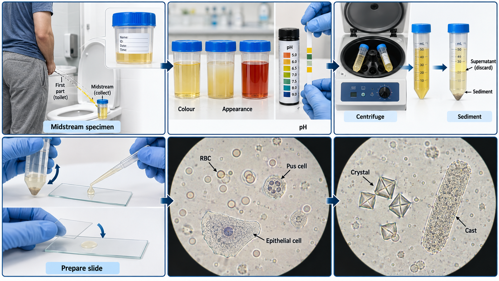
  2. Collect the specimen: Explain clean-catch midstream collection; the patient cleans the area as instructed, begins voiding, then collects the midstream portion without touching the inside of the container or lid.
  3. Inspect the urine: Assess the volume, colour and appearance. Note whether it is clear, cloudy, turbid or visibly blood-stained, and assess pH using the provided method.
  4. Prepare sediment: Transfer an appropriate aliquot to a centrifuge tube, balance the centrifuge correctly, centrifuge the sample, remove most supernatant and gently resuspend the remaining sediment.
  5. Prepare the slide: Place a drop of resuspended sediment onto a clean glass slide and apply a coverslip without introducing excessive bubbles.
  6. Examine systematically: Scan first under low power for casts, crystals and general distribution, then use higher power to identify and assess RBCs, pus cells and epithelial cells.
  7. Report and interpret: Record the physical findings and microscopic constituents systematically, identify pyuria or hematuria when present and state the overall interpretation.

🖼️ Visual Learning

VISUAL 1

Urine Collection, Sediment Preparation and Microscopic Examination
 

📝 Important Viva Questions

1. Why is a clean-catch midstream urine specimen preferred?

Answer: It reduces contamination from urethral, genital and skin flora and provides a more representative urine specimen.

2. What is pyuria?

Answer: Pyuria is an increased number of white blood cells or pus cells in urine and commonly indicates inflammation or infection within the urinary tract.

3. What does the presence of increased RBCs in urine indicate?

Answer: Increased urinary RBCs indicate hematuria and should be interpreted with the patient’s clinical findings and other urine abnormalities.

4. Why is urinary sediment examined first under low power and then under higher power?

Answer: Low power allows systematic scanning and recognition of larger structures such as casts and crystals, while higher power helps assess cells such as RBCs, pus cells and epithelial cells.

5. Which important constituents should you actively look for during urine microscopy?

Answer: Important constituents include RBCs, pus cells, epithelial cells, crystals and urinary casts.

Scroll to Top
💬 WhatsApp Support