FINAL YEAR MBBS • KMU CURRICULUM • AIM CLINICAL LEARNING
🩺 AIM LONG CASE 06
Pregnancy, Delivery & Puerperium — Integrated Maternal Assessment
Endocrine & Reproduction Module | A whole-patient journey through labour, birth and the early postnatal period.
2. Patient Scenario
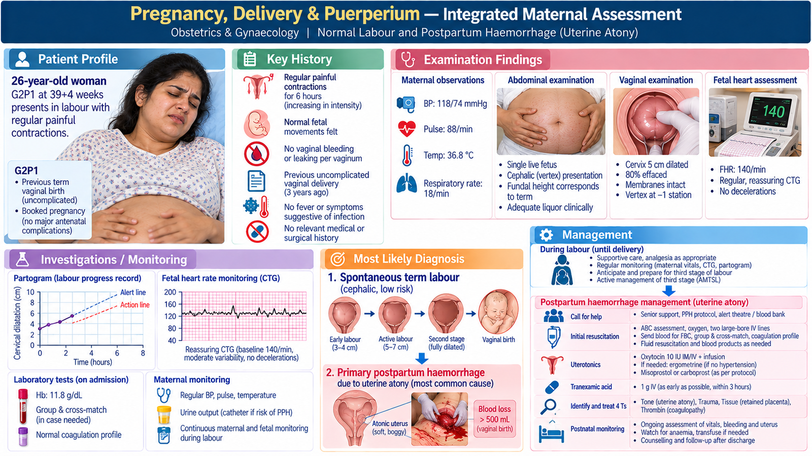
A 26-year-old woman, gravida 2 para 1, at 39 weeks and 4 days of gestation is admitted to the labour ward with painful regular uterine contractions for six hours. She reports that her baby has been moving normally. Her previous delivery was vaginal and uncomplicated. She is anxious about whether labour is progressing and asks about pain relief.
Initial clinical question: Is the mother in established labour, and are the mother and fetus currently well?
3. Student Task
Obtain a focused obstetric and medical history; assess maternal wellbeing, fetal wellbeing and the stage of labour; interpret serial labour observations; recognize indications for escalation or operative delivery; discuss safe delivery and third-stage management; identify postpartum haemorrhage and puerperal warning signs; and formulate a patient-centred postnatal plan.
4. Relevant History
Present illness and labour symptoms
- Contractions: Began six hours ago, became progressively stronger and more frequent; currently about three contractions per 10 minutes. Progressive painful contractions suggest labour but cervical change establishes its progress.
- Membranes and vaginal loss: No gush or persistent leakage of fluid; no vaginal bleeding beyond slight blood-stained mucus. Bleeding or abnormal liquor would change the risk assessment.
- Fetal movements: Normal by maternal report. Reduced movements require prompt fetal assessment.
- Associated concerns: No fever, persistent severe abdominal pain between contractions, headache, visual disturbance or breathlessness. These symptoms may indicate infection or another maternal complication.
Pregnancy and background history
- Antenatal course: Regular visits, routine screening reported as reassuring, an appropriately growing single fetus and no reported hypertensive or diabetic disorder. Verify the antenatal record rather than accepting recollection alone.
- Previous obstetric history: One term spontaneous vaginal birth with no postpartum haemorrhage. Prior vaginal delivery makes spontaneous birth more likely, but current assessment remains essential.
- Risk factors: Ask specifically about previous Caesarean section or uterine surgery, prior PPH, placenta praevia, multiple pregnancy, malpresentation and any known fetal concerns; these influence birth planning.
- Health and treatment: No chronic illness, regular medication other than antenatal supplements, allergies or relevant surgery. Review haemoglobin and blood group/Rh status from antenatal documentation.
- Preferences and support: She wishes to remain involved in decisions, would like a birth companion if permitted, and asks about available analgesia. Respectful communication is part of safe intrapartum care.
5. Physical Examination
- General and vital signs: Alert and comfortable between contractions; pulse 88/min, blood pressure 118/74 mmHg, temperature 36.8°C, respiratory rate 18/min. No pallor, dehydration or signs of acute distress.
- Abdominal assessment: Uterus corresponds to term; longitudinal lie, cephalic presentation; head partly engaged; contractions palpable, approximately three in 10 minutes; no abnormal tenderness between contractions.
- Fetal assessment: Baseline fetal heart rate around 140/min with reassuring findings on the initial assessment. Interpret subsequent monitoring in the context of maternal status and labour progress.
- Vaginal examination: With explanation, consent, privacy and appropriate asepsis, the cervix is 5 cm dilated, well effaced; membranes intact; vertex presenting; no palpable cord. Record cervical dilatation, station, position and membrane status. Avoid unnecessary repeated examinations.
Interpretation: This is established first-stage labour in a clinically stable woman with a cephalic fetus and no current indication for emergency delivery.
6. Clinical Problem Representation
A 26-year-old G2P1 at term has progressive regular contractions, a cervix dilated to 5 cm and a vertex-presenting fetus. Maternal observations and initial fetal assessment are reassuring, with no bleeding, membrane rupture or clinical infection. The immediate problem is to monitor established labour safely, determine progress and provide supportive care while preparing for delivery and postnatal observation.
7. Differential Diagnosis / Competing Clinical Assessments
The initial symptom of painful contractions requires distinction between:
- Established labour (most likely): Regular progressive contractions with significant cervical dilatation and effacement.
- Latent phase of labour: Contractions may be painful but cervical change is less advanced; serial assessment clarifies progression.
- False labour: Irregular contractions without progressive cervical change; inconsistent with the current examination.
- Obstructed or abnormally progressing labour: Not established at admission; consider later if dilatation or descent is inadequate, especially with poor descent, malposition or maternal/fetal concerns.
Key principle: The diagnosis and safety of labour depend on maternal-fetal status and observed progress, not contraction pain alone.
8. Investigations & Interpretation
Uncomplicated labour is primarily a clinical diagnosis. The most useful investigations and observations are those that alter management.
| Assessment / test | Reason | Case finding and interpretation |
|---|---|---|
| Serial maternal pulse, BP, temperature | Detect bleeding, hypertensive disease, infection or dehydration. | Initially normal; trend matters more than one value. |
| Fetal heart monitoring | Assess fetal wellbeing and identify concerns requiring review. | Initially reassuring. Continuous CTG if risk factors or abnormalities arise; interpretation includes baseline, variability, decelerations and clinical context. |
| Labour progress record and clinically indicated vaginal reassessment | Follow cervical change, descent, contractions and maternal/fetal status. | Cervix 5 cm at admission, later 8 cm after several hours, then fully dilated; progressive dilatation and descent support normal progress. |
| Antenatal blood count and blood group/Rh review | Identify anaemia and prepare for haemorrhage risk. | Documented haemoglobin 11.4 g/dL and blood group known; additional sampling depends on clinical risk and local protocol. |
| Bedside ultrasound when clinically indicated | Clarify uncertain presentation or fetal position. | Not routinely necessary here because cephalic presentation is clinically established. |
Subsequent progress and birth
Labour progresses to full dilatation with reassuring maternal and fetal observations. The fetal head descends and she gives birth vaginally to a vigorous term baby. The placenta is delivered complete after active management of the third stage. Soon afterward the midwife observes brisk vaginal bleeding and a soft, enlarged uterus.
Interpretation: This is an acute change after delivery. Assess and treat immediately for primary postpartum haemorrhage, with uterine atony as the leading cause.
9. Final Diagnosis
Term singleton pregnancy in spontaneous labour with normal progress and vaginal delivery, complicated by primary postpartum haemorrhage most consistent with uterine atony.
The brisk bleeding and poorly contracted uterus after birth make atony the most likely immediate mechanism. Placental tissue retention, genital tract trauma and coagulation problems must also be considered during assessment.
10. Management
A. Care during the first and second stages
- Explain findings and obtain informed consent for examinations and interventions; support hydration, comfort, positioning, bladder emptying and choice of labour analgesia.
- Document contractions, cervical change, descent, maternal observations and fetal wellbeing with an appropriate labour care record; reassess if progress slows or clinical status changes.
- Use shared decision-making if operative birth becomes necessary. Caesarean section may be required for persistent non-reassuring fetal status, obstruction or other circumstances where safe vaginal birth is unlikely. Operative vaginal birth requires full dilatation, an appropriately low engaged head, known position, suitable conditions and an experienced operator.
B. Delivery and third stage
- Provide skilled attendance during birth, ensure newborn assessment and support immediate maternal–infant contact if both are stable.
- Use active management of the third stage, including a prophylactic uterotonic according to local protocol, to reduce the risk of postpartum haemorrhage.
- Check placental completeness, assess uterine contraction and observe the volume and pattern of blood loss.
C. Immediate response to postpartum haemorrhage
- Call for help early: Alert the obstetric, anaesthetic, midwifery and blood bank teams; assess airway, breathing, circulation and consciousness.
- Resuscitate and monitor: Quantify blood loss, record frequent vitals, secure large-bore IV access, send blood for CBC, grouping/crossmatch and coagulation tests as indicated, begin appropriate IV fluids and prepare blood products when necessary.
- Treat suspected atony: Perform uterine massage, give oxytocin or another appropriate uterotonic according to protocol, empty the bladder and reassess uterine tone and bleeding.
- Give tranexamic acid promptly: Administer IV tranexamic acid as soon as possible and within three hours of birth in clinically diagnosed PPH, alongside standard care and according to local emergency protocol.
- Assess the cause: Consider the ‘4 Ts’ — tone, tissue, trauma and thrombin. Inspect for trauma and retained placenta/tissue; escalate promptly if bleeding continues.
- Escalate if ongoing: Senior-led measures may include uterine balloon tamponade, surgical treatment and transfusion as clinically required. Do not delay definitive control of bleeding.
D. Monitoring and early puerperium
Following control of bleeding, observe pulse, BP, uterine tone, vaginal loss and urine output; assess symptomatic anaemia and review follow-up haemoglobin as indicated. Check perineal injury, pain, bladder function, breastfeeding and emotional wellbeing. Arrange an individualized discharge and review plan after clinical stability is established.
11. Complications & Red Flags
- Immediate maternal: Ongoing haemorrhage, hypovolaemic shock, severe anaemia, transfusion requirement and organ hypoperfusion.
- Intrapartum: Abnormal fetal heart tracing, obstructed labour, maternal exhaustion, infection and uterine rupture in susceptible patients.
- Early puerperium: Secondary bleeding, endometritis, venous thromboembolism, hypertension-related complications and significant postnatal mental health symptoms.
Urgent escalation: Heavy or persistent bleeding, fainting, tachycardia or hypotension; fever with uterine tenderness or offensive lochia; severe headache or visual disturbance; chest pain or breathlessness; unilateral painful leg swelling; confusion, delusions or thoughts of harm to self or baby.
12. Patient Counselling & Follow-Up
Explain in accessible terms that labour progressed normally and birth was vaginal, but the uterus did not contract firmly immediately after delivery, causing heavier bleeding. Describe the treatment provided, her response and the need to monitor recovery. Invite questions, acknowledge anxiety and involve a chosen support person with permission.
- Recovery: Rest, nutrition, adequate fluids and iron therapy if anaemia is diagnosed; advise on gradual return to activity and review of persistent weakness or dizziness.
- Postnatal care: Discuss normal lochia versus excessive bleeding, perineal hygiene, breastfeeding support, urinary/bowel function and suitable contraception.
- Warning symptoms: Seek immediate medical attention for heavy bleeding, collapse, fever, severe abdominal pain, chest symptoms, severe headache or marked psychological deterioration.
- Next pregnancy: Document the PPH clearly and explain that it should be reported early in a future pregnancy because it affects risk planning.
- Continuity: Arrange postnatal review, assess emotional wellbeing and coordinate community, midwifery and obstetric support as needed.
13. Long Case Presentation to the Examiner
“This is a 26-year-old gravida 2 para 1 at 39 weeks and 4 days who presented with progressive regular painful uterine contractions. She had an uncomplicated antenatal course and previous vaginal delivery. At admission, she was stable, with a longitudinal lie, cephalic presentation, reassuring initial fetal assessment and a cervix 5 cm dilated. There was no vaginal bleeding, membrane rupture or evidence of infection. I assessed her as being in established first-stage labour.”
“She progressed to full dilatation and delivered vaginally. In the third stage/early postpartum period, she developed brisk vaginal bleeding associated with a soft uterus, suggesting primary postpartum haemorrhage due to uterine atony. Priorities are immediate team escalation, resuscitation, uterotonics, early tranexamic acid, assessment of other causes and close maternal monitoring, followed by postnatal counselling and follow-up.”
14. Examiner Discussion / Viva Preparation
Q1. How do you distinguish established labour from false labour?
Established labour involves regular progressive contractions with cervical effacement and dilatation; false labour does not produce progressive cervical change.
Q2. What observations determine whether labour is progressing safely?
Maternal vital signs and symptoms, contraction pattern, cervical dilatation, fetal descent and position, membrane/liquor status and fetal heart assessment, all interpreted together over time.
Q3. What findings would prompt consideration of operative delivery?
Persistent fetal compromise, failure of progress with suspected obstruction, significant maternal complications or another obstetric indication. Choice between operative vaginal birth and Caesarean depends on the stage of labour and whether prerequisites for safe assisted vaginal delivery are satisfied.
Q4. Why is active management of the third stage important?
It reduces the risk of postpartum haemorrhage by promoting uterine contraction after placental delivery.
Q5. What is the likely cause of heavy bleeding with a soft uterus immediately after birth?
Uterine atony. However, retained tissue, genital tract trauma and coagulopathy must be assessed rather than assuming atony is the only cause.
Q6. What are your first management priorities in primary PPH?
Call for help, assess and stabilize circulation, quantify blood loss, give uterotonics, administer early IV tranexamic acid, secure IV access and blood testing, and identify/treat the cause while escalating persistent bleeding.
Q7. Which findings indicate important puerperal complications after discharge?
Excessive bleeding, fever with uterine tenderness, chest pain or breathlessness, severe headache/visual symptoms, and severe mood or behavioural changes requiring urgent review.
Q8. What must be communicated before discharge after PPH?
What happened, treatment and recovery, anaemia assessment, warning symptoms, follow-up arrangements, emotional support and relevance to future pregnancies.
15. AIM Long Case — High-Yield Review
- Regular contractions alone do not confirm established labour; assess cervical progress.
- Review the antenatal record for previous uterine surgery, risk factors and relevant tests.
- Monitor maternal and fetal wellbeing throughout labour, not merely at admission.
- Interpret progress using serial cervical dilatation, descent and the clinical picture.
- Cephalic presentation with reassuring monitoring generally supports continued labour care.
- Operative vaginal delivery requires suitable prerequisites; it is not interchangeable with Caesarean section.
- Active third-stage management reduces PPH risk.
- Brisk postpartum bleeding with a boggy uterus strongly suggests atony.
- Use the 4 Ts to assess the causes of PPH: tone, tissue, trauma, thrombin.
- PPH treatment and resuscitation occur simultaneously; do not delay help or escalation.
- Postnatal review includes bleeding, infection, VTE warning signs, emotional wellbeing and breastfeeding.
- Professional case presentation prioritizes chronology, important findings and decisions.