🩺 Station 4 — Histology of Pituitary Gland
AIM OSPE/OSCE Lab — Practical Station | KMU Style | MBBS Practical + Viva
📋 Complete OSPE Station Content
OSPE Station Name
Histology of Pituitary Gland — Identification of Anterior and Posterior Pituitary
Learning Target
- Identify the pituitary gland under the microscope using key histological features.
- Differentiate anterior pituitary from posterior pituitary on the basis of cellularity, staining, and arrangement.
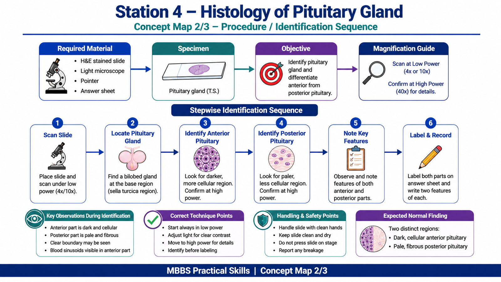
Required Material
- Prepared H&E slide of pituitary gland
- Light microscope
- Pointer / labeled digital slide image
- Student answer sheet
- Pencil
Student Task / Procedure
- Focus the slide under low power.
- Identify the pituitary gland.
- Locate the darker, more cellular area.
- Locate the paler, less cellular area.
- Label anterior pituitary and posterior pituitary.
- Write two identifying features of each part.
- Mention one functional significance of each part.
Observation / Identification Points
The student should identify:
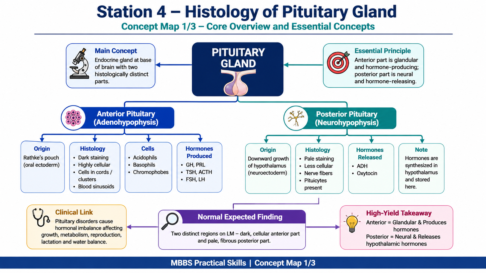
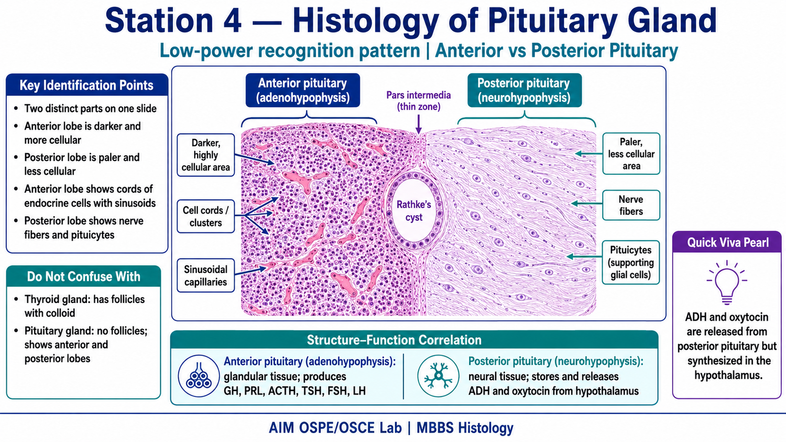
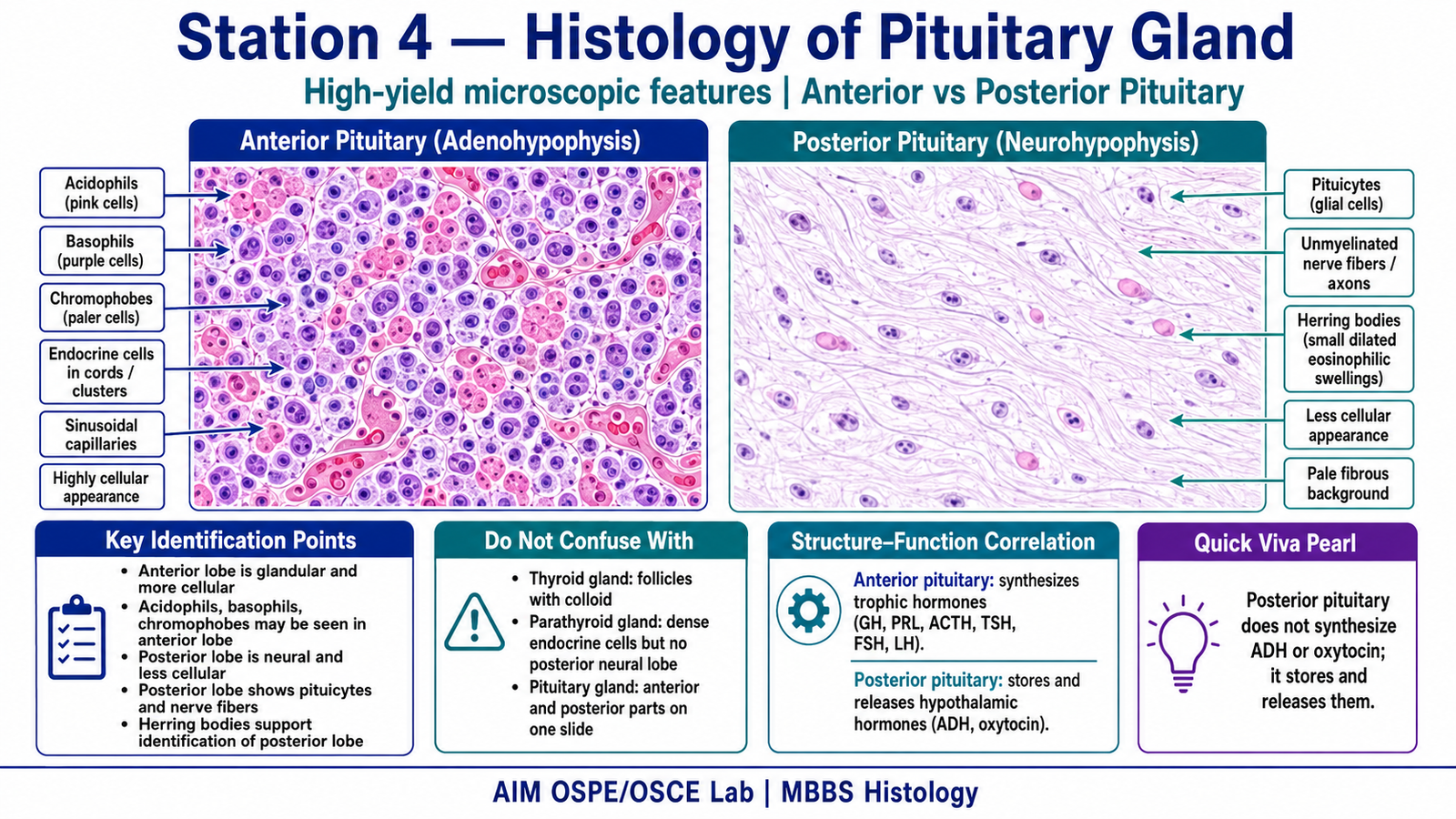
Anterior Pituitary / Adenohypophysis
- Darker staining area
- Highly cellular
- Cells arranged in cords or clusters
- Blood sinusoids present
- Acidophils, basophils, and chromophobes may be seen
- Produces trophic hormones such as GH, ACTH, TSH, FSH, LH, and prolactin
Posterior Pituitary / Neurohypophysis
- Paler staining area
- Less cellular
- Contains nerve fibers and pituicytes
- No obvious endocrine cell cords
- Stores and releases ADH and oxytocin produced by hypothalamic neurons
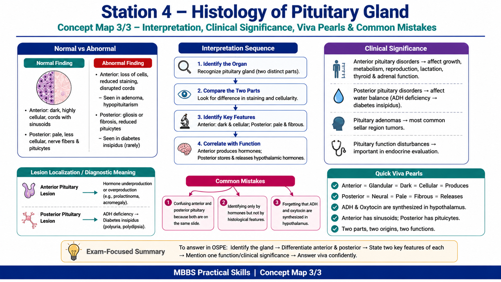
Result / Interpretation
The slide is identified as pituitary gland because it shows two histologically distinct regions: a dark, cellular anterior pituitary and a pale, fibrous posterior pituitary.
The anterior pituitary is glandular and hormone-producing, while the posterior pituitary is neural tissue that stores and releases hypothalamic hormones. Clinically, pituitary lesions may disturb growth, thyroid function, adrenal function, reproduction, lactation, and water balance.
Viva Questions
1. How do you identify the anterior pituitary under the microscope?
It is darker staining, highly cellular, and contains endocrine cells arranged in cords with blood sinusoids.
2. How do you identify the posterior pituitary?
It is paler, less cellular, and contains nerve fibers with pituicytes.
3. Which part of the pituitary produces hormones?
The anterior pituitary produces hormones such as GH, ACTH, TSH, FSH, LH, and prolactin.
4. Which hormones are released from the posterior pituitary?
ADH and oxytocin.
5. Where are ADH and oxytocin actually synthesized?
They are synthesized in the hypothalamus and released from the posterior pituitary.
Common Student Mistakes
- Confusing posterior pituitary with anterior pituitary because both are present on the same slide.
- Writing only hormone names without mentioning histological identification features.
- Forgetting that ADH and oxytocin are synthesized in the hypothalamus, not the posterior pituitary.
AIM Feedback
To improve, first identify the overall slide as pituitary gland, then compare the two regions. Remember: anterior pituitary is dark, cellular, and glandular, while posterior pituitary is pale, fibrous, and neural. In viva, always connect structure with function: anterior pituitary produces hormones; posterior pituitary releases hypothalamic hormones.
🖼️ Visual / Image Support
🧩 Concept Map / Interpretation Support