🩺 Station 5 — Histology of Adrenal Gland
AIM OSPE/OSCE Lab — Practical Station | KMU Style | MBBS Practical + Viva
📋 Complete OSPE Station Content
OSPE Station Name
Station 6 — Histology of Adrenal Gland
Learning Target
By the end of this station, the student should be able to:
- Identify the adrenal gland under the microscope and differentiate adrenal cortex from adrenal medulla.
- Recognize the adrenal cortical zones and relate their structure to basic hormone secretion.
Required Material
- Prepared histology slide of adrenal gland
- Light microscope
- Pointer or marked field image
- Answer sheet / LMS response box
- Labeled reference image for examiner use
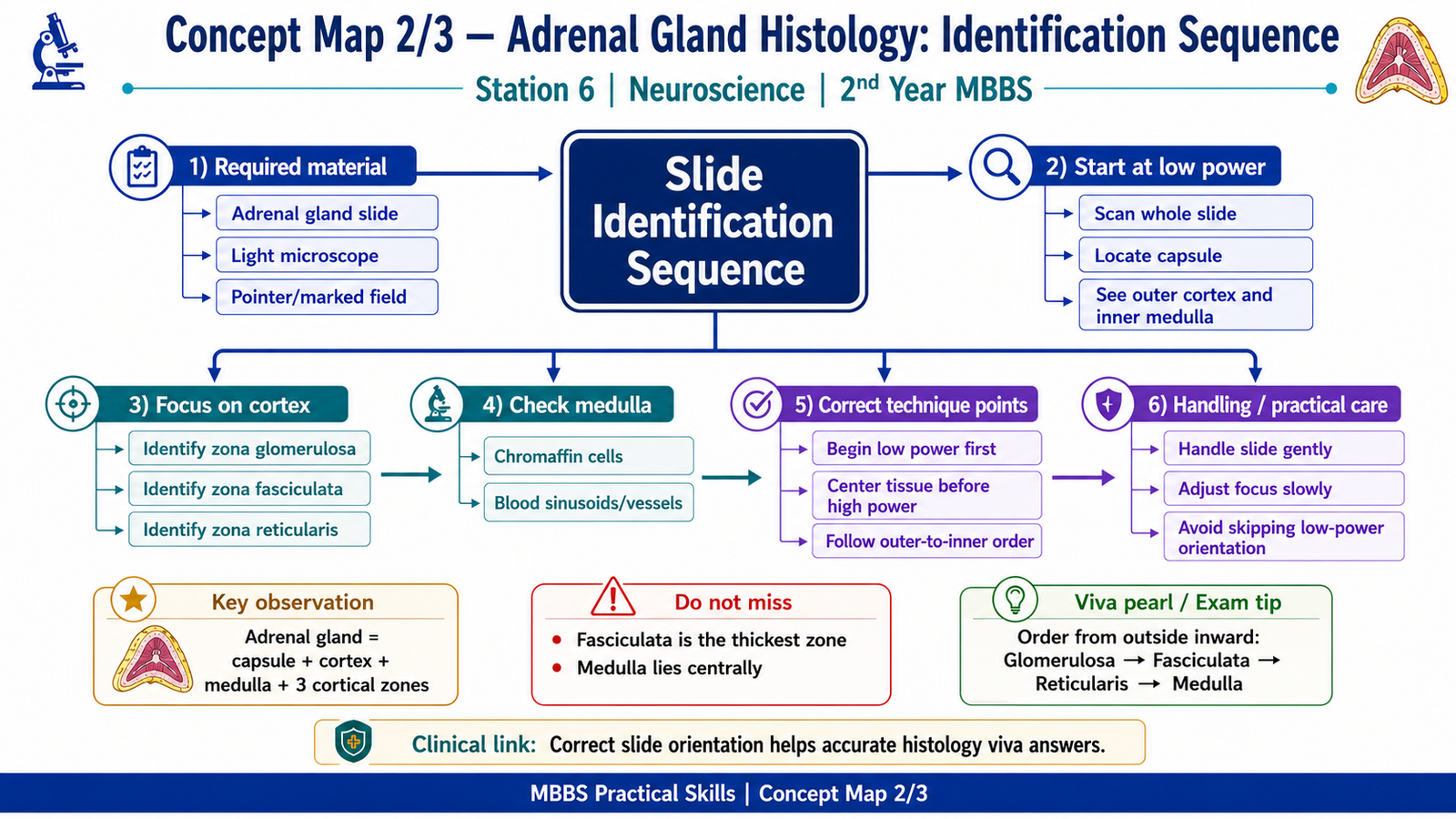
Student Task / Procedure
- Observe the given histology slide under low power.
- Identify the organ shown in the slide.
- Identify the outer cortex and inner medulla.
- Name the three zones of the adrenal cortex.
- State one hormone secreted by each cortical zone.
- Answer the viva questions asked by the examiner.
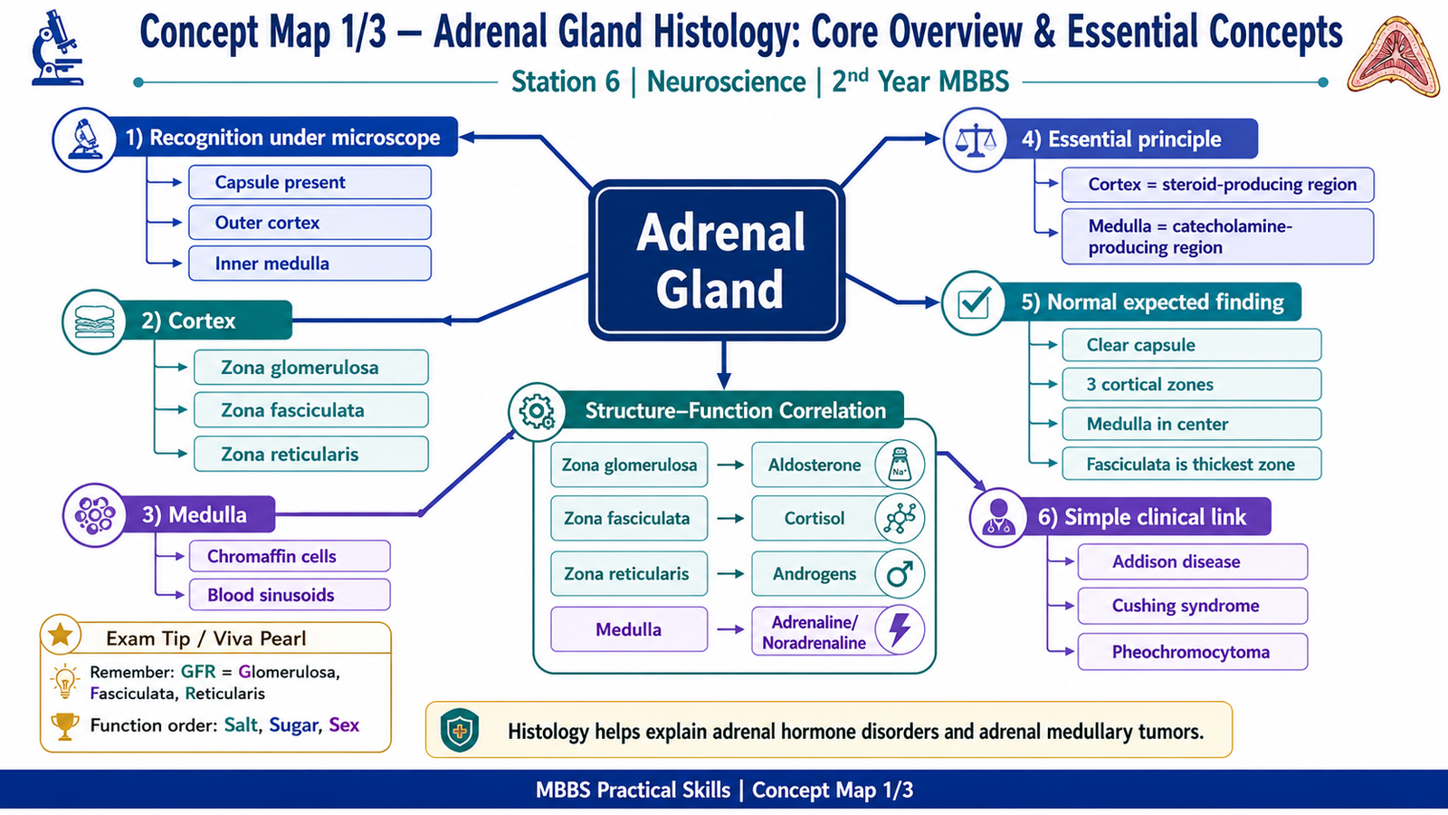
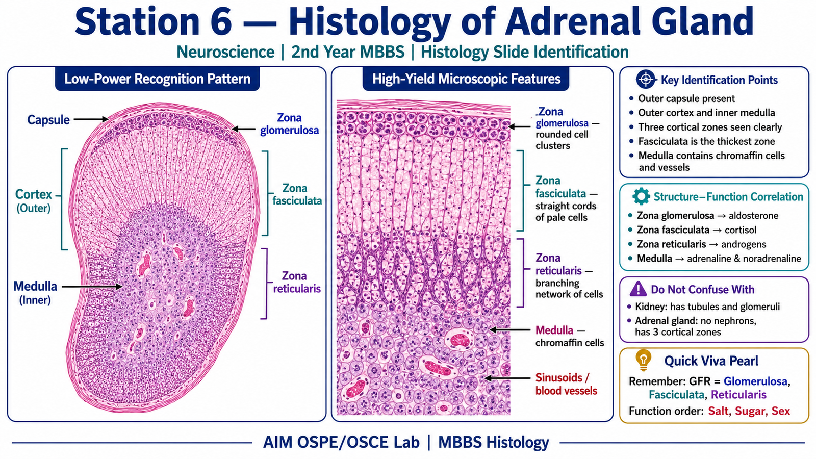
Observation / Identification Points
The student should identify:
- Adrenal gland as an endocrine gland with cortex and medulla.
- Capsule present at the outer surface.
- Adrenal cortex as the outer, darker-staining region.
- Adrenal medulla as the central, lighter-staining region.
- Zona glomerulosa as the thin outer cortical zone with cells arranged in rounded clusters.
- Zona fasciculata as the thick middle cortical zone with cells arranged in straight cords.
- Zona reticularis as the inner cortical zone with irregular anastomosing cords.
- Medulla containing chromaffin cells and blood sinusoids.
Result / Interpretation
The slide shows the adrenal gland, which has two functionally different parts:
- Adrenal cortex produces steroid hormones.
- Zona glomerulosa → mineralocorticoids, mainly aldosterone
- Zona fasciculata → glucocorticoids, mainly cortisol
- Zona reticularis → weak androgens
- Adrenal medulla produces catecholamines, mainly adrenaline and noradrenaline.
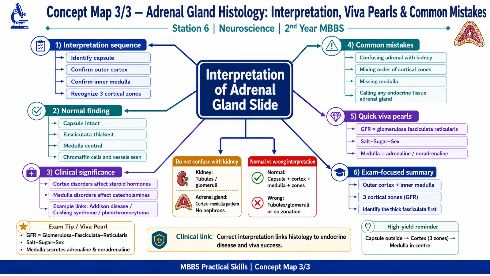
Clinical significance: Correct identification of adrenal cortical zones helps students understand disorders such as Cushing syndrome, Addison disease, hyperaldosteronism, and adrenal medullary tumors such as pheochromocytoma.
Viva Questions
1. How do you identify adrenal gland histologically?
Adrenal gland is identified by the presence of an outer cortex and inner medulla, with cortical zones arranged from outside inward.
2. Name the three zones of adrenal cortex from outside inward.
Zona glomerulosa, zona fasciculata, and zona reticularis.
3. Which cortical zone secretes aldosterone?
Zona glomerulosa secretes aldosterone.
4. Which adrenal cortical zone is the thickest?
Zona fasciculata is the thickest zone.
5. What does the adrenal medulla secrete?
The adrenal medulla secretes catecholamines, mainly adrenaline and noradrenaline.
Common Student Mistakes
- Confusing adrenal cortex with adrenal medulla.
- Forgetting the correct order of cortical zones.
- Mixing up hormones secreted by different adrenal cortical zones.
AIM Feedback
You correctly identify the adrenal gland when you first locate the outer cortex and inner medulla. After that, focus on the cortex from outside inward: glomerulosa, fasciculata, reticularis. Remember the simple functional pattern: salt, sugar, sex — glomerulosa controls salt through aldosterone, fasciculata controls sugar/stress through cortisol, and reticularis produces weak sex steroids.
🖼️ Visual / Image Support
🧩 Concept Map / Interpretation Support