
Rapid Revision • Practical Skills • Viva
🎯 Candidate Task
Examine this patient’s ocular alignment and extraocular movements systematically. Demonstrate the major directions of gaze and report any abnormal ocular-motility finding you identify.
⚡ Model Station Answer
- The eyes are inspected for alignment in the primary position.
- I assess conjugate extraocular movements through the major directions of gaze using an H-pattern.
- I look for restriction or asymmetry of movement and ask about diplopia during testing.
- I also observe for nystagmus in the different gaze positions.
- If abnormal, I would state the affected eye and the specific direction in which movement is limited.
🔎 How to Perform / Approach
- Prepare the patient. Perform hand hygiene, introduce yourself, obtain consent and seat the patient upright at eye level.
- Inspect primary gaze. Ask the patient to look straight ahead and compare the position and alignment of both eyes.
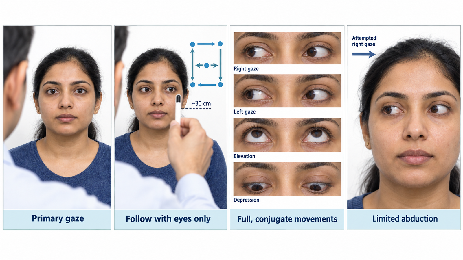
- Position the target. Hold your finger or a small target about 30 cm in front of the patient’s eyes and ask them to keep their head still.
- Test ocular movements. Ask the patient to follow the target with their eyes only while moving it slowly through a large H-pattern to assess the major directions of gaze.
- Observe carefully. Compare both eyes for conjugate movement, asymmetry, restriction or failure of an eye to reach a particular gaze position; ask whether diplopia or pain occurs.
- Check abnormal movements. Observe for nystagmus, particularly when the eyes are held briefly in the lateral gaze positions.
- Report the finding. State whether movements are full and conjugate or specify the affected eye and direction of restricted movement.
🖼️ Visual Learning
Systematic Extraocular Movement Examination
🎥 Practical Demonstration
📝 Important Viva Questions
Answer: It allows systematic assessment of the major directions of gaze and helps reveal restriction or asymmetry of ocular movement.
Answer: Cranial nerves III, IV and VI: the oculomotor, trochlear and abducens nerves.
Answer: The lateral rectus muscle, supplied by the abducens nerve (CN VI).
Answer: Ask the patient to report any double vision or pain while following the target.
Answer: State which eye is affected and the exact direction in which movement is restricted; also mention associated diplopia or nystagmus if present.