
Station 07 — Interpretation of Common Ophthalmic Investigations
Rapid Revision • Practical Skills • Viva
🎯 Candidate Task
You are shown a series of common ophthalmic investigation outputs. Identify each investigation, describe the important information or abnormality shown, and state its principal clinical use or interpretation.
⚡ Model Station Answer
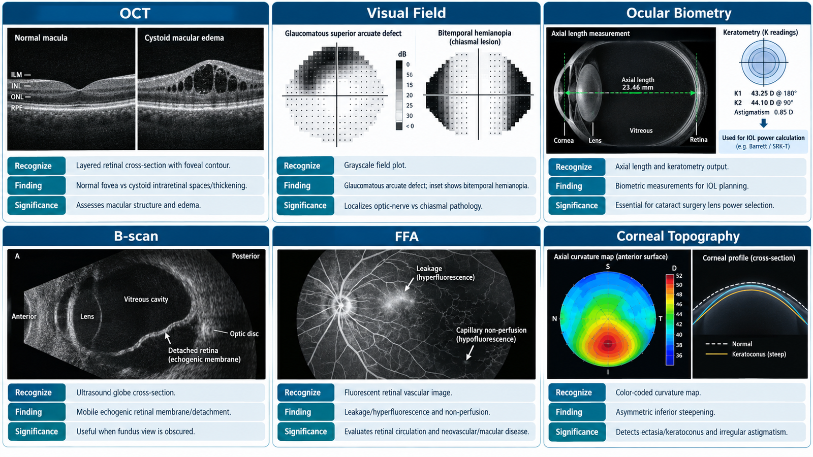
- OCT: provides cross-sectional imaging of retinal layers and can assess macular structure and retinal nerve-fibre layer thickness.
- Visual field: I would identify the pattern of field loss, such as a nasal step/arcuate defect, bitemporal loss or a homonymous defect, and relate it to the likely site of disease.
- Ocular biometry: measures ocular parameters, especially axial length and corneal power, used principally for intraocular-lens power calculation.
- B-scan ultrasonography: demonstrates posterior-segment structures when direct visualization is limited and may show abnormalities such as retinal detachment or vitreous echoes.
- FFA and corneal topography: FFA evaluates retinal vascular filling, leakage and non-perfusion; corneal topography maps corneal curvature and helps detect irregular astigmatism or keratoconus.
🔎 How to Perform / Approach
🖼️ Visual Learning
Recognition of Common Ophthalmic Investigation Outputs
📝 Important Viva Questions
Answer: OCT provides high-resolution cross-sectional imaging of ocular tissues, particularly retinal layers, macular structure and retinal nerve-fibre layer thickness.
Answer: Typical glaucomatous defects include a nasal step and arcuate scotoma corresponding to retinal nerve-fibre bundle loss.
Answer: It measures ocular parameters such as axial length and corneal power and is principally used to calculate intraocular-lens power before cataract surgery.
Answer: It is useful for assessing the posterior segment when the ocular media are opaque, for example with dense cataract or vitreous haemorrhage.
Answer: FFA assesses retinal vascular circulation, leakage and non-perfusion, whereas corneal topography maps corneal curvature and identifies abnormalities such as irregular astigmatism or keratoconus.