
Course Content
👁️ Station 1 — Ophthalmic History Taking
• Take a structured history from a patient presenting with an ocular complaint. • Identify the chief ocular complaint and its duration. • Explore relevant associated ocular symptoms. • Obtain relevant previous ocular, medical and treatment history. • Present the important findings of the ocular history in a concise clinical format.
0/1
🔤 Station 2 — Visual Acuity Examination
• Correctly position the patient and visual-acuity chart. • Assess distance visual acuity in each eye separately. • Record visual acuity correctly. • Use pinhole assessment where appropriate. • Interpret an abnormal visual-acuity result.
0/1
🔦 Station 3 — Pupillary Examination
• Inspect pupil size, shape and symmetry. • Assess direct pupillary light reflex. • Assess consensual pupillary light reflex. • Assess the pupillary response systematically. • Recognize an abnormal pupillary response.
0/1
👀 Station 4 — Visual Field Examination by Confrontation
• Position the patient correctly for confrontation testing. • Examine each eye separately. • Assess the major visual-field quadrants systematically. • Compare the patient's visual field with the examiner's field. • Identify the presence of a gross visual-field defect.
0/1
🔬 Station 5 — Slit-Lamp and Anterior Segment Examination
Integrated KMU activities: 5. Slit-Lamp Examination 6. Anterior Segment Examination Learning Objectives The student should be able to: • Identify the major parts/components of a slit lamp. • Explain the basic purpose of slit-lamp examination. • Position the patient appropriately. • Examine the anterior segment systematically using the slit lamp. • Identify important anterior-segment structures during examination.
0/1
🩺 Station 6 — Direct Ophthalmoscopy and Fundus Examination
• Demonstrate correct handling of the direct ophthalmoscope. • Position themselves and the patient appropriately. • Perform direct ophthalmoscopy systematically. • Visualize the optic disc and retinal background. • Describe important fundus findings seen during examination.
0/1
📊 Station 7 — Interpretation of Common Ophthalmic Investigations
The student should be able to: • Identify a presented ophthalmic investigation. • Describe the basic information provided by OCT. • Interpret common visual-field findings. • Recognize the role/output of ocular biometry. • Interpret basic B-scan findings. • Recognize FFA and corneal-topography images and state their clinical use.
0/1
👁️ Station 8 — Ptosis Examination
• Inspect a patient with suspected ptosis. • Compare eyelid position between both eyes. • Perform a systematic clinical assessment of ptosis. • Identify important abnormal findings. • Record and communicate the findings appropriately.
0/1
👓 Station 9 — Examination of Common Lid Abnormalities
• Examine the eyelids systematically. • Recognize ectropion. • Recognize entropion. • Identify chalazion. • Identify stye. • Differentiate common lid abnormalities from one another on clinical examination or photographs.
0/1
👁️ Station 10 — Proptosis Examination
• Recognize the presence of proptosis. • Compare both eyes for symmetry. • Perform a structured ocular examination in a patient with proptosis. • Assess relevant associated ocular findings. • Present the examination findings systematically.
0/1
💧 Station 11 — Topical Anaesthesia, Fluorescein Staining and Corneal Assessment
• Demonstrate appropriate use of topical ocular anaesthesia. • Demonstrate fluorescein staining of the ocular surface. • Examine the stained cornea appropriately. • Identify a superficial corneal abnormality/foreign body when presented. • Explain the purpose of corneal scraping in suspected corneal infection.
0/1
💦 Station 12 — Lacrimal Regurgitation Test
• Explain the purpose of the lacrimal regurgitation test. • Position the patient appropriately. • Perform the test correctly. • Observe for reflux from the lacrimal puncta. • Interpret a positive or negative result.
0/1
🚨 Station 13 — Ocular Trauma and Chemical Eye Injury First Aid
• Perform an initial assessment of an ocular-trauma patient. • Identify the need for urgent eye first aid. • Demonstrate immediate management of chemical eye injury. • Perform appropriate eye irrigation/eye wash. • Explain essential precautions during initial ocular-trauma management.
0/1
🔍 Station 14 — Optic Disc Examination: Normal, Glaucomatous and Swollen Disc
• Identify and describe a normal optic disc. • Examine or interpret the optic disc systematically. • Recognize features of a glaucomatous disc. • Recognize a swollen optic disc. • Differentiate a normal disc from major abnormal disc appearances.
0/1
🩸 Station 15 — Retinal Lesions and Retinal Vascular Disease
• Inspect a retinal photograph/fundus image systematically. • Detect common retinal abnormalities. • Recognize common retinal vascular abnormalities. • Differentiate common retinal vascular conditions from one another. • Describe the important visual findings presented.
0/1
⚠️ Station 16 — Recognition of Retinal Detachment
• Recognize retinal detachment in a clinical image. • Describe the abnormal retinal appearance. • Distinguish detached retina from a normal retinal appearance. • Identify retinal detachment as a significant cause of visual loss. • Communicate the finding clearly in an OSCE setting.
0/1
📈 Station 17 — Tonometry and Glaucoma Assessment
• Identify the Goldmann applanation tonometer. • Explain the purpose of tonometry. • Describe the basic method of Goldmann tonometry. • Interpret an intraocular-pressure reading in the clinical context. • Relate tonometry to assessment of glaucoma.
0/1
🌫️ Station 18 — Cataract Examination and Recognition
• Perform a basic ocular assessment for cataract. • Recognize the presence of cataract on ocular examination. • Describe the abnormal lens appearance. • Differentiate a clear lens from a cataractous lens. • Recognize adult and congenital cataract when presented clinically or visually.
0/1
↔️ Station 19 — Extraocular Movements and Ocular Motor Examination
• Inspect ocular alignment. • Examine extraocular movements systematically. • Test the major directions of gaze. • Identify limitation of ocular movement. • Recognize an abnormal ocular-motility pattern during examination.
0/1
🔁 Station 20 — Squint Examination: Cover Tests and Squint Pattern
• Assess ocular alignment. • Perform the cover test. • Perform the uncover test. • Perform the alternate-cover test. • Identify the presence of manifest or latent deviation from the examination. • Differentiate an esotropia pattern from an exotropia pattern.
0/1
AIM OSPE/OSCE Lab — Ophthalmology Module | 4th Year MBBS

AIM OSCE / OSPE • MBBS • 4th Year • Eye
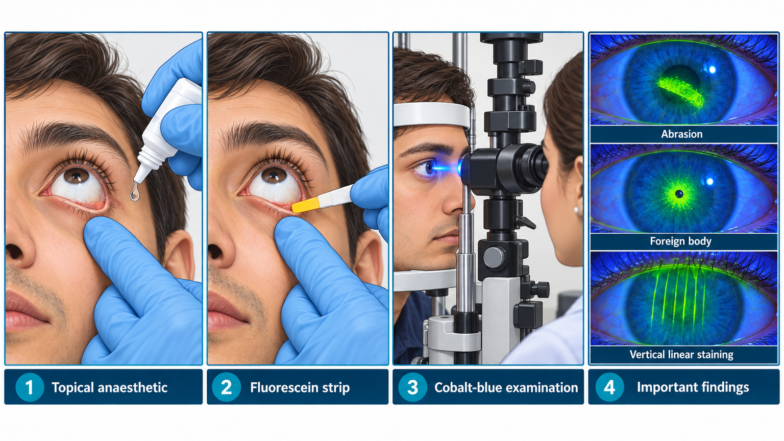
Station 11 — Topical Anaesthesia, Fluorescein Staining and Corneal Assessment
Rapid Revision • Practical Skills • Viva

🎯 Candidate Task

Demonstrate the appropriate use of topical ocular anaesthesia and fluorescein staining, then examine the cornea and identify any superficial abnormality presented. Explain the purpose of corneal scraping when corneal infection is suspected.

⚡ Model Station Answer

  • I would explain the procedure, obtain consent and check for relevant allergy before instilling topical ocular anaesthetic safely.
  • I would apply fluorescein to the lower conjunctival fornix without touching the cornea.
  • I would examine the cornea under magnification using cobalt-blue illumination.
  • Areas of epithelial loss stain bright green; I would describe the site, size and pattern and look for a superficial foreign body.
  • Corneal scraping in suspected microbial keratitis obtains material for microscopy and culture to identify the organism and guide antimicrobial treatment.

🔎 How to Perform / Approach

1
Prepare. Perform hand hygiene, introduce yourself, confirm the patient and explain the examination.
2
Position safely. Seat the patient comfortably at the slit lamp or appropriate examination device and obtain consent.
3
Instil topical anaesthetic. Ask the patient to look upward, gently lower the lower lid and place the drop into the lower fornix without allowing the bottle tip to touch the eye or lashes.
4
Apply fluorescein. Moisten a fluorescein strip if required and gently touch it to the lower conjunctival fornix, avoiding direct contact with the cornea.
5
Examine with blue light. Ask the patient to blink, then systematically inspect the corneal surface using cobalt-blue illumination and magnification.
6
State the finding. Identify any bright-green epithelial defect and describe its location, size and pattern; inspect for a visible superficial foreign body.
7
Complete. Thank the patient, dispose of used material safely and state that suspected infectious corneal ulceration may require corneal scraping for microbiological investigation.
Exam point: Never rub the fluorescein strip directly across the cornea. Vertical linear staining should prompt consideration of a foreign body beneath the upper eyelid.

🖼️ Visual Learning

VISUAL 1

Topical Anaesthesia, Fluorescein Staining and Corneal Examination


🎥 Practical Demonstration

Anterior Segment Eye Examination — Geeky Medics
Demonstrates practical anterior-segment examination, fluorescein application and examination using blue illumination.


▶ Watch Practical Demonstration

📝 Important Viva Questions

1. Why is topical ocular anaesthetic used in this station?
Answer: It reduces ocular surface discomfort and facilitates careful examination in a painful eye.
2. Where should a fluorescein strip be applied?
Answer: It should be gently applied to the lower conjunctival fornix rather than rubbed directly on the cornea.
3. What does bright green fluorescein staining of the cornea indicate?
Answer: It indicates a defect in the corneal epithelium, such as an abrasion or ulcerated epithelial surface.
4. What may vertical linear fluorescein staining suggest?
Answer: It may suggest a foreign body trapped beneath the upper eyelid repeatedly scratching the cornea.
5. Why is corneal scraping performed in suspected corneal infection?
Answer: It provides material for microbiological examination and culture so the infecting organism can be identified and antimicrobial therapy appropriately guided.
Scroll to Top
💬 WhatsApp Support