
Course Content
👁️ Station 1 — Ophthalmic History Taking
• Take a structured history from a patient presenting with an ocular complaint. • Identify the chief ocular complaint and its duration. • Explore relevant associated ocular symptoms. • Obtain relevant previous ocular, medical and treatment history. • Present the important findings of the ocular history in a concise clinical format.
0/1
🔤 Station 2 — Visual Acuity Examination
• Correctly position the patient and visual-acuity chart. • Assess distance visual acuity in each eye separately. • Record visual acuity correctly. • Use pinhole assessment where appropriate. • Interpret an abnormal visual-acuity result.
0/1
🔦 Station 3 — Pupillary Examination
• Inspect pupil size, shape and symmetry. • Assess direct pupillary light reflex. • Assess consensual pupillary light reflex. • Assess the pupillary response systematically. • Recognize an abnormal pupillary response.
0/1
👀 Station 4 — Visual Field Examination by Confrontation
• Position the patient correctly for confrontation testing. • Examine each eye separately. • Assess the major visual-field quadrants systematically. • Compare the patient's visual field with the examiner's field. • Identify the presence of a gross visual-field defect.
0/1
🔬 Station 5 — Slit-Lamp and Anterior Segment Examination
Integrated KMU activities: 5. Slit-Lamp Examination 6. Anterior Segment Examination Learning Objectives The student should be able to: • Identify the major parts/components of a slit lamp. • Explain the basic purpose of slit-lamp examination. • Position the patient appropriately. • Examine the anterior segment systematically using the slit lamp. • Identify important anterior-segment structures during examination.
0/1
🩺 Station 6 — Direct Ophthalmoscopy and Fundus Examination
• Demonstrate correct handling of the direct ophthalmoscope. • Position themselves and the patient appropriately. • Perform direct ophthalmoscopy systematically. • Visualize the optic disc and retinal background. • Describe important fundus findings seen during examination.
0/1
📊 Station 7 — Interpretation of Common Ophthalmic Investigations
The student should be able to: • Identify a presented ophthalmic investigation. • Describe the basic information provided by OCT. • Interpret common visual-field findings. • Recognize the role/output of ocular biometry. • Interpret basic B-scan findings. • Recognize FFA and corneal-topography images and state their clinical use.
0/1
👁️ Station 8 — Ptosis Examination
• Inspect a patient with suspected ptosis. • Compare eyelid position between both eyes. • Perform a systematic clinical assessment of ptosis. • Identify important abnormal findings. • Record and communicate the findings appropriately.
0/1
👓 Station 9 — Examination of Common Lid Abnormalities
• Examine the eyelids systematically. • Recognize ectropion. • Recognize entropion. • Identify chalazion. • Identify stye. • Differentiate common lid abnormalities from one another on clinical examination or photographs.
0/1
👁️ Station 10 — Proptosis Examination
• Recognize the presence of proptosis. • Compare both eyes for symmetry. • Perform a structured ocular examination in a patient with proptosis. • Assess relevant associated ocular findings. • Present the examination findings systematically.
0/1
💧 Station 11 — Topical Anaesthesia, Fluorescein Staining and Corneal Assessment
• Demonstrate appropriate use of topical ocular anaesthesia. • Demonstrate fluorescein staining of the ocular surface. • Examine the stained cornea appropriately. • Identify a superficial corneal abnormality/foreign body when presented. • Explain the purpose of corneal scraping in suspected corneal infection.
0/1
💦 Station 12 — Lacrimal Regurgitation Test
• Explain the purpose of the lacrimal regurgitation test. • Position the patient appropriately. • Perform the test correctly. • Observe for reflux from the lacrimal puncta. • Interpret a positive or negative result.
0/1
🚨 Station 13 — Ocular Trauma and Chemical Eye Injury First Aid
• Perform an initial assessment of an ocular-trauma patient. • Identify the need for urgent eye first aid. • Demonstrate immediate management of chemical eye injury. • Perform appropriate eye irrigation/eye wash. • Explain essential precautions during initial ocular-trauma management.
0/1
🔍 Station 14 — Optic Disc Examination: Normal, Glaucomatous and Swollen Disc
• Identify and describe a normal optic disc. • Examine or interpret the optic disc systematically. • Recognize features of a glaucomatous disc. • Recognize a swollen optic disc. • Differentiate a normal disc from major abnormal disc appearances.
0/1
🩸 Station 15 — Retinal Lesions and Retinal Vascular Disease
• Inspect a retinal photograph/fundus image systematically. • Detect common retinal abnormalities. • Recognize common retinal vascular abnormalities. • Differentiate common retinal vascular conditions from one another. • Describe the important visual findings presented.
0/1
⚠️ Station 16 — Recognition of Retinal Detachment
• Recognize retinal detachment in a clinical image. • Describe the abnormal retinal appearance. • Distinguish detached retina from a normal retinal appearance. • Identify retinal detachment as a significant cause of visual loss. • Communicate the finding clearly in an OSCE setting.
0/1
📈 Station 17 — Tonometry and Glaucoma Assessment
• Identify the Goldmann applanation tonometer. • Explain the purpose of tonometry. • Describe the basic method of Goldmann tonometry. • Interpret an intraocular-pressure reading in the clinical context. • Relate tonometry to assessment of glaucoma.
0/1
🌫️ Station 18 — Cataract Examination and Recognition
• Perform a basic ocular assessment for cataract. • Recognize the presence of cataract on ocular examination. • Describe the abnormal lens appearance. • Differentiate a clear lens from a cataractous lens. • Recognize adult and congenital cataract when presented clinically or visually.
0/1
↔️ Station 19 — Extraocular Movements and Ocular Motor Examination
• Inspect ocular alignment. • Examine extraocular movements systematically. • Test the major directions of gaze. • Identify limitation of ocular movement. • Recognize an abnormal ocular-motility pattern during examination.
0/1
🔁 Station 20 — Squint Examination: Cover Tests and Squint Pattern
• Assess ocular alignment. • Perform the cover test. • Perform the uncover test. • Perform the alternate-cover test. • Identify the presence of manifest or latent deviation from the examination. • Differentiate an esotropia pattern from an exotropia pattern.
0/1
AIM OSPE/OSCE Lab — Ophthalmology Module | 4th Year MBBS
AIM OSCE / OSPE • MBBS

Station 12 — Lacrimal Regurgitation Test

4th Year • Ophthalmology • Rapid Revision • Practical Skills • Viva

🎯 Candidate Task

Demonstrate the lacrimal regurgitation test on this patient and state how you would interpret the result. Explain briefly what the test assesses.

⚡ Model Station Answer

Purpose: I would assess the lacrimal drainage system for regurgitation suggestive of nasolacrimal duct obstruction.

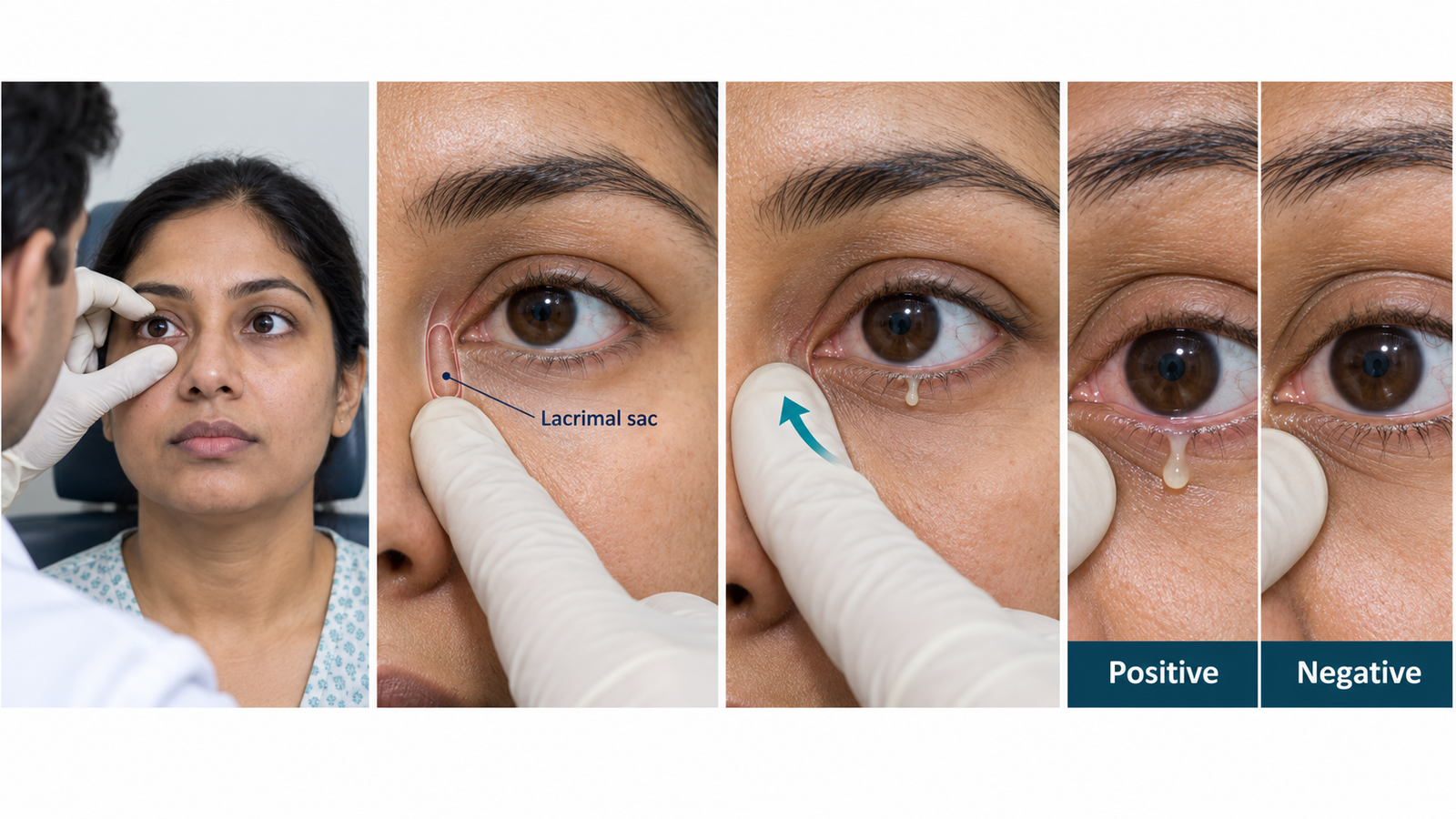
Technique: I would apply gentle, firm pressure over the lacrimal sac area and observe the upper and lower puncta.

Positive result: Reflux of clear, mucoid or mucopurulent material through a punctum is a positive test and supports distal lacrimal drainage obstruction.

Negative result: No reflux is seen on pressure; however, a negative test alone does not exclude partial or functional obstruction.

🔎 How to Perform / Approach

  1. Prepare: Introduce yourself, confirm the patient, explain the test, obtain consent and perform hand hygiene.
  2. Position: Seat the patient comfortably at your eye level with good illumination and expose the medial canthal region.
  3. Inspect: Look for watering, discharge, swelling around the lacrimal sac and the appearance of the puncta.
  4. Locate the sac: Trace the inferior orbital margin medially toward the anterior lacrimal crest; the lacrimal sac lies immediately behind this region near the medial canthus.
  5. Compress: Place the index finger over the lacrimal sac area and apply gentle but firm pressure in an upward and medial direction.
  6. Observe and interpret: Watch both puncta for reflux and note whether any expressed material is clear, mucoid or mucopurulent. State whether the test is positive or negative.
Exam point: The examiner should clearly see that you are pressing over the lacrimal sac region while simultaneously observing the puncta for reflux.

🖼️ Visual Learning

VISUAL 1

Lacrimal Regurgitation Test — ROPLAS Technique
 

🎥 Practical Demonstration

Regurgitation Test (Lacrimal System Examination) — practical demonstration suitable for ophthalmology learners.

▶ Watch Practical Demonstration

📝 Important Viva Questions

1. What is the purpose of the lacrimal regurgitation test?

Answer: It is a simple clinical test used to detect regurgitation from the lacrimal sac and screen for obstruction of the lacrimal drainage pathway.

2. Where do you apply pressure during the test?

Answer: Pressure is applied over the lacrimal sac area behind the anterior lacrimal crest near the medial canthus.

3. What constitutes a positive lacrimal regurgitation test?

Answer: Reflux of fluid, mucus or mucopurulent material through one or both lacrimal puncta when the sac is compressed.

4. What does a positive test commonly suggest?

Answer: It supports nasolacrimal duct obstruction and may be seen in chronic dacryocystitis.

5. Does a negative ROPLAS completely exclude lacrimal drainage obstruction?

Answer: No. Absence of regurgitation does not completely exclude partial obstruction or functional impairment of lacrimal drainage.

Scroll to Top
💬 WhatsApp Support