FINAL YEAR MBBS | KMU CURRICULUM | AIM CLINICAL LEARNING
🩺 AIM LONG CASE 03
Obstructive Jaundice — Suspected Pancreatic Malignancy
GIT Module | Surgery | Related AIM Topic 3
An integrated clinical approach to painless progressive jaundice, biliary obstruction, suspected carcinoma of the pancreatic head, management and patient-centred counselling.
1. Long Case Title
Long Case 03 — Evaluation of a Patient with Obstructive Jaundice and Suspected Pancreatic Malignancy
Discipline: General Surgery
Clinical focus: Assessment of jaundice, differentiation of its causes, interpretation of hepatobiliary investigations, assessment of suspected pancreatic head carcinoma, management of biliary obstruction and counselling.
2. Patient Scenario
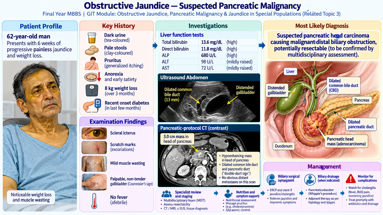
A 62-year-old man presents to the surgical outpatient department with yellow discoloration of his eyes and skin for six weeks. The discoloration began gradually and has progressively increased.
He also reports reduced appetite, generalized weakness and unintentional weight loss over the preceding three months.
He has no history of previous jaundice or known chronic liver disease.
The student is asked to assess this patient and discuss the probable diagnosis and management.
3. Student Task
After completing this long case, the student should be able to:
- Take a comprehensive, clinically focused history of jaundice.
- Perform an appropriate general and abdominal examination.
- Differentiate obstructive jaundice from hepatocellular and hemolytic causes.
- Identify features suggesting malignant biliary obstruction.
- Formulate important differential diagnoses.
- Select and interpret appropriate laboratory and imaging investigations.
- Assess suspected carcinoma of the head of the pancreas and discuss disease extent.
- Explain initial management, biliary decompression and possible treatment options.
- Recognize important complications requiring urgent treatment.
- Provide sensitive, patient-centred counselling and follow-up advice.
4. Relevant History
A. Presenting Complaints
- Yellow discoloration of eyes and skin — six weeks.
- Dark urine and pale stools — approximately five weeks.
- Generalized itching — four weeks.
- Reduced appetite and weight loss — three months.
B. History of Presenting Illness
Progression of jaundice: The patient describes gradual onset and progressive worsening. There have been no previous similar episodes.
Clinical significance: Persistent progressive jaundice in an older adult raises concern for malignant biliary obstruction, particularly when accompanied by weight loss.
Urine and stool changes: The patient reports tea-colored urine and pale, clay-colored stools.
Clinical significance: Conjugated bilirubin is excreted in urine, causing dark urine. Reduced bilirubin reaching the intestine results in pale stools.
Pruritus: He has troublesome generalized itching, particularly at night.
Clinical significance: Pruritus supports cholestasis and can substantially affect sleep and quality of life.
Abdominal pain: There is no significant right upper quadrant colicky pain. He occasionally experiences mild upper abdominal discomfort.
Clinical significance: Painless progressive jaundice is concerning for malignant obstruction, although absence of pain does not establish malignancy.
Constitutional symptoms: He has lost approximately 8 kg over three months, with reduced appetite and increasing fatigue.
Clinical significance: Unintentional weight loss and anorexia strengthen suspicion of malignancy.
C. Important Associated Symptoms
- Fever or rigors: Absent. Their presence with jaundice and abdominal pain would suggest acute cholangitis.
- Vomiting or persistent early satiety: Absent. These may suggest gastric outlet or duodenal obstruction in advanced disease.
- Bleeding or easy bruising: Absent. Cholestasis may impair vitamin K absorption and contribute to coagulopathy.
- New-onset diabetes symptoms: Ask about polyuria, polydipsia and recent hyperglycemia, which may accompany pancreatic disease.
D. Relevant Past and Risk-Factor History
- No known hepatitis B or C infection.
- No previous gallstone disease or biliary surgery.
- No history of recurrent pancreatitis.
- Type 2 diabetes diagnosed approximately one year earlier.
- History of cigarette smoking for 25 years.
- No known family history of pancreatic malignancy.
Clinical significance: Older age, smoking, weight loss and recently diagnosed diabetes increase clinical suspicion of pancreatic malignancy. None is independently diagnostic.
E. Medication and Social History
Review prescribed medications, herbal preparations and potentially hepatotoxic drugs. Assess alcohol intake, nutritional status, occupational activities, functional independence and family support.
The patient lives with his family and is concerned that the jaundice may indicate a serious disease. His concerns should be acknowledged without making a premature diagnosis.
Progressive painless jaundice + dark urine + pale stools + pruritus + significant weight loss in an older patient strongly suggests malignant extrahepatic biliary obstruction.
5. Physical Examination
A. General Examination
The patient is alert, cooperative and visibly jaundiced. He appears thin, with evidence of recent weight loss.
- Temperature: 36.8°C — no current fever.
- Pulse: 88/min, regular.
- Blood pressure: 122/76 mmHg.
- Respiratory rate: 16/min.
- Scleral icterus: Marked.
- Scratch marks: Present over the limbs and trunk.
- Muscle wasting: Mild generalized wasting.
- Peripheral lymph nodes: No palpable left supraclavicular lymphadenopathy.
Interpretation: Jaundice confirms clinically significant hyperbilirubinemia. Excoriations support cholestatic pruritus, while wasting raises concern for chronic illness or malignancy.
B. Abdominal Examination
- Abdomen is soft and non-distended.
- No significant right upper quadrant tenderness.
- A smooth, non-tender, palpable gallbladder is noted beneath the right costal margin.
- No clinically detectable ascites.
- No obvious hepatosplenomegaly.
- No palpable abdominal mass apart from the gallbladder.
A palpable, distended, non-tender gallbladder in a jaundiced patient suggests obstruction distal to the cystic duct, often from malignancy such as pancreatic head carcinoma. It is not diagnostic and may be absent in pancreatic cancer.
C. Relevant Negative Findings
- No fever, hypotension or altered mental status suggesting severe cholangitis.
- No stigmata of established chronic liver disease such as spider angiomas, palmar erythema or ascites.
- No abdominal guarding or rigidity.
- No clinically evident distant metastatic disease, although examination cannot exclude it.
Clinical interpretation: The examination supports extrahepatic biliary obstruction with a possible distal malignant cause. Imaging is essential to establish the site, cause and extent of obstruction.
6. Clinical Problem Representation
A 62-year-old man with smoking history and recently diagnosed diabetes presents with six weeks of progressive painless jaundice, dark urine, pale stools and pruritus, accompanied by significant weight loss and anorexia. Examination reveals marked icterus, excoriations and a palpable non-tender gallbladder without fever, abdominal tenderness or clinical signs of chronic liver disease.
Main clinical problem: Progressive extrahepatic obstructive jaundice, highly suspicious for malignant distal biliary obstruction, particularly carcinoma of the pancreatic head.
7. Differential Diagnosis
| Diagnosis | Supporting Features | Distinguishing Features |
|---|---|---|
| Carcinoma of pancreatic head | Progressive painless jaundice, weight loss, palpable gallbladder | Pancreatic head mass on pancreatic-protocol CT |
| Distal cholangiocarcinoma | Progressive obstructive jaundice and weight loss | Distal bile duct lesion without a primary pancreatic mass |
| Periampullary carcinoma | Obstructive jaundice in an older patient | Ampullary lesion on endoscopic evaluation; jaundice may fluctuate |
| Choledocholithiasis | Extrahepatic biliary obstruction | More commonly associated with biliary pain; ductal stone on imaging |
| Benign distal biliary stricture | Cholestatic jaundice and duct dilatation | Previous pancreatitis, biliary instrumentation or surgery may support it |
Important distinction: Hepatocellular jaundice remains part of the initial clinical differential, but pale stools, marked pruritus, gallbladder distension and subsequent duct dilatation favor mechanical obstruction.
8. Investigations and Interpretation
A. Initial Laboratory Investigations
| Investigation | Representative Result | Clinical Interpretation |
|---|---|---|
| Total bilirubin | 14.2 mg/dL | Significant hyperbilirubinemia |
| Direct bilirubin | 11.8 mg/dL | Predominantly conjugated hyperbilirubinemia |
| ALP | 680 U/L | Marked cholestatic enzyme elevation |
| GGT | 510 U/L | Supports hepatobiliary origin of elevated ALP |
| ALT / AST | 85 / 70 U/L | Mild-to-moderate transaminase elevation |
| Albumin | 3.3 g/dL | Mild reduction; assess nutrition and hepatic function |
| INR | 1.4 | Mild coagulopathy; possible vitamin K malabsorption |
| CBC | Hb 11.6 g/dL; WBC 7,800/µL | Mild anemia without leukocytosis |
| Renal function | Creatinine 0.9 mg/dL | Baseline renal function for imaging and treatment planning |
Why these tests? Liver biochemistry helps identify the pattern of jaundice; CBC screens for anemia or infection; coagulation studies assess bleeding risk; and renal function is relevant before contrast imaging and intervention.
Predominantly conjugated hyperbilirubinemia with disproportionately raised ALP and GGT supports cholestasis. This biochemical pattern alone does not prove extrahepatic obstruction; imaging is required.
B. First-Line Imaging — Abdominal Ultrasonography
Why requested: Ultrasound is a useful initial imaging investigation to identify biliary duct dilatation, gallstones, gallbladder abnormalities and possible pancreatic lesions.
Representative findings:
- Dilated intrahepatic biliary radicals.
- Common bile duct diameter approximately 13 mm.
- Distended gallbladder without visible stones.
- Possible hypoechoic lesion in the pancreatic head region.
Interpretation: Dilated biliary ducts confirm a mechanical obstructive pattern. The distal location and suspected pancreatic lesion raise concern for pancreatic head malignancy.
C. Pancreatic-Protocol Contrast-Enhanced CT
Why requested: To characterize the pancreatic lesion, assess its relationship with nearby major vessels, identify regional disease and look for distant metastases.
Representative findings:
- Approximately 3.0 cm hypoenhancing mass in the pancreatic head.
- Upstream dilatation of the common bile duct and pancreatic duct.
- No obvious liver metastases.
- No major arterial involvement or significant portal/superior mesenteric venous involvement identified.
- No radiologically evident distant metastatic disease on the staging study.
Interpretation: These findings are highly suspicious for carcinoma of the pancreatic head causing distal biliary obstruction. The absence of obvious major vascular invasion or metastases suggests potential resectability, subject to multidisciplinary assessment.
D. Additional Relevant Investigations
MRCP: Useful when non-invasive delineation of the biliary and pancreatic ducts is needed or the level and cause of obstruction remain uncertain.
Endoscopic ultrasound with tissue sampling: Considered when histological confirmation is needed, particularly before non-surgical oncological treatment or when imaging is inconclusive.
ERCP: Primarily a therapeutic procedure for biliary drainage and stenting when indicated, rather than the routine first diagnostic investigation.
CA 19-9: May provide supportive baseline information but is not diagnostic. Levels can be elevated in benign obstruction and should be interpreted cautiously, particularly before biliary drainage.
Glucose / HbA1c: Assess diabetes control and perioperative metabolic status.
E. Assessing Disease Extent
The student should understand that management depends on:
- Presence or absence of distant metastases.
- Relationship of the tumor to major nearby vessels.
- Patient’s general condition, nutritional status and fitness for surgery.
- Whether complete surgical removal is technically feasible.
Detailed specialist staging classifications are not necessary for this undergraduate long case.
9. Final Diagnosis
Most likely clinical and radiological diagnosis:
Suspected carcinoma of the head of the pancreas causing malignant distal extrahepatic biliary obstruction, with progressive obstructive jaundice.
Disease extent: Imaging suggests potentially resectable localized disease, pending formal multidisciplinary review.
Associated clinical problems: Cholestatic pruritus, weight loss, nutritional compromise and mild coagulopathy.
Important qualification: Imaging provides strong suspicion, but definitive histological diagnosis, when required, depends on tissue examination. Resectability is not established by the clinical examination alone.
10. Management
A. Immediate Priorities
- Assess hemodynamic stability, hydration, temperature and mental status.
- Look specifically for fever, right upper quadrant pain, hypotension or confusion suggesting cholangitis.
- Arrange timely surgical/hepatopancreatobiliary assessment.
- Review laboratory results, imaging and coagulation status.
Rationale: Stable malignant obstruction requires prompt assessment, whereas associated cholangitis may require emergency antibiotics and biliary drainage.
B. Supportive Management
- Provide adequate hydration and correct electrolyte abnormalities.
- Assess dietary intake and arrange nutritional support where required.
- Manage pruritus with appropriate skin care and clinician-selected medication when necessary.
- Review coagulation abnormalities and consider vitamin K when deficiency is suspected.
- Optimize diabetes control and other comorbidities.
- Address pain, sleep disturbance and psychological distress.
C. Management of Biliary Obstruction
Endoscopic biliary stenting: ERCP-guided stent placement may relieve obstruction when drainage is clinically indicated, particularly in cholangitis, significant symptomatic obstruction, delayed surgery or unresectable disease.
Clinical principle: Routine preoperative biliary drainage is not required for every stable patient with potentially resectable pancreatic cancer. The decision depends on the clinical situation and surgical plan.
Alternative drainage approaches may be considered by the specialist team if ERCP is unsuccessful or unsuitable.
D. Definitive Treatment
Potentially resectable disease: Refer to a specialist hepatopancreatobiliary surgical team for consideration of pancreaticoduodenectomy (Whipple procedure), after appropriate staging and assessment of operative fitness.
Selected patients: The multidisciplinary team may recommend systemic treatment before or after surgery according to disease characteristics and fitness.
Unresectable or metastatic disease: Treatment focuses on symptom relief, biliary drainage when indicated, appropriate oncological treatment and early palliative/supportive care.
E. Monitoring and Follow-Up
- Monitor bilirubin, ALP, liver function and clinical improvement.
- Monitor renal function, hydration and coagulation status.
- Assess for fever, abdominal pain or signs of biliary infection.
- Review weight, oral intake and functional status.
- After stenting, monitor for recurrent jaundice, cholangitis or stent blockage.
- Coordinate follow-up with surgery, oncology and supportive-care services as appropriate.
Management is determined by clinical stability, presence of infection, disease extent, resectability and patient fitness. Biliary stenting relieves obstruction but does not by itself treat the underlying malignancy.
11. Complications and Red Flags
Important Complications
- Ascending cholangitis: Infection developing in an obstructed biliary system.
- Coagulopathy: Reduced absorption of fat-soluble vitamin K during prolonged cholestasis.
- Malnutrition: Associated with anorexia, weight loss and impaired nutritional intake.
- Gastric outlet obstruction: May develop from locally advanced pancreatic disease.
- Progressive malignant disease: Local invasion or distant metastasis may limit treatment options.
- Recurrent biliary obstruction: May occur following stent blockage or disease progression.
New fever or rigors, abdominal pain and jaundice should raise concern for acute cholangitis. Hypotension or altered mental status suggests severe illness.
Action: Urgent hospital assessment, resuscitation, intravenous antibiotics and timely biliary drainage when indicated.
12. Patient Counselling and Follow-Up
A. Explaining the Findings
Explain that the investigations show a blockage in the bile drainage system and an abnormal growth in the region of the pancreatic head. This is concerning for cancer, but the patient should understand what is confirmed and what still requires specialist assessment.
Use clear, non-technical language, allow questions and avoid making unsupported promises about prognosis.
B. Discussing Treatment Options
- Explain that further assessment determines whether surgery is possible.
- Describe the general purpose of pancreatic surgery without unnecessary operative detail.
- Explain that a biliary stent may be used to relieve obstruction when indicated.
- Discuss oncology and supportive-care options if the disease cannot be removed surgically.
- Emphasize that treatment decisions are individualized and made with the patient.
C. Supporting the Patient
- Assess emotional concerns and preferred level of information.
- Offer family involvement with the patient’s permission.
- Provide nutritional advice and encourage adequate intake.
- Advise smoking cessation.
- Encourage attendance at scheduled specialist appointments.
- Discuss symptom control, quality of life and available support.
D. Warning Symptoms and Follow-Up
Advise urgent medical review for fever, rigors, worsening abdominal pain, confusion, fainting, persistent vomiting or rapidly worsening illness.
Follow-up should review imaging and treatment decisions, bilirubin trends, nutrition, symptom burden and the patient’s preferences.
Professional principle: Communicate diagnostic uncertainty honestly, obtain informed consent for investigations and procedures, preserve dignity and involve the patient in shared decision-making.
13. Long Case Presentation
“I examined a 62-year-old gentleman presenting with six weeks of progressive painless jaundice, dark urine, pale stools and generalized pruritus. He also reports anorexia and approximately 8 kg of unintentional weight loss over three months. His relevant history includes smoking and recently diagnosed diabetes. On examination, he is deeply icteric with scratch marks, mild wasting and a palpable non-tender gallbladder. He is afebrile and hemodynamically stable, with no clinical signs of chronic liver disease or peritonitis.”
“My clinical impression is malignant extrahepatic biliary obstruction, most likely due to carcinoma of the pancreatic head. His laboratory findings demonstrate conjugated hyperbilirubinemia with a cholestatic enzyme pattern. Ultrasound shows biliary dilatation, while contrast-enhanced CT identifies a pancreatic head mass without obvious distant metastasis or major vascular involvement. I would arrange specialist multidisciplinary assessment of resectability, provide supportive treatment, monitor for cholangitis and counsel the patient regarding further evaluation and management options.”
14. Examiner Discussion / Viva Preparation
Q1. Which features favor malignant obstructive jaundice in this patient?
Model answer: Older age, painless progressive jaundice, significant weight loss, anorexia and a palpable non-tender gallbladder. The associated pale stools, dark urine and pruritus support cholestasis.
Q2. How would you clinically differentiate obstructive jaundice from hepatocellular jaundice?
Model answer: Obstructive jaundice commonly causes pale stools, dark urine and marked pruritus. A distended gallbladder may be present. Hepatocellular disease may be associated with hepatitis risk factors, constitutional symptoms and signs of chronic liver disease. Liver tests and imaging are required because clinical features overlap.
Q3. What is the significance of Courvoisier sign?
Model answer: A palpable, distended, non-tender gallbladder with jaundice suggests distal biliary obstruction, often malignant. It is not specific for pancreatic cancer and its absence does not exclude malignancy.
Q4. How do the liver function tests support obstructive jaundice?
Model answer: Predominantly direct hyperbilirubinemia with disproportionate ALP and GGT elevation indicates a cholestatic pattern. Ultrasound or other imaging is needed to confirm extrahepatic obstruction.
Q5. What is the role of ultrasound and contrast-enhanced CT?
Model answer: Ultrasound identifies biliary dilatation and may suggest the site or cause of obstruction. Pancreatic-protocol CT characterizes the mass and assesses major vessel involvement, regional spread and distant metastases to guide treatment planning.
Q6. What determines whether pancreatic head carcinoma can be treated surgically?
Model answer: The absence of distant metastases, the relationship of the tumor to major vessels, technical feasibility of complete removal and the patient’s fitness for major surgery. The decision requires specialist multidisciplinary review.
Q7. When is urgent biliary drainage necessary?
Model answer: Acute cholangitis associated with biliary obstruction requires urgent treatment and timely drainage. Drainage may also be indicated for selected patients with significant symptomatic obstruction or when definitive treatment is delayed.
Q8. How would you counsel a patient with suspected pancreatic malignancy?
Model answer: Explain the abnormal findings clearly and sensitively, distinguish suspicion from confirmed diagnosis, discuss further assessment and possible treatment, invite questions, respect preferences and arrange timely specialist and supportive-care follow-up.
15. AIM Long Case — High-Yield Review
- Progressive painless jaundice with weight loss in an older adult raises suspicion of malignant biliary obstruction.
- Dark urine and pale stools are important clues to conjugated hyperbilirubinemia and reduced bile flow into the intestine.
- Pruritus is a characteristic and often troublesome symptom of cholestasis.
- Courvoisier sign suggests distal biliary obstruction but does not independently confirm malignancy.
- Direct bilirubin, ALP and GGT elevation support a cholestatic biochemical pattern.
- Ultrasound is useful as the initial investigation for suspected biliary obstruction.
- Pancreatic-protocol CT helps identify the mass and assess disease extent and potential resectability.
- CA 19-9 is not a diagnostic test and can be elevated in benign biliary obstruction.
- Pancreaticoduodenectomy is a potential curative surgical option for appropriately selected patients with resectable disease.
- ERCP-guided stenting can relieve biliary obstruction when indicated; routine drainage before surgery is not necessary in every stable patient.
- Fever, jaundice and abdominal pain suggest acute cholangitis and require urgent assessment.
- Patient-centred counselling must address diagnostic uncertainty, treatment choices, nutrition, emotional concerns and follow-up.
The strongest long-case presentation connects the history of painless progressive jaundice, cholestatic laboratory findings, biliary dilatation and pancreatic imaging to a clear diagnostic impression and a safe, individualized management plan.
AIM Long Case — Integrated Clinical Visual