FINAL YEAR MBBS · KMU CURRICULUM · AIM CLINICAL LEARNING
🩺 AIM Short Case 02
Jaundice — Focused Clinical Examination
GIT & Hepatobiliary Module · Bedside recognition and interpretation of adult and pediatric jaundice
1. Patient / Clinical Setting
A 62-year-old man presents with yellow discoloration of his eyes and skin for three weeks, associated with itching, dark urine and pale stools. He is clinically stable.
Pediatric extension: A neonate or child with visible jaundice requiring focused assessment for clinically important warning findings.
2. Student Task
Examine the patient for jaundice, identify relevant abdominal and systemic findings, and present your clinical impression, particularly features suggesting biliary obstruction. For a neonatal or childhood patient, identify findings requiring further investigation.
3. Focused Examination Approach
Initial observation and general examination
- Introduce yourself, obtain consent, ensure privacy and use good lighting.
- Assess appearance, consciousness and vital signs.
- Inspect sclerae and skin for yellow discoloration; look for scratch marks, pallor, bruising and edema.
- Look for spider angiomas and palmar erythema suggesting chronic liver disease.
Abdominal examination
- Inspect: distension, scars, visible masses and dilated veins.
- Palpate: liver size, surface and tenderness; spleen; and a possible palpable gallbladder. A painless palpable gallbladder in a jaundiced adult raises concern for distal malignant biliary obstruction.
- Percuss: liver span and assess for ascites if indicated.
- Auscultate: bowel sounds when relevant.
Neonatal and childhood examination
- Establish age in hours or days, gestational maturity, feeding and activity.
- Examine for jaundice, pallor, bruising, cephalohematoma and hepatosplenomegaly.
- Identify pale stools, dark urine, poor feeding, lethargy or abnormal tone.
- Remember that visual examination cannot reliably quantify neonatal bilirubin.
4. Important Clinical Findings
Positive findings
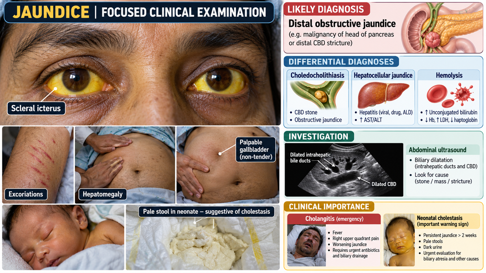
- Scleral icterus: supports clinically apparent hyperbilirubinemia.
- Excoriations: suggest cholestatic pruritus.
- Palpable non-tender gallbladder: raises suspicion for distal malignant obstruction, but is not diagnostic.
- Smooth hepatomegaly: may accompany biliary obstruction.
- Reported dark urine and pale stools: support conjugated hyperbilirubinemia with impaired bile delivery to the bowel.
Relevant negative findings
- No fever or right upper quadrant tenderness: acute cholangitis is less likely at present.
- No prominent chronic liver disease stigmata, ascites or splenomegaly: fewer clinical clues to established cirrhosis and portal hypertension.
Important pediatric findings
- Jaundice in the first 24 hours of life requires prompt assessment.
- Pale or acholic stools with dark urine suggest cholestasis.
- Lethargy, poor feeding or abnormal tone may indicate serious neonatal illness.
5. Presentation of Findings
“This 62-year-old man is conscious, oriented and hemodynamically stable. He has scleral icterus and generalized excoriations. Abdominal examination reveals a palpable, non-tender gallbladder and mild smooth hepatomegaly. There is no clinically detectable ascites, splenomegaly or prominent chronic liver disease stigmata. These findings suggest obstructive jaundice, with distal malignant biliary obstruction an important possibility requiring investigation.”
6. Most Likely Diagnosis
Adult: Obstructive jaundice, clinically suspicious for distal extrahepatic biliary obstruction, possibly related to a pancreatic-head or distal biliary malignancy.
Pediatric extension: Neonatal or childhood jaundice requiring differentiation of unconjugated from conjugated hyperbilirubinemia.
The cause cannot be established by examination alone.
7. Differential Diagnosis
- Choledocholithiasis: episodic right upper quadrant pain, sometimes with fever.
- Hepatocellular jaundice: hepatitis features or chronic liver disease signs; hepatocellular liver-enzyme pattern.
- Hemolytic jaundice: pallor or splenomegaly with predominantly unconjugated bilirubin elevation.
In neonates: consider physiological jaundice, hemolysis and neonatal cholestasis, differentiated by onset, examination and bilirubin fractionation.
8. Relevant Investigations
- Fractionated bilirubin and liver biochemistry: identify conjugated hyperbilirubinemia and a cholestatic ALP/GGT pattern.
- Abdominal ultrasound: assess biliary dilatation, gallstones and possible obstruction.
- Contrast-enhanced CT or MRCP, when indicated: define the level and cause of obstruction.
- CBC and PT/INR: assess anemia, infection and coagulation abnormalities.
Pediatric application: obtain objective bilirubin measurement and interpret using age and gestation; fractionate bilirubin for prolonged jaundice or suspected cholestasis.
9. Management Discussion
- Assess stability and identify infection or hepatic dysfunction.
- Arrange prompt investigations to establish the cause of obstruction.
- Refer suspected malignant obstruction for specialist assessment and appropriate treatment.
- Provide symptomatic care for pruritus and address clinically significant complications.
- In neonates, use age-specific bilirubin thresholds for treatment; urgently investigate suspected conjugated jaundice.
10. Red Flags / Serious Complications
- Jaundice with fever, abdominal pain or hypotension: possible cholangitis.
- Confusion, bleeding or worsening consciousness: possible severe hepatic dysfunction.
- Progressive painless jaundice with weight loss: possible malignancy.
- Neonatal jaundice within 24 hours, lethargy or abnormal tone.
- Persistent neonatal jaundice with pale stools or dark urine: possible cholestasis.
Urgently escalate assessment when any of these warning findings is present.
11. Examiner Viva Preparation
Q1. Why examine the sclerae?
Yellow scleral discoloration is a useful clinical sign of hyperbilirubinemia.
Q2. What does a palpable, non-tender gallbladder suggest?
Distal biliary obstruction, raising concern for malignancy without confirming it.
Q3. Which clues support obstruction?
Pruritus, excoriations, pale stools, dark urine and a palpable gallbladder.
Q4. What is the first-line imaging test?
Abdominal ultrasonography.
Q5. What neonatal finding suggests cholestasis?
Pale or acholic stools, especially with dark urine.
Q6. Why is visual neonatal jaundice assessment insufficient?
Skin color does not reliably estimate bilirubin concentration or treatment requirements.
Q7. What findings suggest acute cholangitis?
Fever, right upper quadrant pain and jaundice; hypotension or altered consciousness suggests severe disease.
12. AIM Short Case High-Yield Review
- Examine sclerae and skin in good lighting.
- Look for excoriations and chronic liver disease stigmata.
- A painless palpable gallbladder with jaundice raises concern for malignant obstruction.
- Assess hepatomegaly, splenomegaly and ascites.
- Present relevant positive and negative findings.
- Confirm the bilirubin fraction and liver-enzyme pattern.
- Ultrasound is the usual initial imaging test for suspected obstruction.
- Interpret neonatal bilirubin by age and gestation.
- Pale neonatal stools are an urgent warning clue.
- Common mistake: diagnosing obstruction from scleral icterus alone.
13. AIM Short Case Visual Placeholder