FINAL YEAR MBBS · KMU CURRICULUM · AIM CLINICAL LEARNING
🩺 AIM Short Case 01
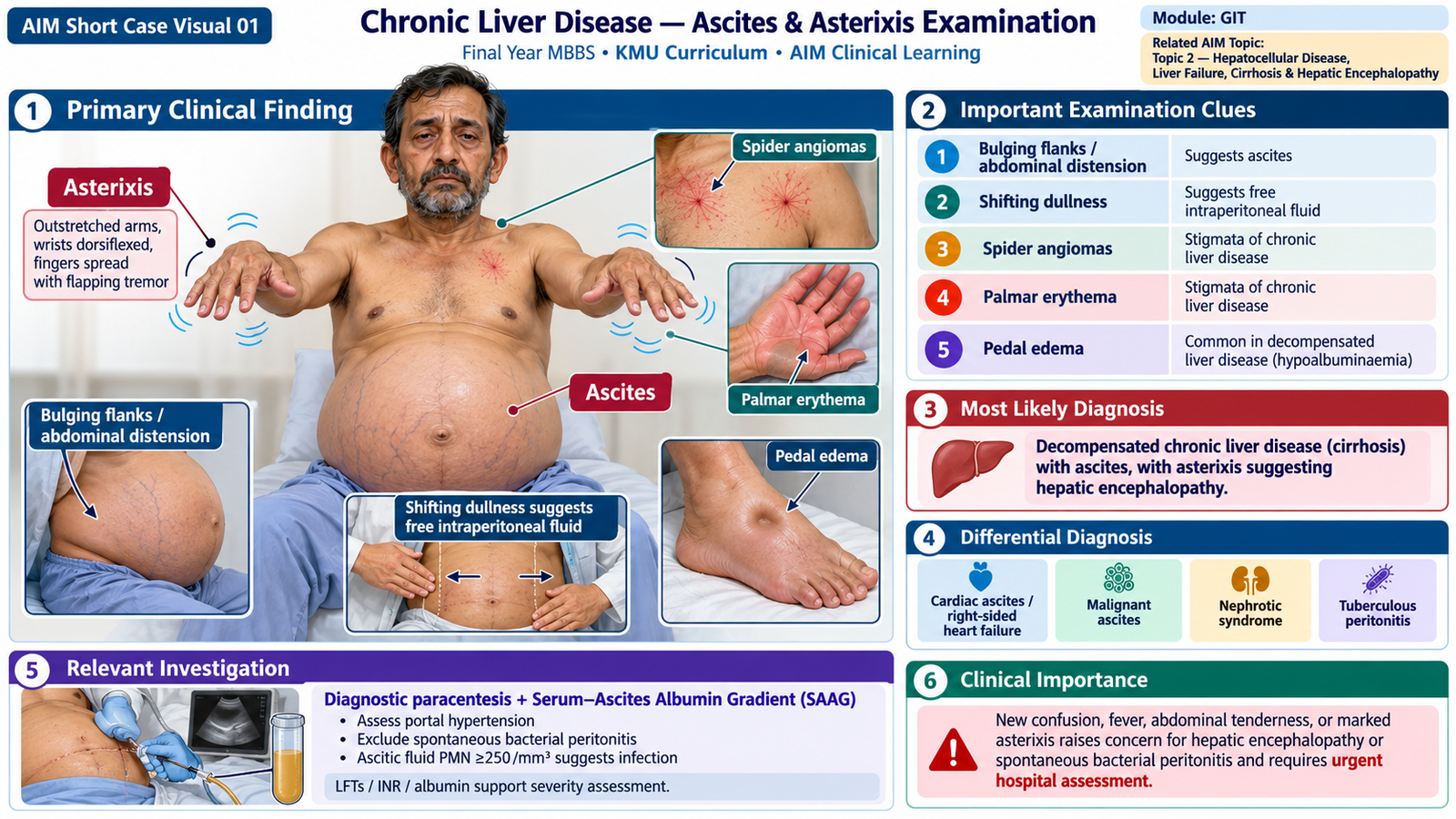
Chronic Liver Disease — Ascites & Asterixis Examination
GIT Module | Focused assessment of cirrhosis, ascites and possible hepatic encephalopathy.
1. Patient / Clinical Setting
A 54-year-old man attends the medical ward with progressive abdominal distension and ankle swelling. He is alert, cooperative and able to undergo a focused bedside examination. His previous records mention a chronic liver disorder.
2. Student Task
“Examine this patient for stigmata of chronic liver disease, assess for ascites, demonstrate how you would elicit asterixis, and present your clinical findings.”
3. Focused Examination Approach
Introduce yourself, obtain consent, provide adequate draping, position the patient at approximately 45° initially, and ensure comfortable exposure of the abdomen.
- Observe from the end of the bed: assess alertness, orientation, jaundice, muscle wasting, abdominal distension and peripheral edema. Altered attention raises concern for encephalopathy; distension suggests ascites.
- Inspect hands, arms, face and chest: look for palmar erythema, spider angiomas, bruising, scratch marks, scleral icterus, parotid enlargement and gynecomastia. These support chronic hepatic dysfunction but are not individually diagnostic.
- Test for asterixis: ask the patient to extend both arms, dorsiflex the wrists and spread the fingers; observe for several seconds. Brief, irregular lapses of maintained wrist extension indicate asterixis (negative myoclonus). It is a sign of metabolic encephalopathy, not specific to liver disease.
- Examine the abdomen: with the patient supine, inspect the contour, flanks, umbilicus, scars and abdominal wall veins. Gently palpate for tenderness, hepatomegaly and splenomegaly; avoid forceful palpation of a tense abdomen.
- Assess for ascites: percuss from the central tympanic abdomen toward each flank for dependent dullness. If present, perform shifting dullness by turning the patient onto one side, allowing fluid to settle, and repeating percussion. A fluid thrill may be useful only in marked ascites; an assistant or the patient places a hand along the midline to reduce skin-transmitted movement.
- Complete with relevant signs: check bilateral pitting ankle edema and briefly assess mental status, speech and attention, especially if asterixis is detected.
Bedside interpretation: Shifting dullness supports clinically detectable free peritoneal fluid; ultrasound is more sensitive for small-volume ascites. Asterixis suggests encephalopathy but also occurs in renal failure, hypercapnia and other metabolic disorders.
4. Important Clinical Findings
Positive findings in this patient
- Spider angiomas and palmar erythema: peripheral stigmata supporting chronic liver disease.
- Distended abdomen, bilateral flank dullness and positive shifting dullness: support ascites.
- Mild splenomegaly: suggests possible portal hypertension.
- Bilateral pitting ankle edema: supports extracellular fluid retention.
- Intermittent bilateral asterixis: suggests a metabolic encephalopathy; assess attention, orientation and possible precipitants.
Relevant negative findings
- No fever, marked abdominal tenderness or guarding on examination; this does not exclude spontaneous bacterial peritonitis.
- No marked disorientation or reduced consciousness at the time of examination; clinically important encephalopathy may still be present.
5. Presentation of Findings
“I examined a 54-year-old man who is alert and cooperative. He has palmar erythema, spider angiomas, a distended abdomen with flank dullness and shifting dullness, mild splenomegaly and bilateral pitting pedal edema. I also elicited bilateral asterixis. There is no guarding or marked abdominal tenderness, and he is not overtly disoriented. These findings suggest chronic liver disease with ascites, likely related to portal hypertension, and possible hepatic encephalopathy. I would confirm the ascites and evaluate for precipitants and infection.”
6. Most Likely Diagnosis
Decompensated chronic liver disease (probable cirrhosis) with clinically detectable ascites and suspected hepatic encephalopathy. Asterixis alone does not establish or grade hepatic encephalopathy; clinical assessment and exclusion of other causes are necessary.
7. Differential Diagnosis
- Cardiac ascites: consider with raised jugular venous pressure, prominent peripheral edema and signs of right heart failure.
- Malignant ascites: consider with weight loss, an abdominal or pelvic mass, and absent typical stigmata of cirrhosis.
- Nephrotic syndrome: generalized edema and heavy proteinuria may dominate; chronic liver stigmata and portal-hypertensive splenomegaly are less typical.
8. Relevant Investigations
- CBC, liver panel, albumin and PT/INR: assess hepatic dysfunction and complications; thrombocytopenia may support portal hypertension, while low albumin and prolonged INR suggest impaired synthetic function.
- Ultrasound abdomen with portal Doppler when indicated: confirm ascites and assess liver morphology, splenomegaly and portal circulation.
- Diagnostic paracentesis with ascitic cell count/differential, albumin and total protein: assess the cause and exclude infection. A serum–ascites albumin gradient (SAAG) ≥1.1 g/dL supports portal hypertension; ascitic PMN ≥250 cells/mm³ suggests spontaneous bacterial peritonitis in the appropriate setting.
- Electrolytes, renal function and glucose: identify reversible contributors to altered mentation, including kidney injury, hyponatremia and hypoglycemia.
9. Management Discussion
- Assess clinical stability and identify reversible precipitants of possible encephalopathy, particularly infection, GI bleeding, constipation and electrolyte disturbances.
- For uncomplicated cirrhotic ascites, use dietary sodium restriction and clinician-guided diuretics, with monitoring of weight, renal function and electrolytes.
- Arrange diagnostic paracentesis for new-onset ascites or appropriate hospital presentations; consider therapeutic drainage for tense, symptomatic ascites.
- If hepatic encephalopathy is clinically suspected, treat precipitants and initiate standard therapy such as lactulose under supervision.
10. Red Flags / Serious Complications
- New confusion, marked drowsiness or impaired airway protection.
- Fever, abdominal pain or tenderness, or unexplained deterioration: possible spontaneous bacterial peritonitis.
- Hematemesis, melena, hypotension or circulatory instability.
- Oliguria or rapidly worsening renal function.
These findings require urgent senior assessment and hospital escalation.
11. Examiner Viva Preparation
Q1. What is shifting dullness, and what does it indicate?
It is a change in the distribution of abdominal percussion dullness with change of patient position, supporting free intraperitoneal fluid.
Q2. How do you demonstrate asterixis?
Ask the patient to hold both arms out with wrists dorsiflexed and fingers spread. Observe for brief, irregular lapses of posture.
Q3. Is asterixis diagnostic of hepatic encephalopathy?
No. It supports metabolic encephalopathy but may occur in uremia, hypercapnia and drug-related toxicity; interpret it with mental status and clinical context.
Q4. Name three signs supporting chronic liver disease.
Spider angiomas, palmar erythema and muscle wasting; jaundice or gynecomastia may also be present.
Q5. Which ascitic fluid results are most useful initially?
Cell count with differential, ascitic albumin (paired with serum albumin for SAAG) and total protein. PMN ≥250 cells/mm³ suggests spontaneous bacterial peritonitis.
Q6. Why is diagnostic paracentesis important in cirrhotic ascites?
It helps establish the cause of ascites and detect spontaneous bacterial peritonitis, which may occur without classic peritoneal signs.
Q7. State the broad principle of managing suspected hepatic encephalopathy.
Identify and correct precipitants, assess severity and safety, and use treatments such as lactulose when clinically indicated.
12. AIM Short Case High-Yield Review
- Look: spider angiomas, palmar erythema, jaundice, wasting and edema.
- Percuss: flank dullness and shifting dullness support ascites.
- Examine hands: asterixis is a brief loss of sustained posture.
- Interpret: asterixis is not specific to hepatic encephalopathy.
- Present: positive findings, key negatives and a cautious clinical impression.
- Investigate: ultrasound and diagnostic ascitic fluid analysis are central.
- Remember: ascitic PMN ≥250 cells/mm³ raises concern for spontaneous bacterial peritonitis.
- Act: confusion, infection, GI bleeding and renal deterioration are red flags.
- Avoid the common error: do not diagnose or grade hepatic encephalopathy from asterixis alone.