FINAL YEAR MBBS | KMU CURRICULUM | AIM CLINICAL LEARNING
🩺 AIM LONG CASE 04
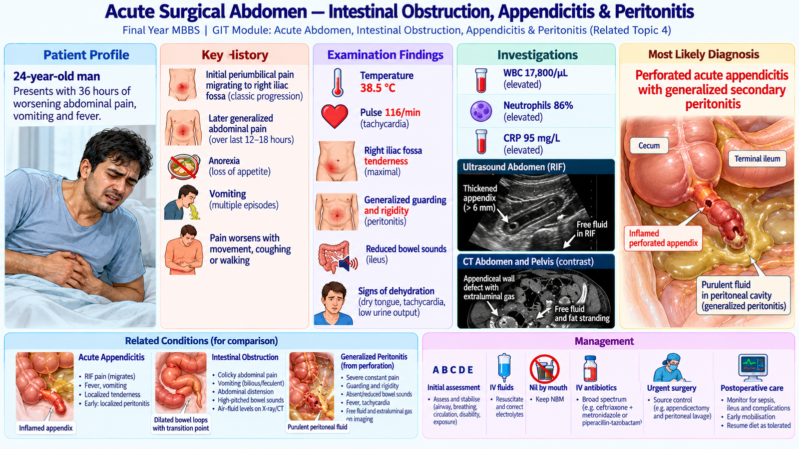
Acute Surgical Abdomen — Intestinal Obstruction, Appendicitis & Peritonitis
GIT Module | Surgery & Family Medicine | Related AIM Topic 4
An integrated clinical approach to acute abdominal pain, recognition of complicated appendicitis, differential diagnosis of intestinal obstruction and peritonitis, emergency investigation, resuscitation, surgical management and safe referral.
1. Long Case Title
Long Case 04 — Evaluation of a Patient with an Acute Surgical Abdomen: Suspected Complicated Appendicitis and Peritonitis
Discipline: General Surgery with Family Medicine integration.
Clinical focus: Comprehensive assessment of acute abdominal pain, recognition of appendicitis and generalized peritonitis, differentiation from intestinal obstruction and other surgical emergencies, interpretation of relevant investigations, initial resuscitation, urgent surgical referral and definitive management.
This representative patient illustrates a common surgical emergency. Intestinal obstruction and other major causes of acute abdominal pain are considered through clinical reasoning and differential diagnosis.
2. Patient Scenario
A 24-year-old man presents to the emergency department with severe abdominal pain for approximately 36 hours.
The pain initially began around the umbilicus but has become more severe and widespread. He reports repeated vomiting, loss of appetite and fever.
His family states that he has been unable to walk comfortably because abdominal movements worsen the pain.
He has no previous history of abdominal surgery or known chronic gastrointestinal disease.
The student is asked to assess the patient, identify the important clinical problems and discuss further investigation and management.
3. Student Task
By completing this long case, the student should be able to:
- Take a focused but comprehensive history of acute abdominal pain.
- Identify important symptoms indicating appendicitis, intestinal obstruction or peritonitis.
- Assess vital signs, hydration and clinical stability.
- Perform an appropriate general and focused abdominal examination.
- Recognize important signs of acute appendicitis and peritoneal irritation.
- Formulate a prioritized differential diagnosis of an acute surgical abdomen.
- Select and interpret relevant laboratory and abdominal imaging investigations.
- Recognize suspected perforation, generalized peritonitis and evolving sepsis.
- Discuss initial resuscitation and definitive surgical management.
- Identify indications for emergency referral from primary healthcare.
- Explain the condition, treatment, possible complications and follow-up to the patient and family.
4. Relevant History
A. Presenting Complaints
- Abdominal pain — 36 hours.
- Vomiting — approximately 24 hours.
- Fever — approximately 18 hours.
- Loss of appetite — since onset of illness.
- Increasing abdominal discomfort and difficulty moving — several hours.
B. History of Presenting Illness
Onset and location of pain: The pain began gradually around the umbilicus and was initially poorly localized.
Clinical significance: Early appendicitis commonly produces visceral pain perceived around the umbilicus.
Migration of pain: After several hours, the pain moved to the right lower abdomen and became more intense.
Clinical significance: Migration of periumbilical pain to the right iliac fossa is a characteristic history feature of acute appendicitis, reflecting involvement of the parietal peritoneum.
Progression: During the last several hours, the patient has experienced increasingly widespread abdominal pain, aggravated by movement, coughing and changing position.
Clinical significance: Progressive generalized pain following localized right iliac fossa pain raises suspicion of appendiceal perforation with peritoneal contamination. The pattern is concerning but not independently diagnostic.
Vomiting: He has vomited three times, initially containing recently eaten food. There is no blood in the vomitus.
Clinical significance: Nausea and vomiting commonly accompany appendicitis. Persistent bilious vomiting, marked distension and absolute constipation would increase suspicion of intestinal obstruction.
Fever: The patient reports fever and chills associated with worsening abdominal pain.
Clinical significance: Fever suggests an inflammatory or infectious process. High fever with diffuse peritoneal signs raises concern for complicated intra-abdominal infection.
C. Bowel and Urinary History
Bowel movements: The patient passed stool the previous day and has passed small amounts of flatus. He has not noticed progressive abdominal distension.
Clinical significance: The absence of marked distension and absolute constipation makes established complete mechanical intestinal obstruction less likely, although early obstruction is not excluded.
Urinary symptoms: No dysuria, urinary frequency or hematuria.
Clinical significance: This makes urinary tract infection and ureteric calculi less likely as explanations for the abdominal pain.
D. Important Past History
- No previous abdominal surgery.
- No known abdominal hernia.
- No previous similar episodes of severe abdominal pain.
- No known inflammatory bowel disease.
- No history of chronic peptic ulcer disease or previous ulcer complications.
- No known diabetes or major chronic illness.
Clinical significance: Previous abdominal surgery would raise suspicion of adhesive intestinal obstruction. A known hernia increases concern for incarceration or strangulation. Previous ulcer disease may suggest perforated peptic ulcer as an alternative cause of generalized peritonitis.
E. Medication and Social History
- No regular medications or known drug allergies.
- Ask about recent analgesic or antibiotic use, as these may modify clinical findings.
- Review recent oral intake and fluid losses to assess dehydration.
- Assess functional limitations, access to healthcare and support at home.
Periumbilical pain migrating to the right iliac fossa, followed by worsening generalized pain, fever and movement-related pain suggests acute appendicitis with possible perforation and peritonitis. This requires prompt surgical evaluation.
5. Physical Examination
A. Initial General Assessment
The patient appears acutely unwell and lies relatively still with his knees slightly flexed. He is conscious, cooperative and visibly uncomfortable.
- Temperature: 38.5°C.
- Pulse: 116/min, regular.
- Blood pressure: 104/66 mmHg.
- Respiratory rate: 24/min.
- Oxygen saturation: 97% on room air.
- Hydration: Dry oral mucosa and reduced oral intake.
- Peripheral perfusion: Extremities warm with mildly delayed capillary refill.
Interpretation: Fever, tachycardia, tachypnea and evidence of dehydration indicate significant acute illness. In the presence of suspected intra-abdominal infection, they raise concern for systemic inflammatory response and evolving sepsis. Blood pressure must be monitored closely.
B. Abdominal Inspection
- Abdominal movements with respiration are reduced.
- Mild generalized abdominal fullness is present.
- No visible surgical scars.
- No obvious external hernia or visible intestinal peristalsis.
Interpretation: Reduced abdominal movement may reflect peritoneal irritation. The absence of previous surgical scars reduces suspicion of postoperative adhesive obstruction but does not exclude intestinal obstruction.
C. Abdominal Palpation
- Marked tenderness over the right iliac fossa.
- Generalized abdominal tenderness.
- Involuntary guarding and rigidity, most pronounced in the lower abdomen.
- Pain worsens with gentle percussion or movement.
- No clearly palpable abdominal mass.
Interpretation: Localized right iliac fossa tenderness is compatible with appendicitis. Generalized guarding and rigidity indicate peritoneal irritation and raise suspicion of perforation or another serious intra-abdominal inflammatory process.
Generalized involuntary guarding, rigidity and pain aggravated by movement strongly suggest peritoneal inflammation. These findings require urgent surgical assessment; repeated painful examinations should be avoided.
D. Percussion and Auscultation
- Gentle percussion produces abdominal pain.
- Bowel sounds are reduced.
- No clear evidence of massive abdominal distension.
Interpretation: Reduced bowel sounds may occur with paralytic ileus secondary to peritonitis. In contrast, mechanical intestinal obstruction may initially produce increased, high-pitched bowel sounds, although auscultatory findings are not sufficiently reliable to establish or exclude obstruction.
E. Focused Examination for Other Surgical Causes
- Examine the inguinal regions for a tender or irreducible hernia.
- Assess for major abdominal distension and features of mechanical obstruction.
- Look for evidence of shock or deteriorating peripheral perfusion.
- Reassess the abdominal findings and vital signs during management.
Clinical interpretation: The combined findings indicate an acute surgical abdomen with generalized peritoneal irritation. Complicated appendicitis is the leading clinical diagnosis, but other causes of perforation and intestinal obstruction remain important alternatives.
6. Clinical Problem Representation
A previously healthy 24-year-old man presents with 36 hours of abdominal pain that began centrally, migrated to the right iliac fossa and subsequently became generalized. This is associated with anorexia, vomiting and fever. Examination reveals tachycardia, tachypnea, dehydration, marked right iliac fossa tenderness, generalized involuntary guarding and rigidity, with reduced bowel sounds.
Main clinical problem: Acute surgical abdomen with generalized peritonitis, most likely due to complicated acute appendicitis with suspected perforation.
Immediate concern: Intra-abdominal infection with risk of worsening sepsis and circulatory compromise.
7. Differential Diagnosis
| Diagnosis | Supporting Features | Features Helping Differentiate |
|---|---|---|
| Perforated acute appendicitis | Migratory pain, anorexia, fever, initial right iliac fossa localization followed by generalized peritoneal signs | Appendiceal inflammation, perforation and surrounding contamination on imaging or at surgery |
| Perforated peptic ulcer | Severe abdominal pain, guarding and generalized rigidity | Typically sudden severe epigastric pain; free intraperitoneal air may be seen on imaging |
| Acute mechanical intestinal obstruction | Vomiting and abdominal discomfort | Colicky pain, distension, constipation or obstipation and dilated bowel loops; peritonitis suggests a complication |
| Strangulated intestinal obstruction | Severe continuous pain, tachycardia and possible peritonitis | Obstruction with signs of ischemia, an irreducible tender hernia or a compromised bowel segment on imaging |
| Inflammatory ileocecal disease | Right lower abdominal pain and systemic inflammatory features | A longer history of altered bowel habit, diarrhea or recurrent symptoms may suggest inflammatory bowel disease |
Clinical reasoning: The history of pain migration strongly favors appendicitis. Generalized rigidity changes the assessment from uncomplicated localized appendicitis to suspected peritonitis, requiring prompt resuscitation and surgical review.
Mechanical intestinal obstruction remains an important alternative. The clinician must actively look for abdominal distension, absolute constipation, previous surgery and hernias rather than assuming that vomiting alone indicates obstruction.
8. Investigations and Interpretation
Investigations should support diagnosis, assess severity and guide intervention. In a patient with clear generalized peritonitis, laboratory testing or imaging must not unnecessarily delay emergency resuscitation and surgical management.
A. Initial Laboratory Investigations
| Investigation | Representative Result | Clinical Interpretation |
|---|---|---|
| CBC | WBC 17,800/µL; neutrophils 86% | Neutrophilic leukocytosis supports acute inflammation or infection but is not specific for appendicitis. |
| C-reactive protein | 95 mg/L | Elevated inflammatory marker, supporting a significant inflammatory process. |
| Serum electrolytes | Sodium 134 mmol/L; potassium 3.4 mmol/L | Mild electrolyte disturbances consistent with vomiting and reduced intake. |
| Renal function | Creatinine 1.2 mg/dL | Provides a baseline for hydration assessment, monitoring and contrast imaging. |
| Serum lactate | 2.1 mmol/L | Mild elevation may reflect physiological stress or impaired perfusion; interpret with clinical findings. |
| Urinalysis | No significant pyuria or hematuria | Makes some urinary causes less likely without excluding them completely. |
Additional tests: Obtain coagulation studies, blood group and crossmatch as appropriate before emergency surgery. Blood cultures may be obtained when sepsis is suspected, without delaying antibiotic administration.
Clinical significance: No single blood test confirms perforated appendicitis. Results must be interpreted alongside history, examination and imaging.
B. Abdominal Ultrasonography
Why requested: Ultrasound can support appendicitis diagnosis, assess fluid collections and identify some alternative abdominal pathologies. It is particularly useful in children and selected younger patients.
Representative findings:
- Thickened, non-compressible appendix in the right iliac fossa, measuring approximately 10 mm in outer diameter.
- Inflammatory change in adjacent tissues.
- Small amount of free fluid in the right lower abdomen.
Interpretation: These findings support acute appendicitis with surrounding inflammation. Free fluid raises concern for complicated disease but does not independently prove perforation.
C. Contrast-Enhanced CT of Abdomen and Pelvis
Why requested: When the patient is sufficiently stable and imaging will guide management without causing harmful delay, CT can define the suspected pathology, assess perforation and collections, and distinguish appendicitis from intestinal obstruction or another cause of peritonitis.
Representative findings:
- Enlarged, inflamed appendix arising from the cecum.
- Focal discontinuity of the appendiceal wall.
- Prominent periappendiceal fat stranding.
- Small adjacent extraluminal gas locules.
- Free intraperitoneal fluid with associated inflammatory changes.
- No discrete, drainable periappendiceal abscess.
- No clear mechanical transition point or markedly dilated bowel loops.
Interpretation: The findings strongly support perforated acute appendicitis with peritoneal contamination. The absence of a mechanical transition point makes primary mechanical intestinal obstruction less likely.
Safety principle: CT is not mandatory before surgical intervention when clinical findings indicate generalized peritonitis and imaging would delay necessary treatment.
D. Imaging for Important Alternative Diagnoses
Suspected intestinal obstruction: Abdominal imaging may reveal dilated bowel loops, proximal fluid accumulation and a transition point. CT can help identify the site, cause and complications such as ischemia.
Suspected hollow-viscus perforation: An erect chest radiograph may demonstrate free subdiaphragmatic air. However, absence of free air does not exclude perforation, and CT is more sensitive when appropriate.
Important interpretation: Perforation and obstruction are clinical emergencies when associated with peritonitis or circulatory compromise. Diagnostic investigation should not postpone resuscitation and timely definitive care.
Migratory abdominal pain + right iliac fossa tenderness + generalized guarding + inflammatory markers + imaging evidence of appendiceal wall disruption and adjacent extraluminal gas strongly supports perforated appendicitis with generalized peritonitis.
9. Final Diagnosis
Most likely diagnosis:
Perforated acute appendicitis causing generalized secondary bacterial peritonitis, with dehydration and systemic inflammatory features.
Underlying mechanism: Progressive inflammation of the appendix may lead to ischemia, necrosis and perforation. Escape of intestinal bacteria and inflammatory contents into the peritoneal cavity produces secondary peritonitis.
Clinical severity: The patient is currently conscious and not frankly hypotensive but has tachycardia, fever, tachypnea and peritoneal signs. He requires emergency surgical management and monitoring for sepsis and circulatory deterioration.
10. Management
A. Immediate Priorities
- ABCDE assessment: Evaluate airway, breathing, circulation, disability and exposure, and identify immediate threats.
- Urgent surgical involvement: Inform the surgical team immediately because generalized peritonitis suggests a condition requiring source control.
- Keep the patient nil by mouth: Prepare for potential emergency anesthesia and surgery.
- Intravenous access: Establish reliable access for fluids, medications and blood sampling.
- Resuscitation: Administer intravenous isotonic crystalloid, guided by perfusion, hydration and reassessment.
- Antibiotics: Start prompt intravenous broad-spectrum antimicrobial therapy covering enteric gram-negative organisms and anaerobes.
- Analgesia and antiemetics: Relieve pain and vomiting while continuing clinical assessment.
Rationale: Perforated appendicitis with peritonitis may lead to sepsis, fluid loss into the peritoneal cavity and circulatory deterioration. Initial stabilization and infection control must proceed alongside preparation for definitive treatment.
B. Supportive Management
- Correct fluid and electrolyte deficits.
- Monitor urine output and renal function.
- Use a urinary catheter if accurate output monitoring is clinically needed.
- Consider nasogastric decompression when persistent vomiting, significant distension or associated ileus warrants it.
- Review medication allergies and relevant pre-anesthetic risks.
- Maintain appropriate fasting and perioperative support.
C. Definitive Surgical Treatment
Emergency operative management is indicated for suspected perforated appendicitis with generalized peritonitis.
The surgical approach may be laparoscopic or open depending on the patient’s condition, available expertise and operative findings.
Major treatment objectives:
- Remove the diseased appendix when feasible.
- Control the source of intra-abdominal contamination.
- Remove infected material and manage peritoneal contamination as appropriate.
- Identify and address unexpected associated pathology.
Detailed operative technique is beyond the scope of this undergraduate long case.
D. Management if Another Cause Is Identified
Mechanical intestinal obstruction: Initial management includes resuscitation, nil by mouth, electrolyte correction, appropriate decompression and surgical assessment. Urgent surgery is required when strangulation, ischemia, perforation or generalized peritonitis is suspected.
Perforated peptic ulcer: Requires resuscitation, antibiotics, appropriate supportive treatment and urgent surgical evaluation for source control.
Appendiceal inflammatory mass or localized abscess: Management may differ from generalized peritonitis. Selected stable patients may be treated with antibiotics and, when appropriate, image-guided drainage following surgical assessment.
E. Role of Family Medicine and Primary Healthcare
If this patient initially presents to a primary healthcare facility, the clinician should:
- Recognize severe persistent abdominal pain and peritoneal signs as red flags.
- Assess vital signs and hydration status.
- Initiate appropriate stabilization within available resources.
- Keep the patient nil by mouth if urgent surgery is suspected.
- Arrange immediate transfer to an emergency surgical facility.
- Communicate relevant clinical findings and treatment already provided to the receiving team.
Clinical principle: Do not delay transfer for extensive outpatient investigations when generalized peritonitis is suspected.
F. Postoperative Monitoring
- Monitor temperature, pulse, blood pressure, respiratory rate and oxygenation.
- Assess pain, hydration and urine output.
- Monitor for persistent fever or abdominal tenderness indicating ongoing infection.
- Evaluate return of bowel function and oral intake.
- Review wound healing and any signs of surgical-site infection.
- Adjust antibiotics according to clinical progress, operative findings and local practice.
G. Follow-Up
Arrange postoperative review to assess wound healing, bowel function, recovery of activity and any complications. Explain warning symptoms and review the histopathology result when available.
The three essential priorities in complicated appendicitis with generalized peritonitis are resuscitation, antimicrobial treatment and timely surgical source control. Antibiotics alone should not substitute for necessary emergency surgery.
11. Complications and Red Flags
A. Important Complications
- Generalized peritonitis: Infection and inflammation involving the peritoneal cavity.
- Intra-abdominal abscess: Localized collection of infected fluid, potentially presenting with persistent postoperative fever.
- Sepsis and septic shock: Severe infection-related systemic illness with possible circulatory and organ dysfunction.
- Paralytic ileus: Temporary impairment of intestinal motility due to peritoneal inflammation or surgery.
- Wound infection: A postoperative complication, particularly in contaminated surgical cases.
- Dehydration and acute kidney injury: May result from vomiting, reduced intake, third-space fluid losses and poor perfusion.
Urgent escalation is required when a patient develops:
- Generalized involuntary guarding or abdominal rigidity.
- Persistent severe pain with rapidly worsening abdominal findings.
- Hypotension, marked tachycardia or poor peripheral perfusion.
- Confusion, reduced consciousness or oliguria.
- Severe distension, persistent vomiting and inability to pass stool or flatus, particularly with continuous pain or peritoneal signs.
Action: Immediate emergency assessment, resuscitation, urgent surgical consultation and timely definitive intervention.
Important clinical mistake: Do not reassure a patient with worsening generalized abdominal pain simply because earlier findings suggested uncomplicated appendicitis.
12. Patient Counselling and Follow-Up
A. Explaining the Condition
Explain that the patient has findings suggesting severe inflammation of the appendix, with possible perforation and spread of infection into the abdominal cavity.
Describe why urgent surgical evaluation and treatment are necessary, using understandable language and acknowledging any diagnostic uncertainty.
B. Explaining the Treatment Plan
- Explain the need for intravenous fluids to correct dehydration and maintain circulation.
- Describe the purpose of antibiotics in treating intra-abdominal infection.
- Explain that surgery is expected to remove the diseased appendix and control infection.
- Discuss that the exact surgical approach may depend on operative findings.
- Inform the patient about anesthesia and important procedural risks in an appropriate, non-alarming manner.
C. Consent and Patient Dignity
Confirm that the patient understands the diagnosis, expected benefits, important risks, alternatives and consequences of delaying treatment. Support informed consent and allow questions. Involve family members only with the patient’s permission, except where applicable emergency consent arrangements are required.
D. Postoperative Advice
- Follow medication and wound-care instructions.
- Gradually resume oral intake and physical activity as medically advised.
- Attend scheduled surgical follow-up.
- Report persistent or recurrent fever, worsening abdominal pain, repeated vomiting, wound discharge or increasing abdominal distension.
- Understand that recovery after perforation may take longer than after uncomplicated appendicitis.
E. Family Medicine Follow-Up
After discharge, primary healthcare review may support wound assessment, medication adherence, nutritional recovery and early recognition of delayed complications.
Professional principle: Counselling should be clear, compassionate, individualized and focused on safe recovery.
13. Long Case Presentation
“I examined a 24-year-old gentleman presenting with 36 hours of abdominal pain that initially started around the umbilicus, migrated to the right iliac fossa and later became generalized. The pain is associated with vomiting, anorexia and fever. There is no history of previous abdominal surgery, hernia or similar episodes. On examination, he appears acutely unwell, with a temperature of 38.5°C, pulse of 116 per minute, respiratory rate of 24 per minute and blood pressure of 104/66 mmHg. He is mildly dehydrated, with marked right iliac fossa tenderness, generalized involuntary guarding, rigidity and reduced bowel sounds.”
“My clinical impression is an acute surgical abdomen with generalized peritonitis, most likely secondary to perforated acute appendicitis. Important alternative diagnoses include perforated peptic ulcer and complicated intestinal obstruction. Laboratory findings support acute inflammation, and CT demonstrates an inflamed appendix with focal wall disruption, extraluminal gas and peritoneal fluid. The patient requires urgent surgical management with intravenous fluid resuscitation, broad-spectrum antibiotics, analgesia and timely operative source control, followed by close monitoring for sepsis and postoperative complications.”
14. Examiner Discussion / Viva Preparation
Q1. Why does the pain of acute appendicitis often begin around the umbilicus and later migrate to the right iliac fossa?
Model Answer: Early inflammation produces poorly localized visceral pain referred to the periumbilical region. As inflammation involves the adjacent parietal peritoneum, the pain becomes more localized to the right iliac fossa.
Q2. Which findings suggest that appendicitis has progressed to generalized peritonitis?
Model Answer: Worsening generalized abdominal pain, involuntary guarding, rigidity, pain with movement, fever, tachycardia and reduced bowel sounds suggest peritoneal inflammation. Imaging may identify perforation, extraluminal gas or inflammatory fluid.
Q3. How can acute appendicitis be clinically differentiated from intestinal obstruction?
Model Answer: Appendicitis commonly produces migratory pain, right iliac fossa tenderness, anorexia and fever. Mechanical intestinal obstruction more typically presents with colicky abdominal pain, vomiting, abdominal distension and constipation or obstipation. Clinical overlap occurs, particularly in complicated disease, and imaging may be necessary.
Q4. What is the significance of generalized involuntary abdominal rigidity?
Model Answer: It indicates peritoneal irritation and raises suspicion of generalized peritonitis, perforation or another serious intra-abdominal process. It requires urgent surgical evaluation and should not be treated as a routine uncomplicated abdominal complaint.
Q5. Which investigations are most useful in suspected complicated appendicitis?
Model Answer: CBC, inflammatory markers, electrolytes and renal function help assess inflammation and physiological status. Ultrasound may identify appendiceal inflammation. CT can define perforation, collections and alternative diagnoses when appropriate and when imaging does not delay emergency treatment.
Q6. What are the immediate management priorities in suspected perforated appendicitis with generalized peritonitis?
Model Answer: Perform ABCDE assessment, establish intravenous access, provide fluid resuscitation, maintain nil by mouth, administer analgesia and appropriate broad-spectrum antibiotics, monitor clinical status and arrange urgent operative source control.
Q7. When does intestinal obstruction require urgent surgical intervention?
Model Answer: Urgent surgery is required when strangulation, bowel ischemia, perforation or generalized peritonitis is suspected, or when clinical deterioration indicates a threatened bowel or failed appropriate conservative management.
Q8. How should a family physician manage a patient presenting with suspected generalized peritonitis?
Model Answer: Recognize the emergency, assess vital signs, initiate appropriate stabilization within available resources, keep the patient nil by mouth if urgent surgery is suspected and arrange immediate transfer to a surgical emergency facility. Outpatient investigations must not delay referral.
15. AIM Long Case — High-Yield Review
- Migratory periumbilical-to-right-iliac-fossa pain is a characteristic feature of acute appendicitis.
- Anorexia, nausea, vomiting and fever support the clinical diagnosis when combined with appropriate abdominal findings.
- Generalized rigidity and involuntary guarding raise concern for peritonitis and possible perforation.
- Tachycardia, fever, tachypnea and poor perfusion require careful assessment for infection-related physiological deterioration.
- Mechanical intestinal obstruction should be considered when colicky pain, distension, vomiting and obstipation occur.
- Previous abdominal surgery and hernias are important historical clues when assessing suspected obstruction.
- Leukocytosis and elevated CRP support inflammation but do not independently diagnose perforated appendicitis.
- Ultrasound and CT help identify appendicitis, perforation, collections and alternative surgical pathology.
- Free intraperitoneal air, bowel ischemia and generalized peritonitis are major warning findings requiring urgent surgical consideration.
- Initial management includes stabilization, fluid and electrolyte correction, analgesia, antibiotics and timely surgical involvement.
- Perforated appendicitis with generalized peritonitis generally requires urgent operative source control.
- Primary healthcare responsibility is early recognition, appropriate stabilization and safe emergency referral without unnecessary delay.
The most important long-case skill is to recognize when an initially localized abdominal condition has become a life-threatening surgical emergency. History, examination and investigations should lead to timely resuscitation and definitive management—not delay them.
AIM Long Case — Integrated Clinical Visual