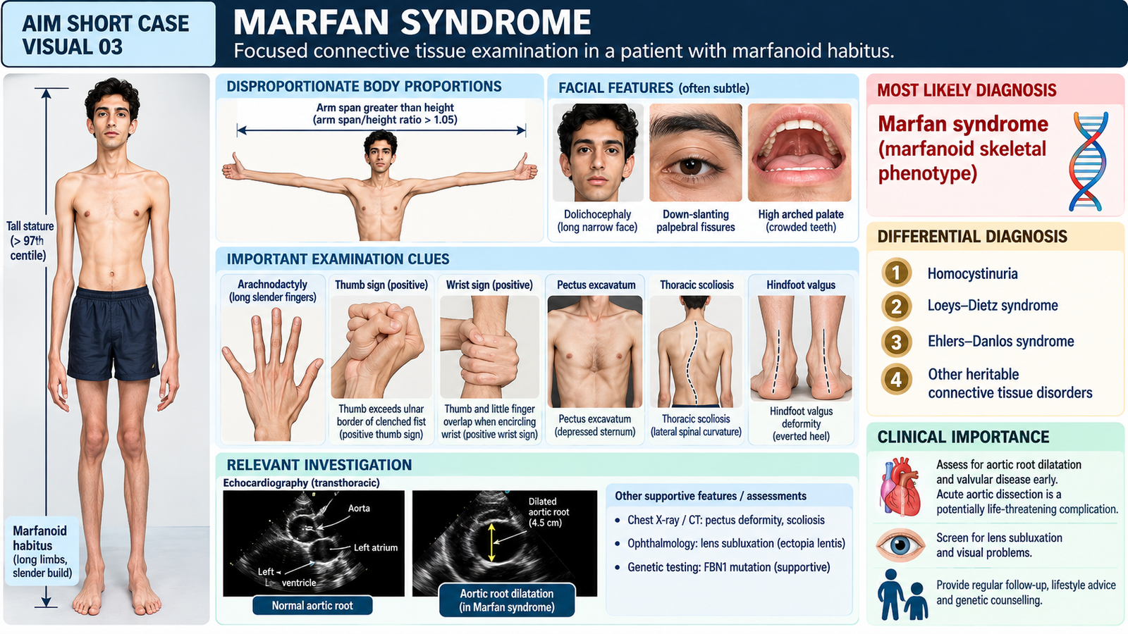
Marfan Syndrome — Focused Connective Tissue Examination
MS Module | Medicine | Final Year MBBS
Focused recognition of skeletal and connective tissue abnormalities, assessment of body proportions and identification of potentially serious cardiovascular and ocular complications.
1. Patient / Clinical Setting
A 19-year-old male university student attends the medical outpatient department because of increasing upper back deformity and difficulty maintaining an upright posture. He is unusually tall and slender, with long fingers and arms.
He reports that his father has a similar tall body build. He is comfortable at rest and has no current chest pain or breathlessness.
2. Student Task
Perform a focused examination for connective tissue abnormalities, assess body proportions, joint hypermobility and skeletal deformities, identify important cardiovascular and ocular findings, and present your clinical impression.
3. Focused Examination Approach
Introduce yourself, obtain consent and ensure appropriate exposure while maintaining patient dignity. Begin with general inspection, followed by focused examination of body proportions, hands, joints and spine. Complete the examination with relevant cardiovascular and ocular screening.
A. General inspection and body proportions
- Body habitus: Observe tall stature, slender build and disproportionately long limbs (dolichostenomelia).
- Arm span: Measure fingertip-to-fingertip distance with both arms extended horizontally and compare it with standing height.
- Upper-to-lower segment ratio: Assess whether the lower body segment is disproportionately long relative to the trunk.
- Facial appearance: Look for an elongated narrow face and a high-arched palate where appropriate.
Clinical interpretation: Increased arm-span-to-height ratio and reduced upper-to-lower segment ratio support disproportionate long-limb growth. These findings must be interpreted using age- and population-appropriate reference values.
B. Hands and fingers
- Arachnodactyly: Inspect for unusually long, slender fingers.
- Thumb sign: Ask the patient to fold the thumb across the palm and close the fingers over it. A positive sign occurs when the entire distal phalanx of the thumb projects beyond the ulnar border of the clenched hand.
- Wrist sign: Ask the patient to encircle the opposite wrist with the thumb and little finger. A positive sign occurs when the distal phalanges overlap.
- Joint mobility: Assess hypermobility of selected peripheral joints without forcing movement.
A positive thumb sign and wrist sign in a tall, slender patient with disproportionate limb length strongly support a marfanoid skeletal phenotype. These signs are supportive but are not independently diagnostic of Marfan syndrome.
C. Chest wall and spinal examination
- Anterior chest wall: Inspect for pectus excavatum or pectus carinatum.
- Spine: Examine from behind for scoliosis, asymmetry of the shoulders and unequal waist contours.
- Forward bending: Observe for rib prominence suggesting structural scoliosis.
- Posture: Look for excessive thoracic kyphosis and other visible spinal deformities.
- Feet: Assess for hindfoot valgus deformity or pes planus.
Clinical interpretation: Chest wall deformity, scoliosis and hindfoot abnormalities provide additional evidence of systemic connective tissue involvement.
D. Focused cardiovascular examination
- Pulse and blood pressure: Record heart rate, rhythm and blood pressure.
- Precordial examination: Inspect and palpate for abnormal cardiac activity.
- Auscultation: Listen for an early diastolic murmur of aortic regurgitation, particularly along the left sternal border with the patient leaning forward.
- Mitral valve: Listen for a midsystolic click or late systolic murmur suggesting mitral valve prolapse.
Clinical interpretation: Cardiovascular involvement is particularly important because aortic root dilatation may progress without prominent symptoms or audible murmurs.
E. Focused ocular assessment
- Ask briefly about blurred vision, significant refractive error and sudden visual changes.
- Assess visual acuity where appropriate.
- Observe ocular alignment and gross abnormalities.
- Recognize that ectopia lentis (lens displacement) requires formal ophthalmological assessment, usually with slit-lamp examination.
Clinical interpretation: Lens displacement, particularly upward and temporal displacement, is an important ocular manifestation of Marfan syndrome.
4. Important Clinical Findings
Positive findings
- Tall, slender habitus with disproportionately long limbs: Suggests an underlying connective tissue disorder affecting skeletal growth.
- Arm span greater than height: Supports disproportionate limb length when interpreted against appropriate reference values.
- Positive thumb and wrist signs: Support arachnodactyly and a marfanoid skeletal phenotype.
- Mild pectus excavatum: Suggests connective tissue-related anterior chest wall deformity.
- Thoracic scoliosis: Indicates spinal skeletal involvement.
- Increased joint mobility: Supports ligamentous laxity, although hypermobility is variable in Marfan syndrome.
Important relevant negative findings
- No current chest pain, acute breathlessness or cardiovascular instability.
- No audible diastolic murmur of aortic regurgitation.
- No obvious focal neurological abnormality.
- No acute visual loss or painful red eye.
The combination of disproportionate long-limb growth, arachnodactyly, positive wrist and thumb signs, and chest or spinal deformities is strongly suggestive of a marfanoid connective tissue phenotype. Marfan syndrome should be suspected, but cardiovascular imaging and ophthalmological assessment are required before diagnostic confirmation.
5. Presentation of Findings
“I examined a 19-year-old male with a tall, slender body habitus and disproportionately long limbs. He has arachnodactyly, positive thumb and wrist signs, mild pectus excavatum and thoracic scoliosis. There is increased peripheral joint mobility. Cardiovascular examination reveals no audible aortic regurgitation murmur, and there is no acute ocular complaint. These findings are suggestive of a marfanoid connective tissue phenotype, with Marfan syndrome as the leading clinical possibility. I would recommend echocardiographic assessment of the aortic root and formal ophthalmological examination.”
6. Most Likely Diagnosis
Suspected Marfan Syndrome — Marfanoid Skeletal Phenotype
Marfan syndrome is a heritable connective tissue disorder commonly associated with pathogenic variants in the FBN1 gene. It particularly affects the skeletal, cardiovascular and ocular systems. The skeletal findings in this case justify diagnostic evaluation but do not alone establish the syndrome.
7. Differential Diagnosis
- Homocystinuria: Can produce a marfanoid habitus and lens displacement. Intellectual or developmental difficulties, thromboembolic disease and elevated plasma homocysteine may help distinguish it.
- Loeys–Dietz syndrome: May cause skeletal abnormalities and aortic aneurysms. Hypertelorism, bifid uvula or cleft palate and arterial tortuosity favor this diagnosis.
- Ehlers–Danlos syndrome: May cause joint hypermobility and connective tissue findings; prominent skin hyperextensibility, tissue fragility and abnormal scarring favor certain Ehlers–Danlos subtypes.
8. Relevant Investigations
| Investigation | Purpose and expected finding |
|---|---|
| Transthoracic echocardiography | Assess aortic root diameter, possible dilatation and aortic regurgitation; also assess mitral valve prolapse. |
| Ophthalmological slit-lamp examination | Identify ectopia lentis and evaluate associated ocular abnormalities. |
| FBN1 genetic testing | May support diagnostic confirmation and guide family evaluation when clinically indicated; results require appropriate interpretation. |
| Standing spinal radiograph | Assess and quantify scoliosis when clinically significant spinal deformity is identified. |
The revised Ghent diagnostic framework integrates aortic root involvement, ectopia lentis, family history, pathogenic FBN1 variants and systemic features. A tall habitus and positive hand signs alone do not confirm Marfan syndrome.
9. Management Discussion
- Arrange cardiovascular assessment and echocardiography to identify aortic root dilatation or valvular disease.
- Where aortic involvement is confirmed, specialist-directed beta-blocker or angiotensin receptor blocker therapy may be appropriate.
- Advise avoidance of strenuous isometric exertion and activities that produce marked blood pressure surges until cardiovascular risk is assessed.
- Arrange ophthalmological and orthopedic evaluation as indicated, with appropriate genetic counselling and long-term cardiovascular surveillance.
10. Red Flags / Serious Complications
- Sudden severe chest, back or abdominal pain: Possible acute aortic dissection.
- Syncope or acute neurological deficit with severe pain: May indicate aortic catastrophe.
- Sudden breathlessness with pleuritic chest pain: Possible spontaneous pneumothorax.
- Sudden visual loss or flashes and floaters: Possible retinal detachment or another acute ocular complication.
Urgent escalation: Suspected aortic dissection, pneumothorax or acute vision-threatening disease requires immediate emergency assessment.
11. Examiner Viva Preparation
Q1. What are the characteristic skeletal manifestations of Marfan syndrome?
Tall slender habitus, disproportionately long limbs, arachnodactyly, positive thumb and wrist signs, pectus deformities, scoliosis and hindfoot abnormalities.
Q2. How do you demonstrate the wrist and thumb signs?
The wrist sign is positive when the thumb and fifth finger overlap while encircling the opposite wrist. The thumb sign is positive when the entire distal phalanx projects beyond the ulnar border of the clenched hand.
Q3. What is the most important life-threatening cardiovascular complication?
Aortic root aneurysmal dilatation with risk of ascending aortic dissection or rupture.
Q4. Which ocular manifestation is particularly associated with Marfan syndrome?
Ectopia lentis, commonly with superotemporal displacement of the crystalline lens, which is assessed by ophthalmological examination.
Q5. Which investigation should be prioritized after recognizing the marfanoid phenotype?
Transthoracic echocardiography to evaluate aortic root dimensions and associated valvular abnormalities.
Q6. How can homocystinuria be differentiated from Marfan syndrome?
Homocystinuria may present with thromboembolic events, developmental difficulties and elevated plasma homocysteine. Its lens displacement is often inferonasal, although direction alone is not diagnostic.
Q7. What are the basic principles of management?
Cardiovascular surveillance, appropriate medication for aortic disease, avoidance of excessive strenuous exertion, assessment of ocular and skeletal complications, and family evaluation.
12. AIM Short Case High-Yield Review
- Primary bedside clue: Disproportionately long limbs and arachnodactyly in a tall, slender patient.
- Important hand signs: Positive thumb and wrist signs.
- Body proportions: Assess arm span relative to height and upper-to-lower segment ratio.
- Skeletal involvement: Look for pectus deformity, scoliosis and hindfoot abnormalities.
- Major cardiovascular concern: Aortic root dilatation and dissection risk.
- Important ocular finding: Ectopia lentis.
- Key investigation: Echocardiography for aortic root and valve assessment.
- Important differential: Homocystinuria, especially with thromboembolic manifestations.
- Clinical priority: Arrange cardiovascular evaluation even when the patient is asymptomatic.
- Common examination mistake: Do not diagnose Marfan syndrome from tall stature or arachnodactyly alone, and do not assume a normal cardiac auscultation excludes aortic disease.
AIM Short Case Visual 03 — Bedside Recognition