FINAL YEAR MBBS | KMU CURRICULUM | AIM CLINICAL LEARNING
🩺 AIM SHORT CASE 06
Inflammatory Myopathy — Proximal Muscle Weakness
MS Module — Final Year MBBS
Related AIM Topic 8 — Systemic Autoimmune & Connective Tissue Diseases
Perform a focused proximal muscle examination, identify functional weakness and characteristic dermatomyositis findings, distinguish myopathic from neurological weakness, and interpret relevant investigations.
1. Patient / Clinical Setting
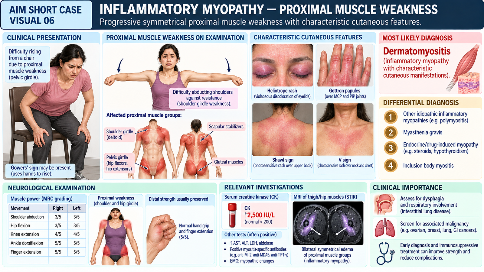
A 38-year-old woman attends the medical outpatient department with gradually progressive difficulty climbing stairs, rising from a low chair and lifting her arms above shoulder level over the past three months.
She also reports a reddish-purple rash around her eyelids and scaly lesions over her knuckles. She has been referred for a focused muscle examination.
2. Student Task
“Examine this patient’s proximal muscle strength, assess relevant neurological findings, identify any characteristic skin manifestations and present your findings with the most likely diagnosis.”
The examination should emphasize symmetrical proximal muscle weakness, functional impairment, preserved sensation and reflexes, and features suggesting inflammatory myopathy.
3. Focused Examination Approach
A. Initial observation
Introduce yourself, obtain consent, assess whether the patient can safely stand and walk, and expose the upper and lower limbs appropriately while maintaining privacy.
- Observe posture, spontaneous movements and difficulty changing position.
- Look for muscle wasting, asymmetry or abnormal involuntary movements.
- Observe the patient rising from a chair, with assistance if required.
- Note whether the patient uses her arms to push herself upward.
- Inspect exposed skin for characteristic inflammatory lesions.
B. Functional assessment of proximal muscles
- Chair-rise test: Ask the patient to rise from a seated position without using her hands, if safe.
- Shoulder function: Ask her to lift both arms above the head and maintain the position.
- Overhead activity: Ask about difficulty combing hair or placing objects on a high shelf.
- Lower limb function: Observe standing and walking; ask about stair climbing.
- Floor-rise maneuver: Consider only if clinically appropriate and safe; do not require it in a patient at risk of falling.
C. Upper limb muscle power
Compare both sides and assess strength using the Medical Research Council (MRC) grading scale.
- Shoulder abduction: Test deltoid strength against resistance.
- Shoulder flexion: Ask the patient to raise the arm against resistance.
- Elbow flexion and extension: Assess biceps and triceps power.
- Distal power: Test wrist extension and finger grip to determine whether distal muscles are relatively preserved.
D. Lower limb muscle power
- Hip flexion: Ask the patient to lift each thigh against resistance.
- Hip extension and abduction: Assess additional proximal pelvic girdle muscle groups as appropriate.
- Knee extension and flexion: Test quadriceps and hamstring strength.
- Distal power: Test ankle dorsiflexion and plantarflexion.
- Document whether weakness is symmetrical and predominantly proximal.
E. Reflexes, tone and sensation
- Assess muscle tone in the upper and lower limbs.
- Test biceps, triceps, knee and ankle reflexes.
- Assess light-touch sensation in a representative distribution.
- Look for abnormal plantar responses or other neurological signs when indicated.
In uncomplicated inflammatory myopathy, sensation is generally preserved, tone is usually normal or reduced, and tendon reflexes are commonly preserved unless weakness is severe.
F. Skin examination for dermatomyositis
- Heliotrope rash: Look for violaceous discoloration around the eyelids, sometimes associated with swelling.
- Gottron papules: Inspect the extensor surfaces of MCP, PIP and DIP joints for erythematous or violaceous papules or plaques.
- Gottron sign: Look for similar lesions over elbows or knees.
- Shawl sign: Inspect the upper back, shoulders and posterior neck for photosensitive erythema when appropriate.
- V-sign: Inspect the anterior neck and upper chest for a photosensitive rash.
- Periungual changes: Look for nailfold erythema and abnormal capillaries.
G. Relevant systemic assessment
- Ask briefly about difficulty swallowing, choking or nasal regurgitation.
- Assess voice quality and cough effectiveness when bulbar weakness is suspected.
- Ask about breathlessness and examine the respiratory system if indicated.
- Review for symptoms suggesting associated connective tissue disease or systemic illness.
4. Important Clinical Findings
Positive Findings in This Patient
| Clinical finding | Clinical interpretation |
|---|---|
| Difficulty rising from a chair without using arms | Functional pelvic girdle weakness |
| Bilateral shoulder abduction power 4-/5 | Symmetrical proximal upper limb weakness |
| Bilateral hip flexion power 3+/5 | Predominant proximal lower limb weakness |
| Distal limb strength 5/5 | Relative preservation of distal muscles |
| Violaceous eyelid discoloration | Heliotrope rash |
| Violaceous scaly papules over finger knuckles | Gottron papules |
Important Relevant Negative Findings
- Normal light-touch sensation in all four limbs.
- Preserved tendon reflexes and normal plantar responses.
- No significant distal muscle weakness.
- No ptosis, diplopia or fluctuating fatigable weakness.
- No clinical dysphagia or respiratory distress at this assessment.
Interpretation: Symmetrical proximal weakness with preserved sensation and characteristic cutaneous lesions strongly suggests dermatomyositis.
5. Presentation of Findings
“I examined a 38-year-old woman with symmetrical proximal muscle weakness affecting both shoulder and pelvic girdles. Shoulder abduction power is approximately 4-/5 bilaterally, and hip flexion is 3+/5. She has difficulty rising from a chair without upper limb support. Distal muscle power, sensation and deep tendon reflexes are preserved. Examination also reveals a heliotrope rash and Gottron papules over the finger joints. These findings are strongly suggestive of dermatomyositis. I would assess muscle enzymes, myositis-associated antibodies and relevant muscle investigations, and screen for swallowing, respiratory and other systemic complications.”
6. Most Likely Diagnosis
Suspected dermatomyositis presenting with symmetrical proximal muscle weakness and characteristic cutaneous manifestations.
The combination of proximal myopathic weakness, heliotrope rash and Gottron papules is highly suggestive of dermatomyositis.
Confirmation requires appropriate clinical evaluation, laboratory testing and, where indicated, muscle imaging or other specialized investigations.
7. Differential Diagnosis
| Differential | Distinguishing clue |
|---|---|
| Other idiopathic inflammatory myopathies | May produce proximal weakness without classic heliotrope rash or Gottron papules. |
| Myasthenia gravis | Fluctuating fatigable weakness, often with ptosis, diplopia or bulbar symptoms; characteristic dermatomyositis skin lesions are absent. |
| Endocrine or drug-induced myopathy | Proximal weakness may occur with thyroid disease, glucocorticoid exposure or myotoxic medication use, generally without characteristic inflammatory skin lesions. |
| Inclusion body myositis | Typically occurs in older adults, with prominent finger flexor and quadriceps weakness, often asymmetrical. |
8. Relevant Investigations
| Investigation | Purpose / expected finding |
|---|---|
| Serum creatine kinase (CK), aldolase, AST and ALT | Assess muscle injury; CK is commonly elevated but may be normal in some patients. |
| ANA and myositis-specific antibody panel | Support characterization of inflammatory myopathy; relevant antibodies include anti-Mi-2, anti-MDA5 and anti-TIF1-gamma. |
| MRI of affected proximal muscles | May demonstrate muscle edema or inflammation and help select a biopsy site. |
| Electromyography (EMG) | May demonstrate a myopathic pattern and assist in differentiating muscle from neurogenic disorders. |
| Muscle biopsy, when indicated | Provides histopathological support, particularly when the diagnosis is uncertain. |
| Organ and malignancy assessment | Evaluate for interstitial lung disease, dysphagia and associated malignancy according to individual risk. |
Investigation selection should be guided by the clinical phenotype and specialist assessment. A normal CK level alone does not exclude dermatomyositis.
9. Management Discussion
- Arrange rheumatology or neuromuscular specialist assessment to confirm the diagnosis and assess disease severity.
- Systemic glucocorticoids are commonly used for active muscle disease, with steroid-sparing immunosuppressive therapy selected according to severity and organ involvement.
- Introduce supervised physiotherapy and graded rehabilitation to preserve strength and function.
- Provide sun protection and appropriate treatment of inflammatory skin lesions.
- Assess swallowing and respiratory function, and undertake individualized malignancy screening.
10. Red Flags / Serious Complications
- New dysphagia, choking or recurrent aspiration — possible bulbar muscle involvement.
- Rapidly worsening breathlessness or hypoxemia — possible interstitial lung disease or respiratory muscle weakness.
- Rapidly progressive severe weakness or inability to mobilize — significant active muscle disease.
- Unexplained weight loss or other concerning systemic symptoms — assess for associated malignancy.
Acute swallowing difficulty, respiratory compromise or rapidly progressive weakness requires urgent assessment.
11. Examiner Viva Preparation
Q1. What is the typical pattern of weakness in inflammatory myopathy?
Usually symmetrical, predominantly proximal muscle weakness affecting the shoulder and pelvic girdles, with difficulty rising from a chair, climbing stairs and lifting the arms.
Q2. How do you distinguish myopathic weakness from peripheral neuropathy at the bedside?
Myopathy commonly produces proximal weakness with preserved sensation and relatively preserved reflexes. Peripheral neuropathy more often causes distal weakness, sensory abnormalities and reduced reflexes.
Q3. What are Gottron papules?
They are erythematous or violaceous papules or plaques, typically over the extensor surfaces of the finger joints, and are characteristic of dermatomyositis.
Q4. What is a heliotrope rash?
A violaceous rash affecting the upper eyelids, sometimes associated with periorbital swelling, classically seen in dermatomyositis.
Q5. Which laboratory investigation is commonly elevated in inflammatory myopathy?
Serum creatine kinase is commonly elevated due to muscle injury. Aldolase and muscle-associated transaminases may also be elevated.
Q6. Why should swallowing and respiratory symptoms be assessed?
Inflammatory myopathy may involve pharyngeal muscles, respiratory muscles or the lungs, resulting in dysphagia, aspiration, respiratory weakness or interstitial lung disease.
Q7. Why is malignancy assessment important in adult dermatomyositis?
Adult-onset dermatomyositis is associated with an increased risk of malignancy, especially around the time of disease onset. Screening should be individualized according to clinical risk factors and current recommendations.
12. AIM Short Case High-Yield Review
- Typical muscle pattern: Symmetrical proximal weakness affecting shoulder and pelvic girdles.
- Functional clue: Difficulty rising from a chair, climbing stairs and performing overhead activities.
- Power examination: Test shoulder abduction and hip flexion bilaterally using MRC grading.
- Neurological pattern: Sensation is usually preserved, with relatively preserved tendon reflexes.
- Characteristic skin sign: Heliotrope rash around the eyelids.
- Characteristic hand finding: Gottron papules over extensor finger joints.
- Most likely diagnosis: Dermatomyositis when proximal myopathy coexists with characteristic cutaneous lesions.
- Important investigations: CK, myositis antibodies, muscle MRI and selected EMG or biopsy.
- Major complications: Dysphagia, aspiration, interstitial lung disease and associated malignancy.
- Common examination mistake: Focusing only on muscle strength without examining the skin, reflexes, sensation and functional movements.
13. AIM Short Case Clinical Visual