FINAL YEAR MBBS | KMU CURRICULUM | AIM CLINICAL LEARNING
🩺 AIM SHORT CASE 04
Systemic Lupus Erythematosus — Focused Clinical Examination
MS Module — Final Year MBBS
Related AIM Topic 8 — Systemic Autoimmune & Connective Tissue Diseases
Recognize characteristic mucocutaneous and musculoskeletal findings, assess systemic involvement and interpret relevant autoimmune investigations.
1. Patient / Clinical Setting
A 24-year-old woman attends the medical outpatient department with intermittent joint pain, facial redness and fatigue for four months. She reports that the facial rash becomes more noticeable after sun exposure.
She is clinically stable and has been referred for a focused examination of her skin, joints and relevant systemic findings.
2. Student Task
“Perform a focused examination of this patient’s skin, oral cavity, hands and joints. Look for evidence of systemic involvement and present your findings with the most likely clinical diagnosis.”
The examination should be respectful, systematic and directed toward identifying characteristic clinical signs rather than obtaining a detailed medical history.
3. Focused Examination Approach
A. Initial observation and general examination
Introduce yourself, obtain consent, ensure appropriate exposure and examine the patient in adequate lighting.
- Observe general appearance, nutritional status and visible skin lesions.
- Look for pallor, which may suggest anemia.
- Record blood pressure, particularly when renal involvement is suspected.
- Inspect the lower limbs for peripheral edema.
B. Examination of the face and skin
- Malar rash: Inspect the cheeks and nasal bridge for erythema, typically sparing the nasolabial folds.
- Photosensitive lesions: Examine exposed areas of the neck, upper chest and forearms.
- Discoid lesions: Look for well-defined scaly plaques with possible scarring or pigmentary change.
- Alopecia: Inspect the scalp for nonscarring hair loss.
- Purpura: Look for petechiae or bruising suggesting possible thrombocytopenia.
C. Oral and mucosal examination
- Inspect the oral mucosa and hard palate for ulcers.
- Note whether ulcers are painful or painless.
- Inspect the lips and visible mucosal surfaces for associated lesions.
D. Hands and musculoskeletal examination
- Inspect the metacarpophalangeal, proximal interphalangeal and wrist joints for swelling.
- Palpate for tenderness, warmth and synovial thickening.
- Assess active and passive movements of the affected joints.
- Look for symmetrical small-joint involvement.
- Identify any reducible deformities suggestive of Jaccoud arthropathy.
E. Focused systemic examination
- Renal: Assess blood pressure, pedal edema and periorbital puffiness.
- Cardiovascular: Auscultate for a pericardial friction rub when clinically indicated.
- Respiratory: Look for signs of pleural effusion or pleuritic involvement.
- Hematological: Assess pallor, purpura and relevant lymphadenopathy.
4. Important Clinical Findings
Positive Findings in This Patient
| Clinical finding | Clinical interpretation |
|---|---|
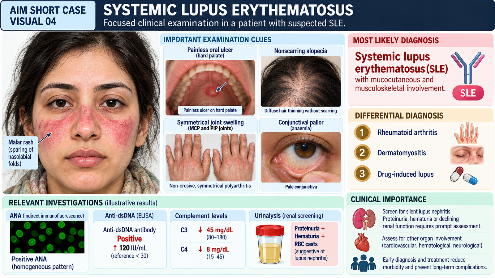
| Erythematous rash across cheeks and nasal bridge, sparing nasolabial folds | Typical malar rash supporting cutaneous lupus |
| Small painless palatal ulcer | Characteristic mucosal manifestation |
| Bilateral MCP and PIP joint tenderness with mild swelling | Inflammatory small-joint involvement |
| Diffuse nonscarring hair thinning | Possible lupus-associated alopecia |
| Mild conjunctival pallor | Possible anemia requiring CBC assessment |
Important Relevant Negative Findings
- No fixed joint deformities or obvious joint destruction.
- No peripheral edema or periorbital puffiness.
- Blood pressure is 112/72 mmHg.
- No clinically detectable pleural effusion or pericardial rub.
- No petechiae or significant bruising.
Interpretation: These negative findings reduce suspicion of certain overt complications but do not exclude lupus nephritis, hematological abnormalities or other internal organ involvement.
5. Presentation of Findings
“I examined a 24-year-old woman who has a malar erythematous rash sparing the nasolabial folds, a painless palatal ulcer, diffuse nonscarring alopecia and symmetrical tenderness with mild swelling of the small joints of both hands. There are no fixed joint deformities, peripheral edema or obvious cardiopulmonary abnormalities. Her blood pressure is normal. These findings are suggestive of systemic lupus erythematosus with mucocutaneous and musculoskeletal involvement. I would recommend autoimmune investigations, a complete blood count and renal assessment to support the diagnosis and evaluate systemic involvement.”
6. Most Likely Diagnosis
Suspected systemic lupus erythematosus (SLE) with mucocutaneous and inflammatory musculoskeletal manifestations.
The combination of malar rash, oral ulceration, nonscarring alopecia and symmetrical inflammatory small-joint involvement strongly supports SLE.
Clinical findings alone do not establish the diagnosis. Autoimmune serology and assessment of organ involvement are required.
7. Differential Diagnosis
| Differential | Distinguishing clue |
|---|---|
| Rheumatoid arthritis | Persistent symmetrical inflammatory polyarthritis; erosive disease and anti-CCP positivity favor RA. |
| Dermatomyositis | Heliotrope rash, Gottron papules and proximal muscle weakness. |
| Drug-induced lupus | Relevant drug exposure, frequent antihistone antibodies and generally less renal involvement. |
8. Relevant Investigations
| Investigation | Purpose / expected finding |
|---|---|
| ANA | Sensitive screening test; frequently positive in SLE but not specific. |
| Anti-dsDNA, anti-Sm and C3/C4 | Support autoimmune diagnosis; anti-dsDNA elevation and low complement may suggest active disease. |
| Complete blood count | Detect anemia, leukopenia or thrombocytopenia. |
| Urinalysis, urine protein quantification and serum creatinine | Screen for proteinuria, hematuria and impaired renal function suggestive of lupus nephritis. |
A positive ANA alone is insufficient to diagnose SLE. Results must be interpreted alongside clinical manifestations and other immunological findings.
9. Management Discussion
- Arrange physician or rheumatology assessment to confirm SLE and evaluate disease activity.
- Advise sun protection and avoidance of excessive ultraviolet exposure.
- Hydroxychloroquine is commonly recommended for confirmed SLE unless contraindicated, with appropriate monitoring.
- Use anti-inflammatory or immunosuppressive treatment according to severity and organ involvement under specialist supervision.
- Monitor blood pressure, CBC, urinalysis and renal function to detect complications early.
10. Red Flags / Serious Complications
- New edema, hypertension, significant proteinuria or hematuria — possible lupus nephritis.
- Chest pain, breathlessness or hypoxia — possible cardiopulmonary involvement.
- Seizures, confusion or new neurological deficits — possible neuropsychiatric involvement.
- Marked pallor, bleeding or purpura — possible serious hematological involvement.
Any suspected major organ involvement requires urgent clinical assessment and escalation.
11. Examiner Viva Preparation
Q1. What is the characteristic distribution of the malar rash in SLE?
It involves the cheeks and nasal bridge, typically sparing the nasolabial folds. It is often aggravated by sunlight.
Q2. How does lupus arthritis differ from rheumatoid arthritis?
Lupus arthritis is usually nonerosive, whereas rheumatoid arthritis may produce persistent erosive joint damage and fixed deformities.
Q3. What is the significance of a painless oral ulcer in this patient?
Oral ulceration is a recognized mucocutaneous manifestation of SLE and supports the diagnosis when accompanied by other characteristic findings.
Q4. Which investigation is commonly used as an initial immunological screening test for SLE?
Antinuclear antibody (ANA). It has high sensitivity but limited specificity and cannot independently confirm SLE.
Q5. Which autoimmune investigations support the diagnosis more specifically?
Anti-dsDNA and anti-Sm antibodies. Complement levels may help assess disease activity.
Q6. How would you assess possible lupus nephritis in this patient?
Check blood pressure and edema, perform urinalysis, quantify urine protein and assess serum creatinine. Renal disease may be present despite a normal physical examination.
Q7. What is the basic treatment principle for uncomplicated SLE?
Sun protection, hydroxychloroquine when appropriate, symptom control and regular monitoring. Treatment is escalated according to disease activity and organ involvement.
12. AIM Short Case High-Yield Review
- Malar rash: Erythema over cheeks and nasal bridge with typical nasolabial fold sparing.
- Oral ulcers: May be painless, particularly on the hard palate.
- Joint involvement: Symmetrical inflammatory polyarthritis, usually nonerosive.
- Alopecia: Nonscarring hair loss is a recognized manifestation.
- Clinical impression: Combined skin, mucosal and joint findings strongly suggest SLE.
- Important differential: Rheumatoid arthritis, dermatomyositis and drug-induced lupus.
- Core investigations: ANA, anti-dsDNA, anti-Sm, complement, CBC and renal screening.
- Management principle: Sun protection, hydroxychloroquine when indicated and organ-specific assessment.
- Red flag: Proteinuria or hematuria may indicate lupus nephritis even without edema.
- Common examination mistake: Diagnosing SLE from a positive ANA alone or failing to screen for silent renal involvement.
13. AIM Short Case Clinical Visual