
Station 10 — Adult Pneumonia Respiratory Examination
🎯 Candidate Task
Perform a focused respiratory examination on this adult patient with an acute respiratory illness. State the important findings, interpret their clinical significance, and outline appropriate investigations and initial management.
⚡ Model Station Answer
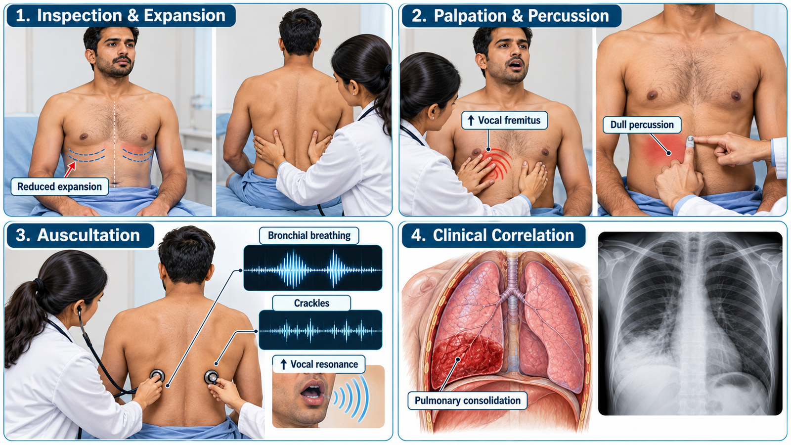
Key findings: I would look for fever, tachypnea, reduced movement of the affected side, increased tactile vocal fremitus, dullness to percussion and bronchial breathing with inspiratory crackles and increased vocal resonance.
Interpretation: These focal findings are consistent with pulmonary consolidation in the appropriate clinical setting.
Investigations: I would assess oxygen saturation and arrange a chest X-ray, with appropriate laboratory and microbiological investigations according to severity.
Management: I would assess severity, provide supportive care including oxygen when indicated, begin appropriate treatment for pneumonia and monitor closely for deterioration or complications.
🔎 How to Perform / Approach
- Prepare: Introduce yourself, confirm the patient, explain the examination, obtain consent, perform hand hygiene and expose the chest appropriately while maintaining dignity.
- General assessment: From the end of the bed assess respiratory distress, respiratory rate, oxygen saturation where available, fever, cyanosis and the patient’s overall condition.
- Inspect the chest: Assess chest shape, symmetry, respiratory pattern and movement; look particularly for reduced movement of one region or hemithorax.
- Palpate: Assess tracheal position, chest expansion and tactile vocal fremitus, comparing corresponding areas on both sides.
- Percuss: Percuss systematically over corresponding lung fields. A localized dull percussion note supports underlying consolidation when correlated with other findings.
- Auscultate: Compare breath sounds bilaterally and assess for bronchial breathing, inspiratory crackles and increased vocal resonance over the affected area.
- Conclude: Summarize the findings, state whether they suggest pulmonary consolidation, and explain that you would complete assessment with oxygen saturation, relevant investigations and severity-guided initial management.
🖼️ Visual Learning
Pulmonary Consolidation — Focused Respiratory Examination
📝 Important Viva Questions
Answer: Reduced local chest expansion, increased tactile vocal fremitus, dull percussion, bronchial breathing, inspiratory crackles and increased vocal resonance are typical findings.
Answer: Consolidated lung transmits high-frequency airway sounds more effectively from the bronchi to the chest wall.
Answer: A chest X-ray is commonly used to identify pulmonary infiltrates or consolidation and assess their extent.
Answer: Important complications include respiratory failure, pleural effusion or empyema, sepsis and lung abscess.
Answer: Significant hypoxemia, severe respiratory distress, altered mental status, hemodynamic instability or rapidly worsening clinical condition are important red flags.
CVS