
Station 02 — ECG Interpretation: Arrhythmias & Heart Blocks
🎯 Candidate Task
You are provided with a 12-lead ECG from a patient presenting with palpitations. Interpret the ECG systematically, identify the rhythm abnormality or conduction disorder, and state the important immediate management implication if a clinically significant arrhythmia is present.
⚡ Model Station Answer
- Systematic review: I would confirm the ECG quality, then assess rate, rhythm, P waves, PR interval, QRS complexes and the relationship between atrial and ventricular activity.
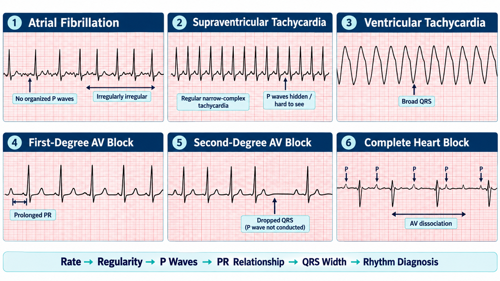
- Atrial fibrillation: An irregularly irregular rhythm with absent organized P waves is consistent with atrial fibrillation.
- SVT: A regular narrow-complex tachycardia, usually with P waves absent or difficult to identify, suggests supraventricular tachycardia.
- Ventricular tachycardia / heart block: A regular broad-complex tachycardia should be treated as ventricular tachycardia when clinically appropriate; heart block is identified by abnormal AV conduction ranging from PR prolongation to dropped beats or AV dissociation.
- Clinical implication: I would immediately assess hemodynamic stability. An unstable significant tachyarrhythmia or bradyarrhythmia requires urgent escalation and appropriate emergency management.
🔎 How to Perform / Approach
- Check the tracing: Confirm patient details where available, ECG calibration and whether the tracing is technically adequate before interpretation.
- Determine the rate: Decide whether the ventricular rate is normal, tachycardic or bradycardic.
- Assess rhythm regularity: Determine whether the R–R intervals are regular or irregular. An irregularly irregular rhythm is an important clue to atrial fibrillation.
- Assess atrial activity and AV conduction: Look for P waves, determine whether each P wave is followed by a QRS complex and assess the PR interval for prolongation, progressive change or dropped beats.
- Assess QRS complexes: Determine whether the QRS complexes are narrow or broad. A regular narrow-complex tachycardia suggests SVT, while a regular broad-complex tachycardia raises strong concern for ventricular tachycardia.
- State the rhythm diagnosis: Combine rate, regularity, P-wave activity, PR relationship and QRS width to identify AF, SVT, VT or the relevant heart block.
- Link ECG to the patient: Assess for hypotension, altered consciousness, ischemic chest pain, shock or acute heart failure. Significant arrhythmia with instability requires urgent management rather than prolonged ECG discussion.
🖼️ Visual Learning
ECG Pattern Recognition — AF, SVT, VT & Heart Blocks
📝 Important Viva Questions
Atrial fibrillation shows an irregularly irregular ventricular rhythm with absence of consistent organized P waves.
SVT typically produces a rapid, regular narrow-complex tachycardia in which P waves may be absent, hidden or difficult to identify.
A regular broad-complex tachycardia should strongly suggest ventricular tachycardia, particularly in a patient with structural or ischemic heart disease.
First-degree AV block has a prolonged PR interval with every P wave conducted. Complete heart block has no consistent relationship between P waves and QRS complexes because atrial and ventricular activity are dissociated.
Immediately assess hemodynamic stability. Hypotension, shock, altered consciousness, ischemic chest pain or acute heart failure indicates the need for urgent escalation and emergency treatment.