
AIM OSCE / OSPE • FINAL YEAR MBBS
OSCE Station 03 — Acute Stroke: NIHSS, Glasgow Coma Scale & Neuroimaging Interpretation
Neuroscience Module • Medicine
Rapid Revision • Clinical Skills • Practical Skills • Viva
🎯 Candidate Task
A patient presents to the emergency department with sudden-onset focal neurological deficits. Demonstrate a structured neurological assessment using the NIH Stroke Scale (NIHSS) and Glasgow Coma Scale (GCS), interpret the supplied CT/MRI findings, and state your immediate diagnostic and management priorities.
⚡ Model Station Answer
Clinical assessment: “I will activate the stroke pathway, assess ABCs, check blood glucose and establish the last-known-well time.”
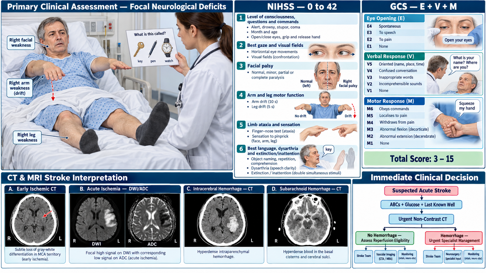
NIHSS: “I will assess consciousness, gaze, visual fields, facial weakness, limb power, ataxia, sensation, language, dysarthria and neglect, then document the total score out of 42.”
GCS: “I will assess eye opening, verbal response and motor response, recording the individual components and total out of 15.”
Imaging: “Urgent non-contrast CT will identify intracranial hemorrhage. MRI diffusion-weighted imaging can demonstrate acute ischemia.”
Priority: “I will arrange immediate specialist stroke assessment, determine eligibility for reperfusion treatment after excluding hemorrhage, and provide appropriate supportive care.”
🔎 How to Perform / Approach
- Recognize and stabilize: Identify sudden hemiparesis, facial weakness or aphasia. Assess ABCs, vital signs, oxygen saturation and capillary glucose; establish last-known-well time and activate the stroke pathway.
- Assess NIHSS — consciousness and cranial functions: Score level of consciousness, orientation questions, commands, gaze, visual fields and facial palsy using the standardized NIHSS assessment.
- Complete NIHSS — motor and cortical functions: Assess right and left arm and leg drift, limb ataxia, sensation, best language, dysarthria and extinction/inattention. Record each item and calculate the NIHSS score (0–42).
- Assess Glasgow Coma Scale: Record eye opening (E1–4), verbal response (V1–5) and motor response (M1–6). State the component scores and total (3–15), documenting untestable components when applicable.
- Interpret urgent neuroimaging: Review non-contrast CT for early ischemic changes, intraparenchymal hemorrhage and subarachnoid blood in sulci or basal cisterns. On MRI, identify acute ischemia by diffusion restriction on DWI with corresponding low ADC.
- Document the clinical interpretation: Record neurological deficits, NIHSS, GCS, last-known-well time and imaging findings. Distinguish likely ischemic stroke from hemorrhagic stroke or subarachnoid hemorrhage.
- State immediate management priorities: Arrange urgent stroke-team assessment and appropriate vascular imaging. Evaluate reperfusion eligibility for ischemic stroke; for hemorrhage, prioritize blood pressure management and specialist/neurosurgical assessment. Monitor neurological status and protect swallowing and airway safety.
🖼️ Visual Learning
Acute Stroke Examination — NIHSS, GCS & CT/MRI Interpretation
🎥 Practical Demonstration
NIH Stroke Scale — Practical Clinical Demonstration
North Dakota Department of Health and Human Services. A practical demonstration of structured NIHSS assessment, useful for observing examination sequence and neurological deficit scoring.
📝 Important Viva Questions
Q1. What is the NIHSS score range, and what does a higher score indicate?
Answer: NIHSS ranges from 0 to 42. Higher scores generally indicate greater stroke-related neurological impairment.
Q2. What are the three components of GCS and their maximum scores?
Answer: Eye opening (4), verbal response (5) and motor response (6), giving a maximum total of 15.
Q3. Why is non-contrast CT brain the initial imaging investigation in suspected acute stroke?
Answer: It rapidly identifies intracranial hemorrhage and helps guide safe decisions regarding acute stroke treatment.
Q4. What neuroimaging findings distinguish acute ischemic stroke, intracerebral hemorrhage and subarachnoid hemorrhage?
Answer: Acute ischemia may show DWI hyperintensity with low ADC; intracerebral hemorrhage appears hyperdense within brain parenchyma on CT, while subarachnoid hemorrhage produces hyperdensity in sulci or basal cisterns.
Q5. What are the immediate management priorities in suspected acute ischemic stroke?
Answer: Stabilize ABCs, check glucose, establish last-known-well time, activate the stroke team, obtain urgent brain imaging and assess eligibility for reperfusion treatment.