
AIM OSCE / OSPE • FINAL YEAR MBBS
OSCE Station 07 — Meningitis: CSF Interpretation & Lumbar Puncture Observation
Neuroscience Module • Medicine
Rapid Revision • Clinical Skills • Practical Skills • Viva
🎯 Candidate Task
A patient with suspected meningitis has undergone cerebrospinal fluid (CSF) examination. Interpret the supplied CSF findings, differentiate the likely types of meningitis, identify relevant radiological complications, and explain important lumbar puncture precautions and procedural steps during observation.
⚡ Model Station Answer
CSF interpretation: “I will assess appearance, opening pressure if available, white cell count and differential, protein, glucose with paired blood glucose, and microbiological results.”
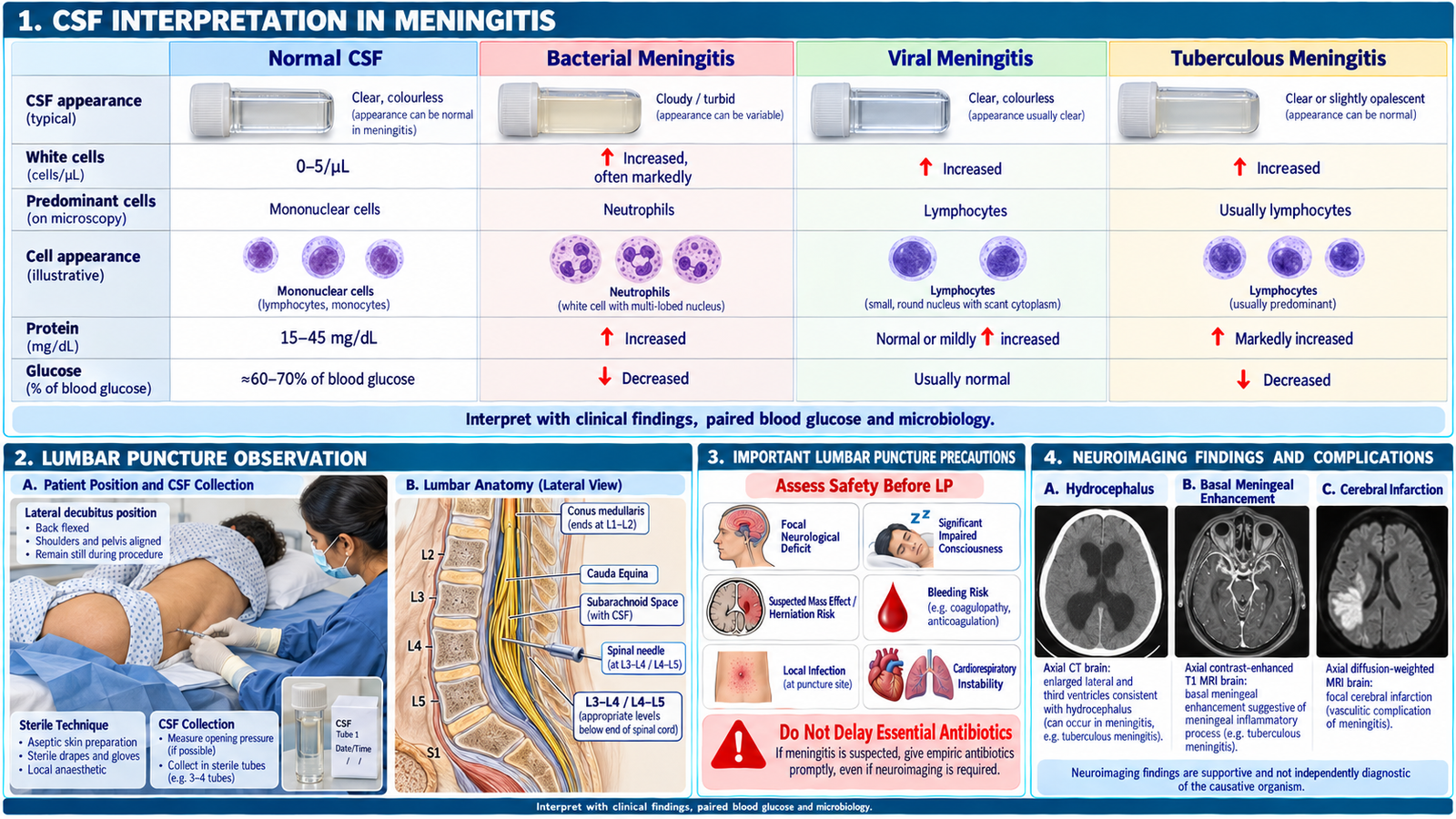
Classification: “Bacterial meningitis usually produces neutrophilic pleocytosis, raised protein and low glucose; viral meningitis usually shows lymphocytes with normal glucose; tuberculous meningitis commonly shows lymphocytes, markedly raised protein and low glucose.”
Imaging: “CT or MRI may identify hydrocephalus, cerebral edema, infarction, abscess or meningeal enhancement.”
Lumbar puncture: “Before the procedure, I will check for contraindications, ensure consent and asepsis, and observe positioning, landmark identification and CSF collection.”
Safety: “Suspected acute bacterial meningitis requires urgent hospital management; investigation must not delay essential antimicrobial treatment.”
🔎 How to Perform / Approach
- Review the CSF report: Confirm the clinical context, CSF appearance, opening pressure if recorded, cell count and differential, protein, glucose with paired serum glucose, Gram stain, culture and relevant molecular tests.
- Differentiate the CSF patterns: Acute pyogenic meningitis typically shows predominantly neutrophils, high protein and low CSF glucose. Viral meningitis usually shows lymphocytic predominance, mildly raised protein and normal glucose. Tuberculous meningitis commonly shows lymphocytes, high protein and low glucose.
- Interpret the likely diagnosis: Relate the CSF pattern to the clinical presentation and microbiological findings. Remember that early viral or tuberculous meningitis may show neutrophils, so no single CSF parameter establishes the diagnosis.
- Recognize imaging findings and complications: Identify hydrocephalus, cerebral edema, infarction, abscess or meningeal enhancement when demonstrated on CT/MRI. Neuroimaging is indicated for selected clinical concerns rather than routinely before every lumbar puncture.
- Check lumbar puncture precautions: Assess for signs of raised intracranial pressure from a mass lesion, focal deficits, significant impaired consciousness, cardiorespiratory instability, infection at the puncture site and bleeding risk. Arrange appropriate senior review or imaging when indicated.
- Observe lumbar puncture technique: Confirm consent, hand hygiene, asepsis and correct positioning. Identify the L3–L4 or L4–L5 interspace, observe local anesthesia, spinal needle insertion, opening-pressure measurement when appropriate, sterile CSF collection and specimen labelling.
- State the clinical priority: Ensure prompt assessment and treatment for suspected bacterial meningitis, without delaying antibiotics for CT or lumbar puncture. Recognize potential procedure complications, including post-dural puncture headache, bleeding and infection.
🖼️ Visual Learning
Meningitis — CSF Pattern Comparison, Lumbar Puncture Landmarks & Precautions
📝 Important Viva Questions
Q1. What CSF findings are typical of acute bacterial meningitis?
Answer: Predominantly neutrophilic pleocytosis, increased protein and decreased CSF glucose relative to blood glucose.
Q2. How does viral meningitis typically differ from tuberculous meningitis on CSF examination?
Answer: Viral meningitis usually shows lymphocytes, mildly elevated protein and normal glucose; tuberculous meningitis usually shows lymphocytes, markedly elevated protein and low glucose.
Q3. At which vertebral levels is lumbar puncture commonly performed in adults?
Answer: Usually at the L3–L4 or L4–L5 interspace, below the termination of the spinal cord.
Q4. What important clinical findings may make lumbar puncture unsafe without further assessment?
Answer: Suspected mass effect or herniation risk, significant impaired consciousness, focal neurological deficits, major bleeding risk, local infection or cardiorespiratory instability.
Q5. Name three important radiological complications of meningitis.
Answer: Hydrocephalus, cerebral infarction and brain abscess; cerebral edema may also occur.