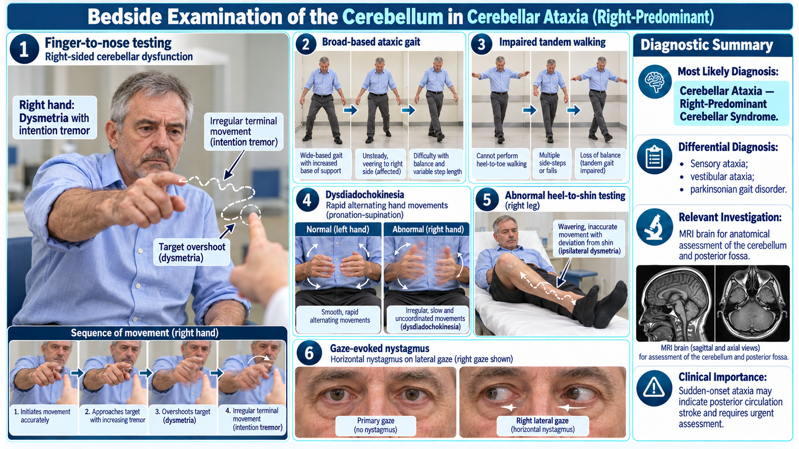
Cerebellar Ataxia — Focused Neurological Examination
Neuroscience Module | Medicine
Focused bedside assessment of gait, balance, limb coordination, dysmetria and intention tremor to recognize cerebellar dysfunction and determine neurological localization.
1. Patient / Clinical Setting
A 56-year-old man attends the medical outpatient department with gradually increasing unsteadiness while walking over six months. He describes difficulty walking in a straight line and occasionally overshooting objects when reaching for them.
Clinical setting: The patient is conscious, cooperative and able to walk with supervision. A focused neurological examination is requested to assess coordination, gait and balance.
2. Student Task
Examine this patient’s gait, balance and limb coordination. Identify the important neurological signs, determine the likely anatomical localization and present your clinical findings to the examiner.
3. Focused Examination Approach
Introduce yourself, obtain consent and ensure the patient is comfortable. Perform coordination tests with the patient seated initially. Assess standing balance and gait only when safe, remaining close enough to prevent falls.
A. Initial Observation
- Posture: Observe the patient while seated for truncal swaying or difficulty maintaining an upright position.
- Spontaneous movements: Look for irregular, poorly coordinated limb movements.
- Speech: Listen for slow, irregular or scanning dysarthria.
- Head and eye movements: Observe for abnormal eye movements or spontaneous nystagmus.
Clinical importance: Truncal instability suggests involvement of midline cerebellar structures, while prominent limb incoordination may indicate cerebellar hemispheric dysfunction.
B. Gait Examination
- Ask the patient to stand with appropriate assistance.
- Observe the width of the base of support.
- Ask the patient to walk forward, turn and return.
- Look for a broad-based, staggering gait with irregular steps and lateral deviation.
- If safe, ask the patient to perform tandem walking, placing heel directly in front of toe.
Interpretation: A broad-based, irregular gait with difficulty performing tandem walking supports cerebellar ataxia.
C. Standing Balance and Romberg Test
- Assess stability while standing with the feet together and eyes open.
- If sufficiently stable, ask the patient to close the eyes while maintaining close supervision.
- Compare the degree of instability with eyes open and closed.
Cerebellar ataxia: Instability is commonly present even with the eyes open and may persist when the eyes are closed.
Sensory ataxia: Balance typically deteriorates markedly after eye closure, producing a positive Romberg sign.
D. Upper Limb Coordination
Finger-to-Nose Test
- Ask the patient to touch their nose and then the examiner’s finger repeatedly.
- Move the target to assess accuracy across different distances.
- Look for overshooting or undershooting the target.
- Observe for increasing tremor as the finger approaches the target.
Interpretation: Dysmetria is impaired judgment of movement distance. Intention tremor becomes more prominent near the intended target.
Rapid Alternating Movements
- Ask the patient to rapidly alternate between palm-up and palm-down movements.
- Observe the rhythm, speed and regularity of movement.
- Compare both hands.
Interpretation: Irregular, poorly coordinated alternating movements demonstrate dysdiadochokinesia.
Finger Tapping
- Ask the patient to rapidly tap the index finger against the thumb.
- Observe irregular timing and poor coordination.
Distinguish irregular cerebellar movements from the progressive decrement characteristic of parkinsonian bradykinesia.
E. Lower Limb Coordination
Heel-to-Shin Test
- With the patient lying comfortably, ask them to place one heel on the opposite knee.
- Ask them to slide the heel down the shin toward the ankle.
- Repeat with the opposite leg.
- Look for wavering, overshooting and inability to maintain a straight movement.
Interpretation: An irregular heel-to-shin movement suggests lower limb cerebellar incoordination, provided significant weakness or sensory loss is not responsible.
F. Eye Movements and Speech
- Nystagmus: Assess eye movements during horizontal and vertical gaze, looking for gaze-evoked nystagmus.
- Saccades: Observe whether eye movements overshoot or undershoot visual targets.
- Speech: Ask the patient to speak a short sentence and listen for irregular articulation or scanning speech.
These findings provide supporting evidence of cerebellar dysfunction.
G. Brief Associated Neurological Examination
- Assess muscle tone for hypotonia where relevant.
- Check limb power to exclude weakness as the principal cause of poor movement.
- Assess joint position and vibration sensation when sensory ataxia is suspected.
- Look for focal cranial nerve abnormalities or pyramidal signs that may suggest an alternative or additional neurological lesion.
4. Important Clinical Findings
Positive Findings
| Clinical Finding | Interpretation |
|---|---|
| Broad-based, staggering gait | Suggests cerebellar gait ataxia. |
| Difficulty with tandem walking | Indicates impaired coordination and dynamic balance. |
| Dysmetria on finger-to-nose testing, more marked on the right | Suggests right cerebellar hemispheric involvement. |
| Intention tremor in the right upper limb | Supports impaired cerebellar control of targeted movement. |
| Irregular rapid alternating movements | Demonstrates dysdiadochokinesia. |
| Impaired heel-to-shin coordination, greater on the right | Supports ipsilateral lower limb cerebellar dysfunction. |
| Gaze-evoked nystagmus | Provides additional evidence of cerebellar or related ocular-motor pathway dysfunction. |
Important Relevant Negative Findings
- No significant limb weakness: Makes a primary motor deficit less likely to explain the incoordination.
- Preserved joint position and vibration sense: Argues against sensory ataxia.
- Imbalance present with eyes open: Supports cerebellar dysfunction rather than dependence on visual compensation.
- No resting pill-rolling tremor or typical rigidity: Makes Parkinson’s disease less likely.
These findings should be interpreted together rather than using any single clinical sign as definitive evidence.
5. Presentation of Findings
“I examined a 56-year-old gentleman who demonstrates a broad-based, unsteady gait with difficulty performing tandem walking. He has dysmetria and intention tremor on finger-to-nose testing, dysdiadochokinesia and impaired heel-to-shin coordination, predominantly on the right side. Gaze-evoked nystagmus is also present. Limb power and joint position sensation are preserved, and instability is evident even with the eyes open. These findings indicate a cerebellar syndrome, with prominent right cerebellar hemispheric involvement.”
6. Most Likely Diagnosis
Cerebellar Ataxia — Right-Predominant Cerebellar Syndrome
The combination of gait ataxia, dysmetria, intention tremor, dysdiadochokinesia and nystagmus supports cerebellar dysfunction. Right-sided limb incoordination suggests right cerebellar hemispheric involvement, while gait instability may reflect additional midline cerebellar dysfunction. The underlying etiology cannot be determined from examination alone.
7. Differential Diagnosis
| Differential | Distinguishing Clinical Clue |
|---|---|
| Sensory ataxia | Impaired proprioception, sensory loss and marked worsening of balance after eye closure. |
| Vestibular ataxia | Prominent vertigo, nausea and imbalance; limb coordination is usually relatively preserved. |
| Parkinsonian gait disorder | Short shuffling steps, bradykinesia, rigidity and reduced arm swing rather than true dysmetria. |
8. Relevant Investigations
Investigations are selected according to onset, progression, associated symptoms and suspected underlying cause.
- MRI brain: Evaluates the cerebellum and posterior fossa for structural lesions, infarction, demyelination or cerebellar atrophy.
- Vitamin B12 level: Helps identify a treatable nutritional cause, particularly when sensory impairment is also suspected.
- Thyroid function tests: Assess for thyroid dysfunction contributing to acquired ataxia.
- Medication and alcohol exposure review: Identifies potentially reversible toxic or drug-related causes of incoordination.
Clinical interpretation: MRI findings must be correlated with the neurological examination; cerebellar signs can occur without a specific structural abnormality visible on imaging.
9. Management Discussion
- Identify and treat the underlying cause where possible, including reversible metabolic, nutritional or medication-related factors.
- Arrange physiotherapy focusing on balance, gait coordination and fall prevention.
- Provide walking assistance or mobility aids when needed for safety.
- Consider occupational or speech therapy if daily functioning or articulation is significantly affected.
- Refer for neurological evaluation when symptoms progress, the cause is uncertain or examination suggests structural pathology.
10. Red Flags / Serious Complications
- Sudden-onset ataxia or inability to walk, suggesting possible posterior circulation stroke.
- Severe headache, repeated vomiting or altered consciousness.
- New cranial nerve deficits, weakness or other focal neurological signs.
- Rapidly progressive ataxia or severe unexplained deterioration.
- Repeated falls causing injury or inability to mobilize safely.
Sudden neurological deterioration or signs of posterior fossa pathology require urgent emergency assessment.
11. Examiner Viva Preparation
Q1. What is ataxia?
Answer: Ataxia is impaired coordination of voluntary movement that is not adequately explained by muscle weakness.
Q2. What is dysmetria, and how is it demonstrated?
Answer: Dysmetria is inaccurate judgment of movement distance, producing overshooting or undershooting of a target. It is demonstrated by finger-to-nose or heel-to-shin testing.
Q3. How does intention tremor differ from resting tremor?
Answer: Intention tremor becomes more pronounced as a movement approaches its target and suggests cerebellar dysfunction. Resting tremor is most evident in a relaxed limb and may decrease during purposeful movement.
Q4. What is dysdiadochokinesia?
Answer: Difficulty performing rapid alternating movements smoothly and rhythmically, commonly demonstrated by repeated pronation and supination of the hands.
Q5. How can cerebellar ataxia be distinguished from sensory ataxia?
Answer: Cerebellar ataxia produces poor coordination and instability even with eyes open. Sensory ataxia is associated with impaired proprioception and marked worsening of balance after eye closure.
Q6. Which side of the cerebellum is suggested by right-sided limb dysmetria?
Answer: The right cerebellar hemisphere because cerebellar hemispheric lesions generally produce ipsilateral limb incoordination.
Q7. Which investigation is most useful when a structural cerebellar lesion is suspected?
Answer: MRI brain, particularly for detailed assessment of the cerebellum, brainstem and posterior fossa. Acute suspected stroke requires emergency stroke imaging according to the clinical pathway.
12. AIM Short Case High-Yield Review
- Key gait sign: Broad-based, staggering gait with impaired tandem walking.
- Dysmetria: Overshooting or undershooting during targeted movement.
- Intention tremor: Tremor increases as the finger approaches a target.
- Dysdiadochokinesia: Irregular rapid alternating movements.
- Lower limb sign: Impaired heel-to-shin coordination.
- Associated clue: Gaze-evoked nystagmus or scanning dysarthria.
- Localization: Cerebellar hemispheric signs are commonly ipsilateral; truncal ataxia suggests midline dysfunction.
- Important differentiation: Sensory ataxia worsens substantially with eye closure and is associated with impaired proprioception.
- Investigation: MRI brain when structural pathology is suspected.
- Common examination mistake: Calling a Romberg test positive solely because a patient with cerebellar ataxia is unstable, without comparing balance with eyes open and closed.
13. AIM Short Case Clinical Visual