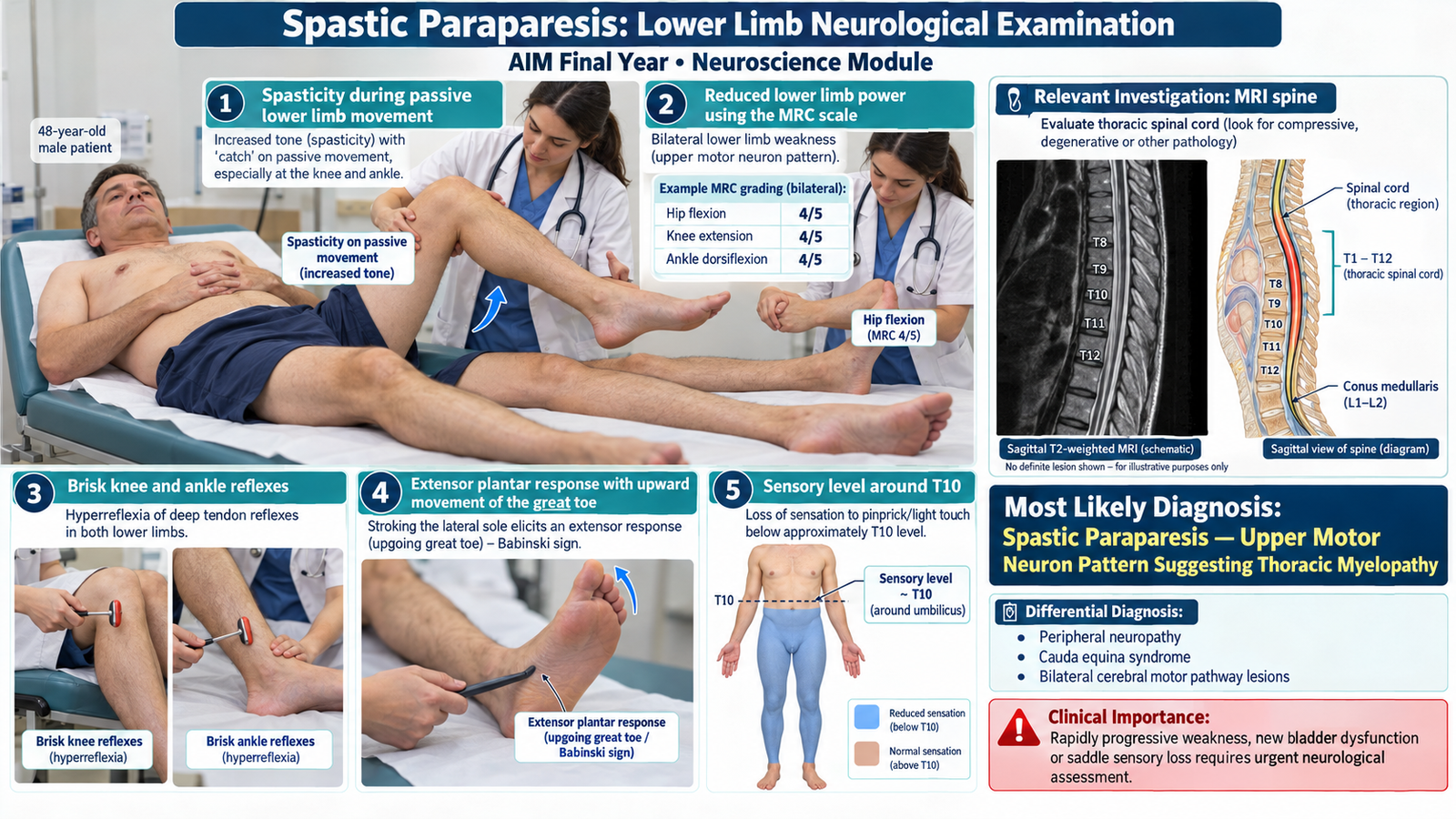
Paraplegia — Lower Limb Neurological Examination
Neuroscience Module | Medicine & Neurosurgery
Focused bedside assessment of lower limb tone, power, reflexes and sensation to identify upper or lower motor neuron signs and localize neurological weakness.
1. Patient / Clinical Setting
A 48-year-old man is admitted to the medical ward with progressive weakness of both lower limbs over three months. He now requires assistance to stand and reports stiffness in both legs with occasional dragging of his feet.
Clinical setting: The patient is conscious, cooperative and clinically stable. A focused lower limb neurological examination is required to identify the pattern of weakness and its likely anatomical localization.
2. Student Task
Examine the lower limbs of this patient, assessing muscle bulk, tone, power, reflexes and sensation. Identify upper or lower motor neuron signs, determine the likely site of neurological involvement and present your findings to the examiner.
3. Focused Examination Approach
Introduce yourself, obtain consent, ask about pain and position the patient comfortably in the supine position. Expose both lower limbs appropriately while maintaining dignity. Examine the two sides systematically and compare findings.
A. Inspection of Both Lower Limbs
- Position and posture: Observe the resting position of both legs and feet, looking for abnormal flexion, extension or deformity.
- Muscle bulk: Look for wasting or asymmetry of the quadriceps, calves and intrinsic foot muscles.
- Involuntary movements: Observe for fasciculations, spasms or spontaneous jerking movements.
- Skin and trophic changes: Look for pressure injuries, abnormal skin changes or contractures in patients with prolonged immobility.
- Associated clues: Note any spinal deformity or visible signs suggesting previous spinal injury or surgery.
Clinical importance: Marked muscle wasting and fasciculations suggest lower motor neuron involvement, whereas relatively preserved muscle bulk with stiffness may support an upper motor neuron pattern.
B. Assessment of Muscle Tone
- Ask the patient to relax completely.
- Support the leg and passively flex and extend the hip and knee.
- Assess passive movements at the ankle.
- Compare resistance to movement in both lower limbs.
- Check whether resistance increases with faster passive movement.
Spasticity: Velocity-dependent increased resistance to passive movement, suggesting upper motor neuron involvement.
Hypotonia: Reduced resistance to passive movement, which may occur with lower motor neuron lesions or during the acute spinal shock phase of spinal cord injury.
C. Assessment of Muscle Power
Test the major muscle groups against resistance, comparing both sides. Grade power using the Medical Research Council (MRC) scale.
| Movement | Principal Muscle Group | Main Root Level |
|---|---|---|
| Hip flexion | Iliopsoas | L2–L3 |
| Knee extension | Quadriceps | L3–L4 |
| Knee flexion | Hamstrings | L5–S1 |
| Ankle dorsiflexion | Tibialis anterior | L4–L5 |
| Great toe extension | Extensor hallucis longus | L5 |
| Ankle plantarflexion | Gastrocnemius and soleus | S1–S2 |
MRC Power Grading: 0/5 = no contraction; 1/5 = flicker; 2/5 = movement with gravity eliminated; 3/5 = movement against gravity; 4/5 = movement against resistance but reduced strength; 5/5 = normal power.
Interpretation: Document the distribution and severity of weakness rather than simply writing “power reduced.” Bilateral weakness with spasticity suggests involvement of descending motor pathways.
D. Deep Tendon Reflexes
- Knee jerk: Assess the patellar tendon reflex, mainly L3–L4.
- Ankle jerk: Assess the Achilles tendon reflex, mainly S1.
- Compare reflexes on both sides.
- Look for increased reflex responses or reflex spread.
- Check for ankle clonus when hyperreflexia or spasticity is present.
Interpretation: Exaggerated tendon reflexes and sustained clonus support an upper motor neuron lesion. Reduced or absent reflexes suggest interruption of the relevant lower motor neuron reflex arc, although acute spinal shock may temporarily suppress reflexes.
E. Plantar Response
- Gently stroke the lateral border of the sole from heel toward the forefoot and curve medially across the ball of the foot.
- Observe movement of the great toe and other toes.
- Compare the responses on both sides.
Normal response: Flexion of the great toe or a neutral response.
Extensor plantar response: Upward movement of the great toe, sometimes with fanning of the remaining toes.
Interpretation: An extensor plantar response in an adult is a significant sign of corticospinal tract dysfunction.
F. Sensory Examination
Assess and compare sensation systematically, preferably with the patient’s eyes closed.
- Light touch: Compare sensation in corresponding regions of both lower limbs.
- Pinprick: Assess pain sensation using an appropriate safe disposable neurological testing device.
- Vibration: Use a 128 Hz tuning fork, beginning at the great toe when appropriate.
- Joint position: Move the great toe slightly upward or downward while holding its sides.
- Sensory level: If spinal cord disease is suspected, assess the trunk and lower limbs to identify a reproducible upper boundary of sensory impairment.
Interpretation: A sensory level across the trunk strongly suggests spinal cord pathology. It should be recorded by dermatome, but the sensory level does not always correspond exactly to the affected vertebral level.
G. Gait and Functional Assessment
If the patient can stand and walk safely:
- Observe rising from the bed or chair.
- Assess standing posture and balance.
- Look for stiff-legged walking, toe dragging or circumduction.
- Observe for a scissoring gait caused by increased hip adductor tone.
- Assess the need for assistance or walking support.
Interpretation: A stiff, scissoring or circumducting gait supports a spastic paraparesis pattern. Do not ask a patient with severe weakness or instability to walk without adequate assistance.
H. Brief Assessment for Associated Spinal Cord Pathology
- Ask specifically about urinary retention, incontinence or altered bowel function.
- Look for a truncal sensory level.
- Check for spinal tenderness or deformity if clinically indicated.
- Perform a brief upper limb neurological assessment to help determine whether neurological involvement extends above the thoracic spinal cord.
- Consider focused sacral neurological assessment when cauda equina syndrome, conus involvement or acute spinal cord disease is suspected, using appropriate privacy, consent and supervision.
4. Important Clinical Findings
Positive Findings
| Clinical Finding | Clinical Interpretation |
|---|---|
| Bilateral lower limb weakness, approximately MRC 3/5 | Indicates significant paraparesis affecting both lower limbs. |
| Increased tone with velocity-dependent spasticity | Supports upper motor neuron involvement. |
| Exaggerated bilateral knee and ankle reflexes | Indicates loss of descending inhibitory control. |
| Bilateral extensor plantar responses | Suggests bilateral corticospinal tract dysfunction. |
| Reduced pinprick sensation below approximately the T10 dermatome | Suggests spinal cord sensory pathway involvement. |
| Stiff, assisted gait | Consistent with spastic paraparesis. |
Important Relevant Negative Findings
- No marked lower limb muscle wasting or fasciculations: Makes a widespread lower motor neuron disorder less likely.
- Upper limb neurological examination is normal: Supports localization below the cervical enlargement, although it does not definitively exclude cervical spinal disease.
- No cranial nerve abnormalities: Makes an associated brainstem lesion less likely.
- No obvious asymmetric cortical neurological signs: Supports a spinal rather than cerebral explanation for the bilateral lower limb findings.
5. Presentation of Findings
“I examined a 48-year-old gentleman who has bilateral lower limb weakness with power approximately 3/5, increased muscle tone, exaggerated knee and ankle reflexes and bilateral extensor plantar responses. There is reduced pinprick sensation below approximately the T10 dermatome. Muscle bulk is relatively preserved, and the upper limb examination is normal. These findings indicate a bilateral upper motor neuron pattern of weakness with an associated sensory level, most consistent with thoracic spinal cord involvement causing spastic paraparesis.”
6. Most Likely Diagnosis
Spastic Paraparesis — Upper Motor Neuron Pattern Suggesting Thoracic Myelopathy
The bilateral lower limb weakness, spasticity, hyperreflexia, extensor plantar responses and sensory level support spinal cord dysfunction. This is an anatomical and clinical diagnosis; the underlying cause, such as compression, inflammation or demyelination, must be established separately. Because measurable voluntary movement remains present, the current examination demonstrates paraparesis rather than complete paraplegia.
7. Differential Diagnosis
| Important Alternative | Distinguishing Clinical Clue |
|---|---|
| Peripheral neuropathy | Usually produces distal weakness, reduced reflexes and length-dependent sensory loss rather than spasticity and a truncal sensory level. |
| Cauda equina syndrome | May cause flaccid weakness, reduced lower limb reflexes, saddle sensory loss and bladder dysfunction. |
| Bilateral cerebral motor pathway lesions | May cause bilateral upper motor neuron weakness, but a distinct truncal sensory level favors spinal cord pathology. |
8. Relevant Investigations
Investigations should identify the anatomical lesion, establish its cause and detect important complications.
- MRI spine: The priority investigation when myelopathy is suspected. It can demonstrate spinal cord compression, intrinsic cord lesions, inflammatory changes or other structural pathology. Image the clinically relevant region, extending coverage when localization is uncertain.
- Complete blood count and inflammatory markers: Consider when infection, inflammation or systemic disease is suspected; abnormalities are supportive rather than diagnostic of myelopathy.
- Vitamin B12 level: May identify a treatable cause of progressive spinal cord dysfunction, particularly when posterior column signs are present.
- Post-void residual bladder volume: Assess when urinary symptoms suggest neurogenic bladder dysfunction or retention.
Clinical interpretation: A definite sensory level and bilateral upper motor neuron findings should prompt evaluation for spinal cord disease; a normal plain spinal radiograph does not exclude significant cord pathology.
9. Management Discussion
- Establish the cause of paraparesis and initiate appropriate treatment after clinical assessment and imaging.
- Arrange urgent MRI and specialist assessment if spinal cord compression or progressive myelopathy is suspected.
- Provide physiotherapy, safe positioning and mobility assistance to preserve function and reduce complications.
- Address spasticity, pain and bladder or bowel dysfunction when present.
- Monitor neurological power, sensory findings and sphincter function for deterioration.
10. Red Flags / Serious Complications
- New or rapidly progressive bilateral lower limb weakness.
- Acute urinary retention, new incontinence or loss of bowel control.
- Saddle sensory loss or rapidly ascending sensory deficit.
- Severe back pain with fever, malignancy history or suspected spinal compression.
- Sudden neurological deterioration after spinal trauma.
New neurological deterioration or sphincter dysfunction requires urgent spinal cord assessment and appropriate emergency imaging.
11. Examiner Viva Preparation
Q1. What is the difference between paraplegia and paraparesis?
Answer: Paraplegia refers to paralysis of both lower limbs, whereas paraparesis indicates partial weakness with some voluntary movement preserved.
Q2. Which examination findings indicate an upper motor neuron lesion?
Answer: Spasticity, exaggerated tendon reflexes, clonus and extensor plantar responses, usually accompanied by weakness.
Q3. How would you differentiate upper and lower motor neuron weakness clinically?
Answer: Upper motor neuron lesions typically cause spasticity, hyperreflexia and extensor plantar responses. Lower motor neuron lesions usually produce hypotonia, reduced reflexes, marked neurogenic wasting and sometimes fasciculations.
Q4. What is the importance of identifying a sensory level?
Answer: A reproducible sensory level across the trunk strongly suggests spinal cord involvement and helps guide anatomical localization and spinal imaging.
Q5. Why can an acute spinal cord lesion initially produce flaccid weakness?
Answer: Acute spinal shock may temporarily suppress spinal reflexes and muscle tone below the lesion, producing flaccidity and areflexia before typical upper motor neuron signs develop.
Q6. Which investigation is most important when spinal cord compression is suspected?
Answer: Urgent MRI of the relevant spinal region, with broader spinal imaging when clinically indicated, to identify compression and guide treatment.
Q7. Which associated symptom makes paraparesis particularly concerning?
Answer: New urinary retention, incontinence or altered saddle sensation suggests important spinal cord, conus or cauda equina involvement and requires urgent assessment.
12. AIM Short Case High-Yield Review
- Core examination: Inspect, assess tone and power, test reflexes and plantar responses, and examine sensation.
- Upper motor neuron signs: Spasticity, hyperreflexia, clonus and extensor plantar response.
- Lower motor neuron signs: Hypotonia, hyporeflexia, marked wasting and fasciculations.
- Power grading: Use the MRC 0–5 scale for each major lower limb movement.
- Important reflexes: Knee jerk predominantly L3–L4; ankle jerk predominantly S1.
- Sensory level: Supports spinal cord localization and guides imaging.
- Clinical diagnosis: Bilateral spastic weakness with a sensory level suggests myelopathy.
- Key investigation: MRI spine to evaluate the cord and identify potentially treatable lesions.
- Emergency finding: Rapid deterioration or new bladder and bowel dysfunction.
- Common examination mistake: Failing to examine sensation and sphincter-related symptoms after identifying bilateral lower limb weakness.
13. AIM Short Case Clinical Visual