
AIM OSCE / OSPE • MBBS
Station 2 — Abnormal RBC Morphology & Identification of Anemia
Rapid Revision • Peripheral Smear Recognition • Viva
🎯 Task
CANDIDATE TASK
Examine the provided peripheral blood smear/image. Differentiate normal from abnormal red blood cells, identify the predominant RBC morphological abnormalities, and suggest the most likely type of anemia represented by the findings.
⚡ Model Station Answer
What to say in the exam
- I would assess RBC size, colour and shape and compare them with normal red cells showing fairly uniform size and central pallor.
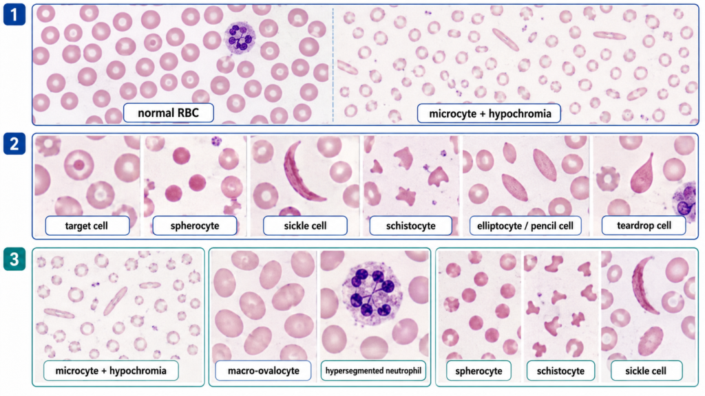
- Microcytic hypochromic cells with marked anisopoikilocytosis and pencil/elliptical cells favour iron-deficiency anemia.
- Macro-ovalocytes, particularly with hypersegmented neutrophils, favour megaloblastic anemia.
- Spherocytes, schistocytes or sickle cells suggest a hemolytic process, while prominent target cells may suggest thalassemia or another hemoglobinopathy.
🔎 Stepwise Approach
- Inspect the smear systematically. If using a microscope, select the well-spread monolayer just behind the feathered edge.
- Establish the normal reference. Normal RBCs are relatively uniform round cells with central pallor occupying roughly one-third of the cell.
- Assess size. Decide whether the predominant cells are normocytic, microcytic or macrocytic; a small lymphocyte nucleus can provide a useful size comparison.
- Assess colour. Look for increased central pallor indicating hypochromia or unusually dense cells with absent central pallor.
- Assess shape. Identify characteristic forms such as target cells, spherocytes, sickle cells, schistocytes, elliptocytes/pencil cells or teardrop cells.
- Look at the overall pattern. Note anisocytosis, poikilocytosis and whether one abnormal morphology predominates.
- Correlate morphology with anemia. State the most likely anemia pattern supported by the dominant smear findings rather than naming a diagnosis from a single nonspecific cell.
Exam tip: Describe the morphology first, then give the likely anemia. Avoid jumping directly to a diagnosis without stating the visible features.
🖼️ Visual Learning
VISUAL 1
Normal vs Abnormal RBC Morphology — Peripheral Smear Recognition
🎥 Practical Video
High Yield Hematology: Blood Smears & Red Blood Cell Morphology
Source: Blueprint Med School
Uses peripheral-smear images to review normal RBC appearance and high-yield abnormal morphologies, helping students practise visual recognition before a hematology spotter.
📝 Important Viva Questions
Q1. What is anisocytosis?
Answer: Variation in the size of circulating red blood cells.
Q2. What is poikilocytosis?
Answer: Variation in RBC shape, with increased numbers of abnormally shaped erythrocytes on the peripheral smear.
Q3. What smear pattern is typical of iron-deficiency anemia?
Answer: Predominantly microcytic hypochromic RBCs with anisocytosis and poikilocytosis; pencil or elliptic cells may be prominent.
Q4. What morphology suggests megaloblastic anemia?
Answer: Macro-ovalocytes with anisopoikilocytosis, often accompanied by hypersegmented neutrophils.
Q5. How do you recognize a spherocyte?
Answer: It is a small, dense, round RBC with absent central pallor. It may occur in hereditary spherocytosis or immune hemolysis.
Q6. What are schistocytes and what do they suggest?
Answer: Schistocytes are fragmented RBCs, often appearing as irregular, triangular or helmet-shaped cells. They suggest mechanical or microangiopathic red-cell destruction.
Q7. Which RBC shapes are particularly associated with hemoglobin disorders?
Answer: Sickle cells strongly suggest sickling disorders, while prominent target cells are commonly seen in thalassemia and other hemoglobinopathies.