
Station 5 — Identification of White Blood Cells on Peripheral Smear
Rapid Revision • Visual Recognition • Viva
🎯 Task
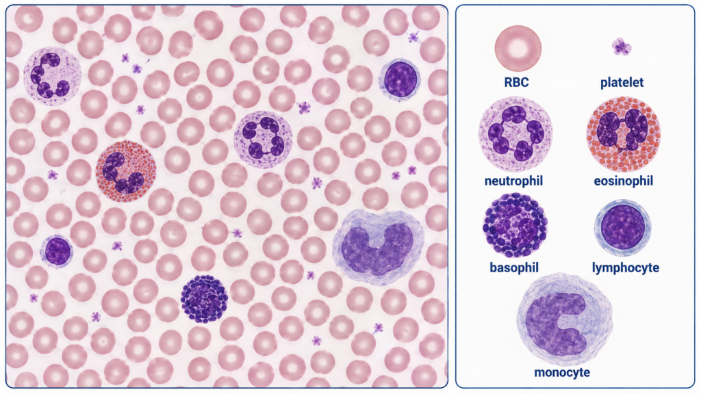
Examine the provided stained peripheral blood smear images. Identify the labelled white blood cells using their morphological features, then compare the smear fields and comment on the white-cell pattern shown.
Neutrophil: segmented 2–5-lobed nucleus with pale cytoplasm and fine granules.
Eosinophil: bilobed nucleus with prominent coarse orange-red granules.
Basophil: coarse deep blue-purple granules, often obscuring the nucleus.
Lymphocyte: dense round nucleus occupying most of the cell with a thin rim of blue cytoplasm. Monocyte: largest cell, with an indented or kidney-shaped nucleus and abundant grey-blue cytoplasm.
Interpretation: the smear with visibly increased numbers of white cells is consistent with leukocytosis.
🔎 Stepwise Procedure / Approach
- Choose the optimal smear area. Assess the monolayer where red cells are well separated with minimal overlap.
- Locate a nucleated cell. Distinguish the larger purple-nucleated leukocyte from surrounding pink-red erythrocytes and small platelets.
- Assess cell size. Decide whether the leukocyte is relatively small, intermediate or large compared with nearby red cells.
- Examine the nucleus. Look for segmentation, bilobing, a dense round nucleus, or an indented/kidney-shaped nucleus.
- Assess cytoplasm and granules. Note the amount and colour of cytoplasm and whether granules are fine, orange-red or deep blue-purple.
- Combine the features. Identify the cell as neutrophil, eosinophil, basophil, lymphocyte or monocyte rather than relying on one feature alone.
- Survey the overall smear. Compare the frequency of leukocytes across representative fields; a conspicuous increase in white cells supports recognition of leukocytosis.
🖼️ Visual Learning
Recognizing the Five Major White Blood Cells and Leukocytosis
🎥 Practical Video
A practical microscope-based demonstration showing stained peripheral blood cells and recognition of neutrophils, eosinophils, basophils, lymphocytes and monocytes.
📝 Important Viva Questions
Answer: A mature neutrophil has a segmented nucleus, typically with about 2–5 lobes connected by thin chromatin strands.
Answer: An eosinophil usually has a bilobed nucleus and prominent coarse orange-red granules, while a neutrophil has a multilobed nucleus with much finer pale granules.
Answer: It contains large coarse deep blue-purple granules that frequently obscure much of the nucleus.
Answer: It has a dense round nucleus occupying most of the cell with only a narrow rim of blue cytoplasm.
Answer: The monocyte. It has abundant grey-blue cytoplasm and an indented, folded or kidney-shaped nucleus.
Answer: Leukocytosis is an increase in the total circulating white blood cell count above the appropriate reference range.
Answer: Assess cell size, nuclear morphology, and cytoplasmic features including granules. The final identification should be based on the combination of these findings.