
Course Content
🔬 Station 1 — Identification of Normal Blood Cells on Peripheral Smear
Learning Outcomes Identify normal RBCs, WBCs and platelets on a provided peripheral blood smear/image. Differentiate the major blood-cell lineages using their morphological features.
0/1
🔴 Station 2 — Abnormal RBC Morphology & Identification of Anemia
Learning Outcomes Differentiate normal from abnormal red blood cells on a peripheral smear. Identify common abnormal RBC shapes. Suggest the likely type of anemia from the provided RBC morphology.
0/1
💊 Station 3 — Prescription Writing for Iron-Deficiency Anemia
Learning Outcomes Write an appropriate prescription for a patient at risk of developing iron-deficiency anemia. Select an appropriate iron preparation and provide clear instructions for its use.
0/1
🩸 Station 4 — Safe Blood Transfusion & Recognition of Transfusion Reactions
Learning Outcomes Demonstrate the essential safety checks required before blood transfusion. Identify common transfusion reactions from a supplied clinical scenario. Describe the appropriate immediate response when a transfusion reaction is suspected.
0/1
⚪ Station 5 — Identification of White Blood Cells on Peripheral Smear
Learning Outcomes Identify the major types of white blood cells on a provided peripheral smear. Differentiate neutrophils, eosinophils, basophils, lymphocytes and monocytes using morphology. Recognize a smear demonstrating leukocytosis.
0/1
🔬 Station 6 — Microscopic Examination of Human and Animal Blood
Learning Outcomes Perform/interpret microscopic examination of provided human and animal blood specimens or images. Differentiate human from animal blood using the supplied microscopic features.
0/1
🔦 Station 7 — Examination of Blood Stains Under Ultraviolet Light
Learning Outcomes Demonstrate the correct approach to examination of a suspected blood stain using ultraviolet light. Recognize findings suggestive of a blood stain on the supplied material.
0/1
🩸 Station 8 — Identification of Blood-Stain Patterns
Learning Outcomes Identify different blood-stain patterns from supplied specimens/images. Differentiate the presented patterns on the basis of their gross appearance.
0/1
💉 Station 9 — EPI Vaccination, Route & Cold-Chain Assessment
Learning Outcomes Identify vaccines included in the Pakistan EPI schedule from provided material. Identify the appropriate route of administration of selected vaccines. Demonstrate/describe correct vaccine storage and maintenance of the cold chain. Recognize important contraindications to vaccination.
0/1
🧪 Station 10 — Interpretation of Coagulation Tests
Learning Outcomes Interpret PT and APTT from provided laboratory results. Interpret bleeding time and clotting time. Use the pattern of abnormalities to identify the likely defect in hemostasis.
0/1
🩸🔬 AIM OSPE/OSCE Lab — Blood & Immunology-II Module | 3rd Year MBBS

AIM OSCE / OSPE • MBBS

🔬 Station 1 — Identification of Normal Blood Cells on Peripheral Smear

Rapid Revision • Visual Recognition • Viva

🎯 Task

CANDIDATE TASK

Examine the provided peripheral blood smear or image. Identify the indicated formed elements of blood and state the key morphological features that allow you to distinguish them.

⚡ Model Station Answer

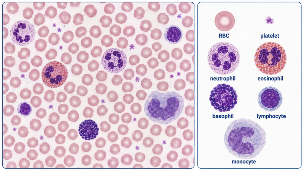
  • RBCs: numerous anucleate pink discs with fairly uniform size and central pallor occupying about one-third of the cell.
  • Platelets: very small purple-staining granular cytoplasmic fragments lying between RBCs.
  • Granulocytes: neutrophils have a multilobed nucleus; eosinophils a bilobed nucleus with coarse orange-red granules; basophils coarse dark blue-purple granules that may obscure the nucleus.
  • Agranulocytes: lymphocytes usually have a dense round nucleus with scant cytoplasm, while monocytes are larger with a folded or kidney-shaped nucleus and abundant pale gray-blue cytoplasm.

🔎 Stepwise Procedure / Approach

  1. Survey the smear systematically. Use a well-spread monolayer area where individual cells are clearly separated.
  2. Recognize the background RBC population. Assess their size, shape, staining and central pallor.
  3. Look for platelets. Identify tiny purple granular fragments between the much larger RBCs.
  4. Locate nucleated cells. Compare their overall size, nuclear shape, chromatin and amount of cytoplasm.
  5. Separate granulocytes from agranulocytes. Granulocytes show conspicuous cytoplasmic granules and characteristic nuclear segmentation.
  6. Differentiate neutrophils, eosinophils and basophils. Use nuclear lobulation together with the colour and prominence of cytoplasmic granules.
  7. Differentiate lymphocytes from monocytes. Use cell size, nuclear shape and nucleus-to-cytoplasm ratio.
  8. State the identification clearly. Name the cell and immediately mention one or two strongest morphological features supporting it.

🖼️ Visual Learning

VISUAL 1

Normal Peripheral Blood Cells — Recognition Guide
🎥 Practical Video

White Blood Cell Identification | WBC Practice | Under Microscope
Source: DK Biosolutions

Microscope-based practice showing stained peripheral-smear cells and the visual features used to recognize neutrophils, eosinophils, basophils, lymphocytes, monocytes, RBCs and platelets.

▶ Watch Practical Video

📝 Important Viva Questions

Q1. What are the main formed elements seen in normal peripheral blood?

Answer: Erythrocytes, leukocytes and platelets.

Q2. What is the normal appearance of an RBC on a stained peripheral smear?

Answer: It is an anucleate pink-red disc with fairly uniform diameter and central pallor measuring approximately one-third of the cell.

Q3. How do you identify a neutrophil?

Answer: A mature neutrophil has a segmented nucleus, usually with 3–5 lobes, and pale cytoplasm containing fine granules.

Q4. How can an eosinophil be distinguished from a basophil?

Answer: Eosinophils usually have a bilobed nucleus with coarse orange-red granules, whereas basophils contain coarse dark blue-purple granules that may obscure the nucleus.

Q5. What features identify a normal small lymphocyte?

Answer: It has a dense round nucleus occupying most of the cell with only a thin rim of basophilic cytoplasm.

Q6. What features help identify a monocyte?

Answer: It is usually the largest normal circulating leukocyte and has a folded or kidney-shaped nucleus with abundant pale gray-blue cytoplasm.

Q7. How are platelets recognized on a peripheral smear?

Answer: They appear as very small anucleate purple-staining granular cytoplasmic fragments scattered between RBCs.

Scroll to Top
💬 WhatsApp Support