
AIM OSCE / OSPE • MBBS
🔬 Station 3 — Identification of Granulation Tissue
Rapid Revision • Practical Skills • Viva
🎯 Task
CANDIDATE TASK
You are shown an H&E-stained histopathology slide/image from a tissue-repair specimen. Identify the tissue, state its characteristic microscopic features, and distinguish it from granulomatous inflammation when asked.
⚡ Model Station Answer What to say in the exam
Identification: This is granulation tissue.
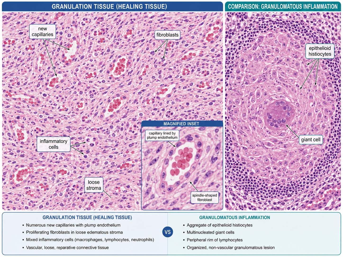
Key features: Numerous newly formed thin-walled capillaries, proliferating fibroblasts and loose edematous connective tissue with inflammatory cells.
Microscopy: The capillaries are lined by plump endothelial cells, with fibroblasts and early collagen deposition between them.
Distinction: Granulomatous inflammation instead shows organized aggregates of epithelioid macrophages, often with multinucleated giant cells and surrounding lymphocytes.
🔎 Stepwise Procedure / Approach
- Start at low power. Assess the overall architecture and look for a loose, highly vascular reparative tissue pattern.
- Assess vascularity. Look for numerous newly formed small capillaries distributed throughout the tissue.
- Examine the vessels. Identify thin vascular channels lined by relatively plump endothelial cells.
- Look between the vessels. Identify proliferating spindle-shaped fibroblasts within a loose edematous extracellular matrix.
- Identify the inflammatory component. Mixed inflammatory cells, particularly macrophages with variable lymphocytes and neutrophils, may be present.
- Search for maturation. Increasing collagen deposition may be visible as the reparative tissue evolves toward scar formation.
- Exclude a granuloma pattern. Absence of a compact epithelioid-histiocyte aggregate with characteristic giant cells supports granulation tissue rather than granulomatous inflammation.
- State the identification clearly. Correlate the vascular proliferation, fibroblasts and loose inflammatory stroma and identify the slide as granulation tissue.
🖼️ Visual Learning
VISUAL 1
Granulation Tissue — Microscopic Recognition and Granuloma Comparison
🎥 Practical Video
🎥 Practical Demonstration — Skin: Granulation Tissue
Source: NUS Pathweb — National University of Singapore
A pathology slide demonstration showing the microscopic appearance of granulation tissue in skin and helping students recognize its practical histological features.
▶ Watch Practical Video
📝 Important Viva Questions
Q1. What is granulation tissue?
Answer: It is newly formed vascular connective tissue produced during tissue repair, composed mainly of proliferating capillaries, fibroblasts and inflammatory cells in a loose extracellular matrix.
Q2. What are the major microscopic features of granulation tissue?
Answer: Numerous newly formed capillaries, proliferating fibroblasts, loose edematous stroma, inflammatory cells and gradually increasing collagen deposition.
Q3. Why does granulation tissue appear highly vascular?
Answer: Because angiogenesis produces numerous newly formed capillaries during the repair process.
Q4. Which connective-tissue cell is particularly important in granulation tissue?
Answer: The fibroblast, which proliferates and produces extracellular matrix and collagen during repair.
Q5. How does granulation tissue differ from a granuloma?
Answer: Granulation tissue is reparative tissue rich in capillaries and fibroblasts, whereas a granuloma is an organized collection of epithelioid macrophages, often with multinucleated giant cells and surrounding lymphocytes.
Q6. Is granulation tissue the same as a mature scar?
Answer: No. Granulation tissue is an early vascular phase of repair; with maturation, vascularity and cellularity decrease while collagen increases to form a scar.
Q7. What is a common examination mistake in this station?
Answer: Confusing granulation tissue with granulomatous inflammation. Look for capillaries and fibroblasts in granulation tissue versus epithelioid macrophage aggregates in a granuloma.