
Course Content
🔬 Station 1 — Microscopic Identification of Acute Inflammation
Learning Outcomes Identify inflammatory cells on a provided microscopic slide. Identify the histopathological changes of acute appendicitis. Relate the demonstrated microscopic findings to acute inflammation.
0/1
🔬 Station 2 — Microscopic Identification of Chronic and Granulomatous Inflammation
Learning Outcomes Identify the morphological changes of chronic inflammation in a provided specimen/slide. Identify the characteristic cells and their arrangement in a granuloma. Distinguish the demonstrated chronic inflammatory pattern from acute inflammation when asked.
0/1
🔬 Station 3 — Identification of Granulation Tissue
Learning Outcomes Identify granulation tissue on a provided histopathology slide/image. Identify its characteristic microscopic features. Distinguish granulation tissue from granulomatous inflammation when asked.
0/1
🧪 Station 4 — Performance and Interpretation of Bacterial Biochemical Tests
Learning Outcomes Perform the bacterial biochemical test provided at the station using the appropriate technique. Observe and interpret the test result. Report the test as positive or negative based on the demonstrated reaction.
0/1
🧫 Station 5 — Identification of Culture Media and Hemolysis on Blood Agar
Learning Outcomes Identify the provided microbiological culture medium. Recognize blood agar, mannitol salt agar, chocolate medium or Cary-Blair transport medium when presented. Identify the demonstrated pattern of hemolysis on blood agar.
0/1
🚑 Station 6 — Demonstration of Gastric Lavage
Learning Outcomes Demonstrate the sequence of steps involved in gastric lavage using the provided model/manikin and equipment. Demonstrate safe handling and appropriate procedural sequence. State the essential precaution(s) relevant to the procedure when asked.
0/1
💊 Station 7 — Prescription Writing for Common Infectious Diseases Acute Tonsillitis / Malaria
Learning Outcomes Construct a complete prescription for the supplied infectious-disease scenario. Select appropriate medication with dose, route, frequency and duration. Demonstrate essential principles of safe and rational prescription writing.
0/1
🦴🔍 Station 8 — Forensic Personal Identification: Bone or Hair/Fibre
Learning Outcomes Determine human sex from a provided bone or identify human hair microscopically, according to the station material. Identify the relevant distinguishing characteristics supporting the conclusion. Differentiate human hair from fibre when that specimen is provided.
0/1
🦠 Station 9 — Laboratory Identification of Common Parasites
Learning Outcomes Identify the supplied parasite/specimen from a laboratory slide or specimen. Recognize the characteristic diagnostic form demonstrated. State the identifying feature supporting the diagnosis.
0/1
🌍 Station 10 — Identification of Communicable Disease Models and Prevention
Learning Outcomes Identify the communicable disease represented by the provided model. Identify the important signs/features demonstrated by the model. State the major complication(s) and relevant preventive measures.
0/1
🧪🔬 AIM OSPE/OSCE Lab — Infection & Inflammation Module | 3rd Year MBBS
AIM OSCE / OSPE • MBBS

🦠 Station 9 — Laboratory Identification of Common Parasites

Hydatid cyst • Leishmania • Malaria • Taenia  |  Rapid Revision • Practical Recognition • Viva

🎯 Task

CANDIDATE TASK

Examine the supplied parasitology slide or specimen. Identify the parasite or diagnostic form demonstrated and state the key morphological feature that supports your identification.

⚡ Model Station Answer — What to say in the exam

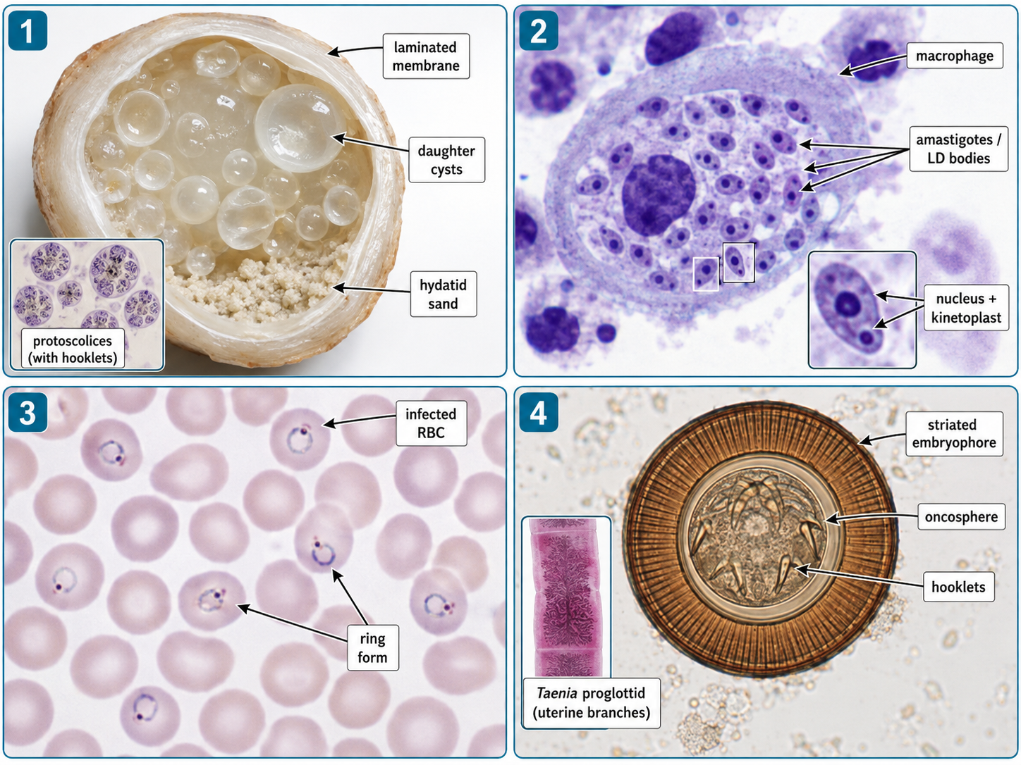
Hydatid cyst: Identified by a cyst with laminated membrane, daughter cysts or hydatid sand; protoscolices and hooklets may be demonstrated microscopically.

Leishmania: Identified by intracellular amastigotes, or LD bodies, within macrophages; each shows a nucleus and kinetoplast.

Malaria: Identified by intraerythrocytic Plasmodium forms on a stained peripheral blood film, commonly including characteristic ring forms.

Taenia: A Taenia egg shows a thick radially striated embryophore surrounding a hexacanth embryo with hooklets.

🔎 Stepwise Procedure / Approach

  1. Identify the material supplied. Decide whether you are looking at a gross specimen, wet preparation, stained tissue preparation or peripheral blood film.
  2. Survey systematically. On microscopy, begin by locating the relevant cells or parasite structures before focusing on fine morphology.
  3. Find the dominant diagnostic form. Look for a cyst component, intracellular body, intraerythrocytic parasite, egg, larval structure or other characteristic parasitic form.
  4. Assess the strongest identifying features. Note wall characteristics, hooklets, daughter cysts, nucleus/kinetoplast, relationship to blood cells or macrophages, and other visible hallmarks.
  5. Match the morphology to the parasite. Use the distinctive combination of features rather than relying on size or colour alone.
  6. State the identification clearly. Name the parasite or diagnostic form first, followed immediately by the strongest supporting feature.
  7. Avoid overcalling species. If the morphology only establishes a genus or group, report only what can be confidently identified from the supplied material.
Exam technique: A strong spotter response is usually: “This is ___, identified by ___.” Give the most distinctive visible feature rather than a long theoretical description.

🖼️ Visual Learning

VISUAL 1

Recognition of Hydatid Cyst, Leishmania, Malaria and Taenia
🎥 Practical Video

🎥 Practical Demonstration — Malaria Microscopy: A Step-by-Step Guide

Source: MCD Global Health

A practical microscopy demonstration showing preparation and examination of malaria blood films. It reinforces recognition of the blood-film component of this station and the systematic approach to microscopic parasite detection.

▶ Watch Practical Video


📝 Important Viva Questions

Q1. What is hydatid sand?

It consists mainly of free protoscolices, hooklets and related parasitic material that settles within a hydatid cyst.

Q2. Which feature helps identify Leishmania amastigotes microscopically?

They are small intracellular bodies, commonly within macrophages, showing a nucleus and a kinetoplast.

Q3. What is another commonly used name for Leishmania amastigotes in tissue?

They are commonly called Leishman-Donovan bodies or LD bodies.

Q4. Which blood films are commonly used for microscopic diagnosis of malaria?

Thick and thin peripheral blood films. Thick films improve parasite detection, while thin films preserve morphology for further identification.

Q5. What is the characteristic early malarial form commonly recognized inside an erythrocyte?

The ring-form trophozoite, appearing as a delicate cytoplasmic ring with a chromatin dot.

Q6. What is the characteristic microscopic appearance of a Taenia egg?

It has a thick radially striated embryophore enclosing a hexacanth oncosphere containing hooklets.

Q7. Can the common human Taenia species be reliably differentiated by egg morphology alone?

No. Their eggs are morphologically very similar, so species identification should not be claimed from the egg alone.

Scroll to Top
💬 WhatsApp Support