
Course Content
🔬 Station 1 — Identification of Tuberculous Osteomyelitis
Learning Outcomes Identify tuberculous osteomyelitis from a provided gross specimen or histopathology image/slide. Identify the key gross and microscopic morphological features supporting the diagnosis. Differentiate the lesion from common pyogenic osteomyelitis when provided with relevant findings.
0/1
🔬 Station 2 — Identification of Common Bone Tumours
Learning Outcomes Identify the provided bone tumour from a gross specimen, image or histopathology slide. Identify the key morphological features supporting the diagnosis. Differentiate osteosarcoma, osteoclastoma and chondrosarcoma using characteristic morphological findings.
0/1
🧪 Station 3 — ASO Test by Latex Agglutination
Learning Outcomes Prepare the specimen and reagents required for the ASO latex agglutination test. Demonstrate the correct steps of the latex agglutination technique. Interpret visible agglutination and report the test as positive or negative.
0/1
🔬 Station 4 — Identification of Squamous Cell Carcinoma and Basal Cell Carcinoma of Skin
Learning Outcomes Identify SCC or BCC from a provided gross specimen/image or histopathology slide. Identify the important morphological features supporting the diagnosis. Differentiate squamous cell carcinoma from basal cell carcinoma on morphological grounds.
0/1
💊 Station 5 — Prescription Writing for Gout and Rheumatoid Arthritis
Learning Outcomes Write a complete and rational prescription for a patient with gout or rheumatoid arthritis based on the supplied clinical scenario. Select an appropriate drug, dose, route, frequency and duration. Demonstrate the essential components of safe prescription writing.
0/1
💊 Station 6 — Prescription Writing for Common Dermatological Disorders
Learning Outcomes Write an appropriate prescription for a supplied case of scabies or psoriasis. Select the appropriate preparation, route, dose/frequency and duration. Include essential patient instructions relevant to safe and effective treatment.
0/1
⚖️ Station 7 — Identification of Mechanical Wounds and Causative Weapon
Learning Outcomes Identify the type of mechanical wound from a provided model, image or injury photograph. Recognize abrasion, bruise, laceration, incised wound or stab wound from its characteristic features. Identify the probable causative weapon/mechanism and relevant medico-legal significance.
0/1
🔫 Station 8 — Examination of Firearm Injury and Identification of Bullet/Ammunition
Learning Outcomes Identify a firearm entry or exit wound from the provided image/model and describe its important features. Interpret features relevant to the probable range or mechanism of firing when demonstrated. Identify a supplied bullet/ammunition specimen and document the important medico-legal findings.
0/1
🩺 Station 9 — Focused Musculoskeletal History and Examination
Learning Outcomes Obtain a focused history relevant to a common musculoskeletal complaint. Perform an appropriate focused general and musculoskeletal examination. Present the relevant positive and negative findings in an organized manner.
0/1
🦴 Station 10 — X-Ray Interpretation of Fracture/Dislocation and Initial Limb Management
Learning Outcomes Identify a fracture or dislocation on a provided X-ray. Describe the site, type, displacement and/or angulation of the injury systematically. Demonstrate or describe appropriate immediate immobilization and initial management of the injured limb.
0/1
AIM OSPE/OSCE Lab — MSK-II Module | 3rd Year MBBS
AIM OSCE / OSPE • MBBS

🧪 Station 3 — ASO Test by Latex Agglutination

Rapid Revision • Practical Skills • Viva

🎯 Task

CANDIDATE TASK

You are provided with a patient’s serum, an ASO latex agglutination kit and the required laboratory materials. Demonstrate the test correctly and report the observed result as positive or negative.

⚡ Model Station Answer What to say in the exam

Specimen: Patient serum with ASO latex reagent and appropriate controls.
Technique: I place the required serum volume on the reaction circle, add ASO latex reagent, mix and rotate the slide for the manufacturer-specified reaction time.
Positive result: Visible agglutination indicates detectable anti-streptolysin O antibodies above the kit’s stated detection limit.
Negative result: A smooth homogeneous suspension without visible agglutination is reported as negative.

🔎 Stepwise Procedure / Approach

  1. Prepare the materials. Arrange patient serum, ASO latex reagent, positive and negative controls, reaction slide/card, micropipette or supplied dropper and separate mixing sticks.
  2. Prepare specimen and reagents. Bring them to the temperature recommended by the supplied kit and gently resuspend the latex reagent before use.
  3. Identify separate reaction circles. Use distinct areas for the patient sample and controls to prevent cross-contamination.
  4. Add the sample. Place the kit-specified volume of serum onto its reaction circle; many teaching kits use approximately 50 µL.
  5. Add ASO latex reagent. Add the quantity specified by the kit and mix it thoroughly with the serum using a clean disposable mixing stick.
  6. Rotate the slide/card. Gently rotate manually or on a rotator for the manufacturer-specified time, commonly around 2 minutes in slide latex methods.
  7. Observe immediately. Look for visible clumping within the correct reading time. Do not interpret a dried reaction after the specified endpoint.
  8. Report the result. Visible agglutination with valid controls is ASO positive; a smooth uniform suspension with valid controls is ASO negative.

🖼️ Visual Learning

VISUAL 1

ASO Latex Agglutination — Setup, Technique and Result
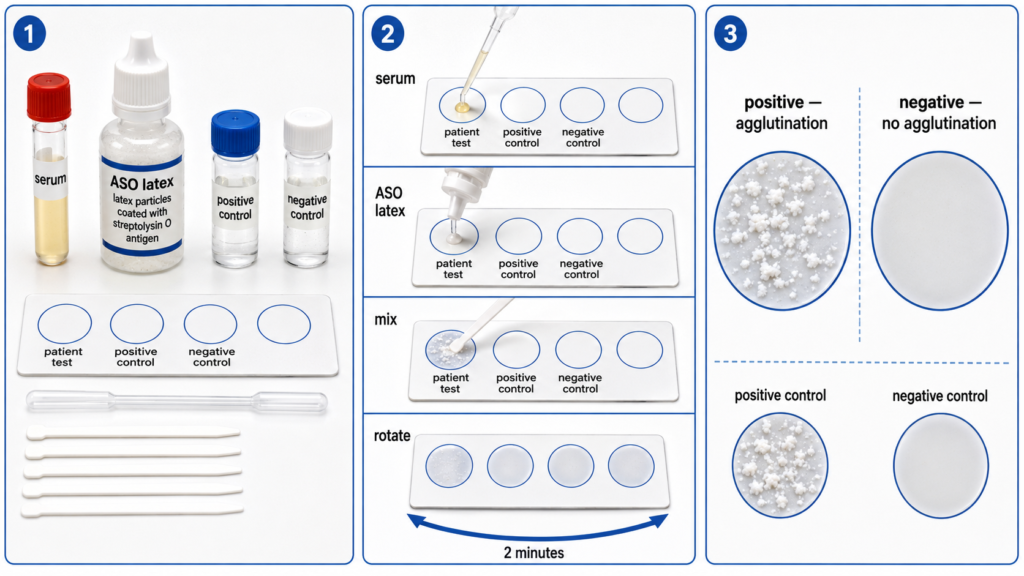
 

🎥 Practical Video

🎥 Practical Demonstration — ASO Latex Test Procedure
Source: Anamol Laboratories Private Limited

This practical demonstration shows the ASO latex kit components, specimen–reagent mixing and interpretation of the latex agglutination reaction.

▶ Watch Practical Video


📝 Important Viva Questions

Q1. What is the principle of the ASO latex agglutination test?

Answer: Latex particles coated with streptolysin O antigen visibly agglutinate when they react with anti-streptolysin O antibodies present in the patient’s serum.

Q2. What specimen is commonly used for this test?

Answer: Patient serum is commonly used for the slide latex agglutination test.

Q3. How does a positive ASO latex reaction appear?

Answer: A positive reaction shows visible agglutination or clumping of the latex particles within the specified reading time.

Q4. How does a negative reaction appear?

Answer: The mixture remains smooth and uniformly dispersed without visible agglutination.

Q5. Why are positive and negative controls used?

Answer: They confirm that the reagent and technique are functioning correctly and that the patient’s reaction can be interpreted reliably.

Q6. Why must the reaction be read within the specified time?

Answer: Delayed reading can produce drying or nonspecific aggregation and may lead to an incorrect interpretation.

Q7. What is the clinical significance of detecting ASO antibodies?

Answer: An elevated ASO response supports evidence of a recent streptococcal infection and can aid assessment of post-streptococcal conditions such as rheumatic fever and acute post-streptococcal glomerulonephritis.

Scroll to Top
💬 WhatsApp Support