
🔬 Station 1 — Identification of Tuberculous Osteomyelitis
Rapid Revision • Practical Pathology • Viva
🎯 Task
You are provided with a gross bone specimen and/or an H&E-stained histopathology image from a case of osteomyelitis. Identify the lesion, state the key morphological features supporting your diagnosis, and distinguish it from common pyogenic osteomyelitis.
⚡ Model Station Answer — What to say in the exam
Identification: This is tuberculous osteomyelitis.
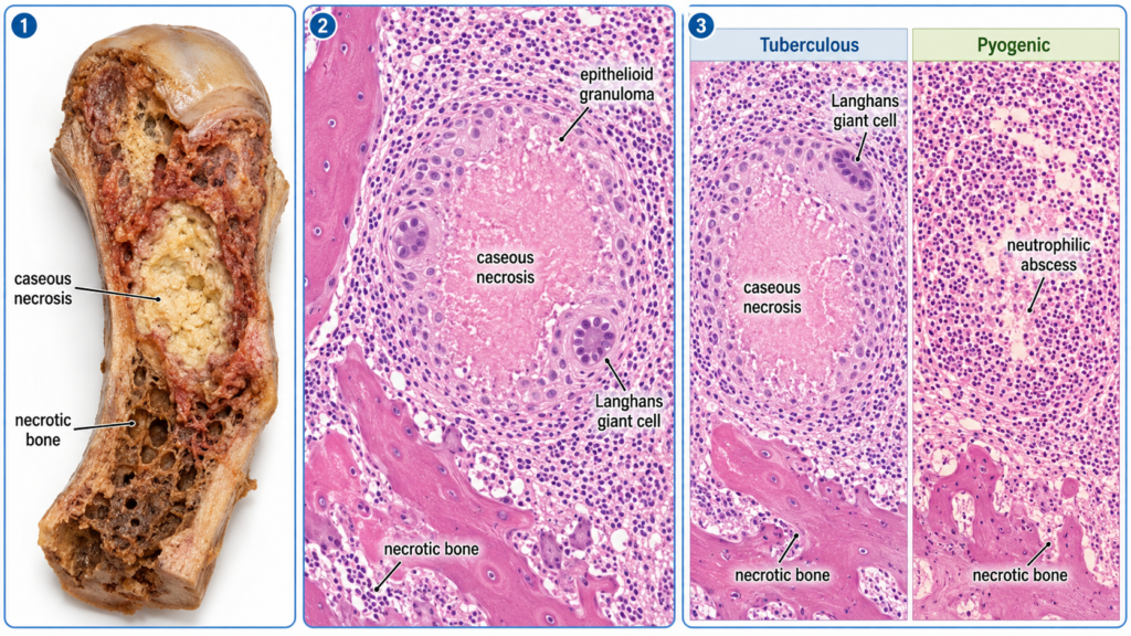
Gross: Destructive bone lesion with pale yellow-white caseous necrotic material and granulation tissue.
Microscopy: Caseating epithelioid granulomas with Langhans-type giant cells, surrounding lymphocytes and areas of necrotic bone.
Differentiation: Tuberculous osteomyelitis is predominantly granulomatous with caseation, whereas pyogenic osteomyelitis shows prominent neutrophilic suppuration and abscess formation.
🔎 Stepwise Procedure / Approach
Identify the tissue. Confirm that the specimen or slide represents bone and look for areas of destruction or necrosis.
- Assess the gross pattern. Look for irregular bone destruction, granulation tissue and pale yellow-white caseous material when visible.
- Scan the slide at low power. Look for a granulomatous inflammatory pattern involving or surrounding bone trabeculae.
- Identify the granuloma. Look for collections of epithelioid histiocytes with central caseous necrosis.
- Confirm supporting cells. Identify Langhans-type multinucleated giant cells and a surrounding rim of lymphocytes.
- Look for bone involvement. Note necrotic or damaged bone trabeculae associated with the inflammatory lesion.
- Differentiate from pyogenic osteomyelitis. Predominant caseating granulomas favor tuberculosis; sheets of neutrophils, pus and suppurative abscesses favor pyogenic infection.
- State the diagnosis clearly. Correlate the dominant morphological features and conclude: tuberculous osteomyelitis.
🖼️ Visual Learning
Tuberculous Osteomyelitis — Gross, Microscopy & Pyogenic Comparison
🎥 Practical Video
🎥 Practical Demonstration — Osteomyelitis Pathology
This MBBS-focused pathology demonstration reviews the gross and microscopic morphology of osteomyelitis and directly compares pyogenic and tuberculous osteomyelitis, helping reinforce the identifying features required at this station.
📝 Important Viva Questions
Answer: A caseating epithelioid cell granuloma containing Langhans-type giant cells and surrounded by lymphocytes.
Answer: It is a form of necrosis producing soft, amorphous, cheese-like material, classically associated with tuberculosis.
Answer: It is a multinucleated giant cell in which the nuclei are typically arranged toward the periphery in a horseshoe or arc-like pattern.
Answer: Tuberculous osteomyelitis shows predominantly caseating granulomatous inflammation, whereas pyogenic osteomyelitis shows prominent neutrophilic suppuration and abscess formation.
Answer: It is caused by Mycobacterium tuberculosis.
Answer: Ziehl–Neelsen stain can demonstrate acid-fast bacilli, although organisms may be sparse in osteoarticular tuberculosis.
Answer: Destruction and necrosis of bone trabeculae may occur, with surrounding granulation tissue and chronic inflammatory changes.