
Station 9 — Focused Musculoskeletal History and Examination
Rapid Revision • Focused History • Musculoskeletal Examination • Viva
🎯 Task
A patient presents with a musculoskeletal complaint affecting a joint or limb. Obtain a focused relevant history, perform an appropriate focused general and musculoskeletal examination, and present the important positive and negative findings to the examiner.
⚡ Model Station Answer
What to say in the exam
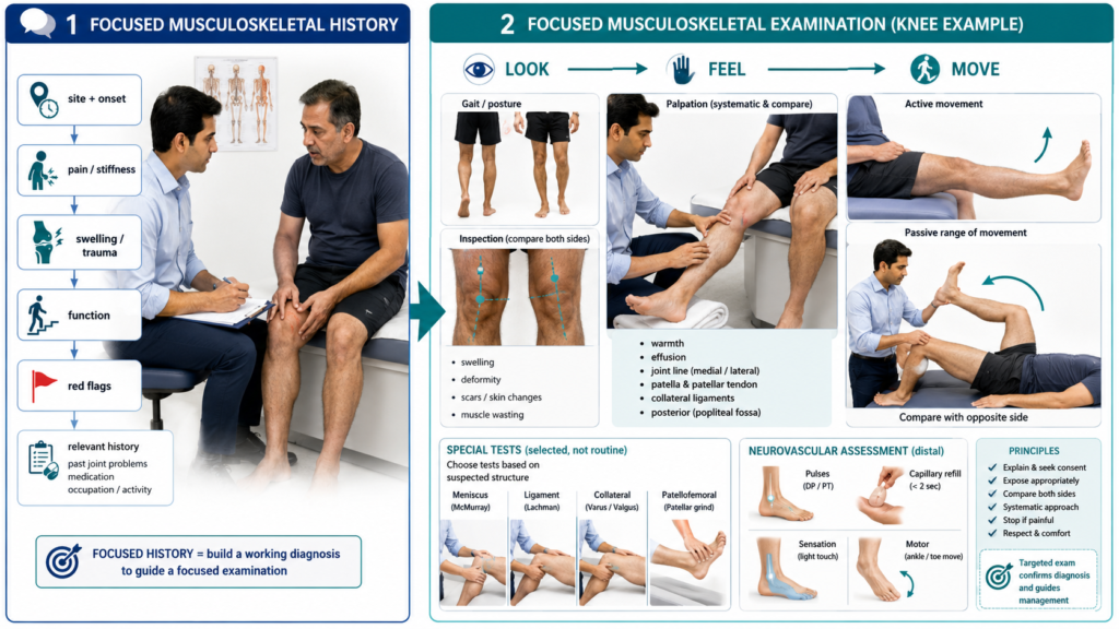
History: I would characterize the pain and stiffness, establish onset and progression, ask about swelling, trauma, functional limitation, associated systemic symptoms and relevant red flags.
Relevant background: I would ask about previous joint disease or injury, medical and drug history, family history and relevant occupation or activity.
Examination: After consent, hand hygiene and appropriate exposure, I would assess the patient generally, observe gait where appropriate, and examine the symptomatic area using look, feel and move, comparing both sides.
Focused assessment: I would perform relevant special tests and neurovascular assessment when indicated.
Presentation: I would summarize the important positive findings, relevant negatives and the likely anatomical or clinical pattern without making unsupported assumptions.
🔎 Stepwise Procedure / Approach
- Introduce yourself and confirm the patient.
Explain that you need to ask about the problem and examine the affected musculoskeletal region. Obtain consent. - Define the main complaint.
Establish the exact site, onset, duration, progression and circumstances in which the symptom began, including any preceding trauma or repetitive activity. - Characterize pain and stiffness.
Ask about pain character, severity, radiation, aggravating or relieving factors, relationship to activity or rest, morning stiffness and night symptoms. - Ask for associated musculoskeletal features.
Enquire about swelling, redness, warmth, reduced movement, instability, locking, weakness or involvement of other joints where relevant. - Screen for important associated symptoms and red flags.
Ask selectively about fever, unexplained constitutional symptoms, neurological symptoms, significant trauma or inability to use the limb. - Assess functional impact and relevant background.
Determine effects on walking, stairs, dressing, work or daily activity. Ask relevant previous joint or bone problems, operations, medical conditions, medications, family history, occupation and activity. - Prepare for examination.
Perform hand hygiene, ensure appropriate exposure, position the patient correctly and ask whether any movement is particularly painful before starting. - Perform general and focused musculoskeletal assessment.
Observe overall posture and gait where appropriate, then examine the symptomatic region systematically using look → feel → move. Compare with the opposite side. - Complete relevant focused tests.
Assess active and passive range of movement and perform only anatomically appropriate special tests. Check distal neurovascular status if injury, neurological symptoms or vascular compromise are relevant. - Complete and present.
Thank the patient, restore comfort and summarize the important positive and negative findings in an organized manner before giving your focused conclusion.
🖼️ Visual Learning
Focused Musculoskeletal Assessment — History to Examination
🎥 Practical Video
🎥 Practical Demonstration — GALS Musculoskeletal Examination
Source: Geeky Medics
A concise OSCE demonstration of musculoskeletal screening using gait, arms, legs and spine, including patient preparation, inspection, movement and presentation of findings. Use a focused regional examination when the history localizes the problem.
📝 Important Viva Questions
Look, feel and move. Appropriate special tests are added according to the joint and suspected pathology.
Comparison helps identify asymmetry in swelling, deformity, tenderness, muscle bulk and range of movement and provides the patient’s own normal reference.
Active movement assesses what the patient can produce, while passive movement helps assess the available joint range independent of voluntary muscle contraction.
The pattern of stiffness and relationship to activity are useful. Prolonged morning stiffness suggests an inflammatory pattern, whereas mechanical symptoms commonly worsen with use.
Ask how symptoms affect activities relevant to the involved region, such as walking, stairs, dressing, gripping, work and usual daily activities.
It is particularly important after trauma or when neurological symptoms, vascular symptoms or possible limb compromise are present.
Give a brief organized summary of the relevant positive findings and important negatives, then state the most likely anatomical or clinical pattern supported by those findings.