
Course Content
🔬 Station 1 — Identification of Tuberculous Osteomyelitis
Learning Outcomes Identify tuberculous osteomyelitis from a provided gross specimen or histopathology image/slide. Identify the key gross and microscopic morphological features supporting the diagnosis. Differentiate the lesion from common pyogenic osteomyelitis when provided with relevant findings.
0/1
🔬 Station 2 — Identification of Common Bone Tumours
Learning Outcomes Identify the provided bone tumour from a gross specimen, image or histopathology slide. Identify the key morphological features supporting the diagnosis. Differentiate osteosarcoma, osteoclastoma and chondrosarcoma using characteristic morphological findings.
0/1
🧪 Station 3 — ASO Test by Latex Agglutination
Learning Outcomes Prepare the specimen and reagents required for the ASO latex agglutination test. Demonstrate the correct steps of the latex agglutination technique. Interpret visible agglutination and report the test as positive or negative.
0/1
🔬 Station 4 — Identification of Squamous Cell Carcinoma and Basal Cell Carcinoma of Skin
Learning Outcomes Identify SCC or BCC from a provided gross specimen/image or histopathology slide. Identify the important morphological features supporting the diagnosis. Differentiate squamous cell carcinoma from basal cell carcinoma on morphological grounds.
0/1
💊 Station 5 — Prescription Writing for Gout and Rheumatoid Arthritis
Learning Outcomes Write a complete and rational prescription for a patient with gout or rheumatoid arthritis based on the supplied clinical scenario. Select an appropriate drug, dose, route, frequency and duration. Demonstrate the essential components of safe prescription writing.
0/1
💊 Station 6 — Prescription Writing for Common Dermatological Disorders
Learning Outcomes Write an appropriate prescription for a supplied case of scabies or psoriasis. Select the appropriate preparation, route, dose/frequency and duration. Include essential patient instructions relevant to safe and effective treatment.
0/1
⚖️ Station 7 — Identification of Mechanical Wounds and Causative Weapon
Learning Outcomes Identify the type of mechanical wound from a provided model, image or injury photograph. Recognize abrasion, bruise, laceration, incised wound or stab wound from its characteristic features. Identify the probable causative weapon/mechanism and relevant medico-legal significance.
0/1
🔫 Station 8 — Examination of Firearm Injury and Identification of Bullet/Ammunition
Learning Outcomes Identify a firearm entry or exit wound from the provided image/model and describe its important features. Interpret features relevant to the probable range or mechanism of firing when demonstrated. Identify a supplied bullet/ammunition specimen and document the important medico-legal findings.
0/1
🩺 Station 9 — Focused Musculoskeletal History and Examination
Learning Outcomes Obtain a focused history relevant to a common musculoskeletal complaint. Perform an appropriate focused general and musculoskeletal examination. Present the relevant positive and negative findings in an organized manner.
0/1
🦴 Station 10 — X-Ray Interpretation of Fracture/Dislocation and Initial Limb Management
Learning Outcomes Identify a fracture or dislocation on a provided X-ray. Describe the site, type, displacement and/or angulation of the injury systematically. Demonstrate or describe appropriate immediate immobilization and initial management of the injured limb.
0/1
AIM OSPE/OSCE Lab — MSK-II Module | 3rd Year MBBS
AIM OSCE / OSPE • MBBS

🔬 Station 2 — Identification of Common Bone Tumours

Osteosarcoma • Osteoclastoma • Chondrosarcoma

Identify • Recognize key morphology • Differentiate

🎯 Task

CANDIDATE TASK

Examine the provided bone tumour gross specimens, images or histopathology slides. Identify each lesion, state the key morphological features supporting your diagnosis, and differentiate the three lesions using their characteristic morphology.

⚡ Model Station Answer

⚡ MODEL STATION ANSWER

What to say in the exam

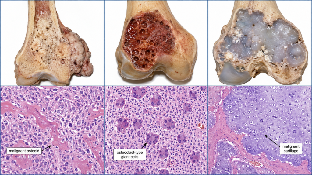
  • Osteosarcoma: A malignant osteogenic tumour characterized microscopically by malignant tumour cells directly producing osteoid. Grossly it is a destructive, firm to gritty grey-white/tan tumour that may breach the cortex.
  • Osteoclastoma / Giant Cell Tumour: An expansile, usually red-brown haemorrhagic lesion. Microscopically there are numerous evenly distributed osteoclast-type multinucleated giant cells among mononuclear stromal cells.
  • Chondrosarcoma: A malignant cartilage-forming tumour with a lobulated, grey-white to bluish translucent cartilaginous cut surface. Microscopically it shows atypical chondrocytes in lacunae within malignant chondroid matrix.
  • Key differentiation: Osteosarcoma produces malignant osteoid, osteoclastoma shows a giant-cell-rich pattern, and chondrosarcoma produces malignant cartilage.

🔎 Stepwise Procedure / Approach

  1. Orient the specimen or slide. Decide whether you are looking at gross pathology, low-power histology or a higher-power microscopic field.
  2. Assess the dominant appearance. On gross specimens look for an expansile versus destructive lesion, colour, margins, cortical involvement and the character of the cut surface.
  3. Look for the tumour matrix. A hard or gritty bone-forming lesion suggests osteoid production, while a lobulated translucent or bluish-grey lesion suggests cartilaginous matrix.
  4. Examine the microscopic architecture. Identify osteoid, abundant multinucleated giant cells or lobules of malignant cartilage before focusing on individual cells.
  5. Identify the hallmark feature. For osteosarcoma confirm malignant cells producing osteoid; for osteoclastoma look for evenly dispersed osteoclast-type giant cells; for chondrosarcoma identify atypical chondrocytes within chondroid matrix.
  6. Assess malignant morphology where relevant. Look for cellular atypia, pleomorphism, destructive growth or invasion in osteosarcoma and chondrosarcoma.
  7. State the diagnosis confidently. Name the tumour and immediately support it with the two or three strongest morphological features rather than giving a long theoretical description.

🖼️ Visual Learning

VISUAL 1

Osteosarcoma vs Osteoclastoma vs Chondrosarcoma — Gross and Microscopic Recognition
🎥 Practical Video

🎥 Practical Demonstration — Osteosarcoma Talking Pot

Source: NUS Pathweb • National University of Singapore

This gross-pathology demonstration shows how to inspect an osteosarcoma specimen and recognize the destructive tumour morphology expected in a practical pathology station.


▶ Watch Practical Video

📝 Important Viva Questions

Q1. What is the defining microscopic feature of osteosarcoma?

Answer: The defining feature is production of osteoid directly by malignant tumour cells.

Q2. What is the characteristic microscopic appearance of osteoclastoma?

Answer: Numerous osteoclast-type multinucleated giant cells are distributed among mononuclear stromal cells, usually in a relatively uniform pattern.

Q3. What is the characteristic gross appearance of a giant cell tumour?

Answer: It is typically an expansile lesion with a soft red-brown, haemorrhagic cut surface, often with cystic change and thinning of the cortex.

Q4. What morphological feature supports a diagnosis of chondrosarcoma?

Answer: Malignant cartilage containing atypical chondrocytes in lacunae, often within a lobulated chondroid matrix, supports the diagnosis.

Q5. How does the gross cut surface of chondrosarcoma commonly appear?

Answer: It is typically lobulated, glistening grey-white to bluish and translucent, with possible myxoid change and calcification.

Q6. What is the quickest morphological way to differentiate these three tumours in an OSPE?

Answer: Identify the dominant diagnostic pattern: malignant osteoid in osteosarcoma, numerous osteoclast-type giant cells in osteoclastoma, and malignant cartilage in chondrosarcoma.

Q7. Is conventional giant cell tumour of bone an ordinary harmless benign tumour?

Answer: No. It is generally considered a locally aggressive/intermediate bone tumour; it can recur locally and, rarely, metastasize.

Scroll to Top
💬 WhatsApp Support