Course Content
🔬 Station 1 — Identification of Tuberculous Osteomyelitis
Learning Outcomes Identify tuberculous osteomyelitis from a provided gross specimen or histopathology image/slide. Identify the key gross and microscopic morphological features supporting the diagnosis. Differentiate the lesion from common pyogenic osteomyelitis when provided with relevant findings.
0/1
🔬 Station 2 — Identification of Common Bone Tumours
Learning Outcomes Identify the provided bone tumour from a gross specimen, image or histopathology slide. Identify the key morphological features supporting the diagnosis. Differentiate osteosarcoma, osteoclastoma and chondrosarcoma using characteristic morphological findings.
0/1
🧪 Station 3 — ASO Test by Latex Agglutination
Learning Outcomes Prepare the specimen and reagents required for the ASO latex agglutination test. Demonstrate the correct steps of the latex agglutination technique. Interpret visible agglutination and report the test as positive or negative.
0/1
🔬 Station 4 — Identification of Squamous Cell Carcinoma and Basal Cell Carcinoma of Skin
Learning Outcomes Identify SCC or BCC from a provided gross specimen/image or histopathology slide. Identify the important morphological features supporting the diagnosis. Differentiate squamous cell carcinoma from basal cell carcinoma on morphological grounds.
0/1
💊 Station 5 — Prescription Writing for Gout and Rheumatoid Arthritis
Learning Outcomes Write a complete and rational prescription for a patient with gout or rheumatoid arthritis based on the supplied clinical scenario. Select an appropriate drug, dose, route, frequency and duration. Demonstrate the essential components of safe prescription writing.
0/1
💊 Station 6 — Prescription Writing for Common Dermatological Disorders
Learning Outcomes Write an appropriate prescription for a supplied case of scabies or psoriasis. Select the appropriate preparation, route, dose/frequency and duration. Include essential patient instructions relevant to safe and effective treatment.
0/1
⚖️ Station 7 — Identification of Mechanical Wounds and Causative Weapon
Learning Outcomes Identify the type of mechanical wound from a provided model, image or injury photograph. Recognize abrasion, bruise, laceration, incised wound or stab wound from its characteristic features. Identify the probable causative weapon/mechanism and relevant medico-legal significance.
0/1
🔫 Station 8 — Examination of Firearm Injury and Identification of Bullet/Ammunition
Learning Outcomes Identify a firearm entry or exit wound from the provided image/model and describe its important features. Interpret features relevant to the probable range or mechanism of firing when demonstrated. Identify a supplied bullet/ammunition specimen and document the important medico-legal findings.
0/1
🩺 Station 9 — Focused Musculoskeletal History and Examination
Learning Outcomes Obtain a focused history relevant to a common musculoskeletal complaint. Perform an appropriate focused general and musculoskeletal examination. Present the relevant positive and negative findings in an organized manner.
0/1
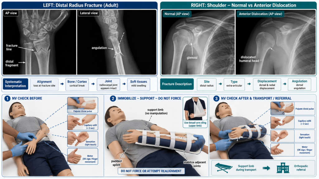
🦴 Station 10 — X-Ray Interpretation of Fracture/Dislocation and Initial Limb Management
Learning Outcomes Identify a fracture or dislocation on a provided X-ray. Describe the site, type, displacement and/or angulation of the injury systematically. Demonstrate or describe appropriate immediate immobilization and initial management of the injured limb.
0/1
AIM OSPE/OSCE Lab — MSK-II Module | 3rd Year MBBS

AIM OSCE / OSPE • MBBS

🦴 Station 10 — X-Ray Interpretation of Fracture/Dislocation and Initial Limb Management

Rapid Revision • X-Ray Interpretation • Initial Limb Care • Viva

🎯 Task

CANDIDATE TASK

You are provided with radiographs of an injured limb. Identify the abnormality, describe the injury systematically including its site, type, displacement or angulation where applicable, and demonstrate or describe the appropriate immediate immobilization and initial management.

⚡ Model Station Answer — What to say in the exam

Interpretation: I would first confirm the patient, side, anatomical region and radiographic views, then inspect alignment, bones, joints and soft tissues systematically.

Injury description: I would identify the affected bone or joint and state the exact site and fracture pattern or direction of dislocation. For a fracture, I would describe displacement, angulation and articular involvement where visible.

Initial management: I would assess and document distal neurovascular status, provide analgesia and immobilize the limb in an appropriate splint while avoiding unnecessary movement.

Completion: I would reassess neurovascular status after immobilization and arrange appropriate orthopedic review and further management.

🔎 Stepwise Procedure / Approach

    1. Confirm the radiograph. Check patient identity, anatomical region, side marker, date and available views; ideally review at least two projections.
    1. Assess image adequacy. Ensure the relevant anatomy is visible and identify whether the image is AP, PA, lateral or another projection.
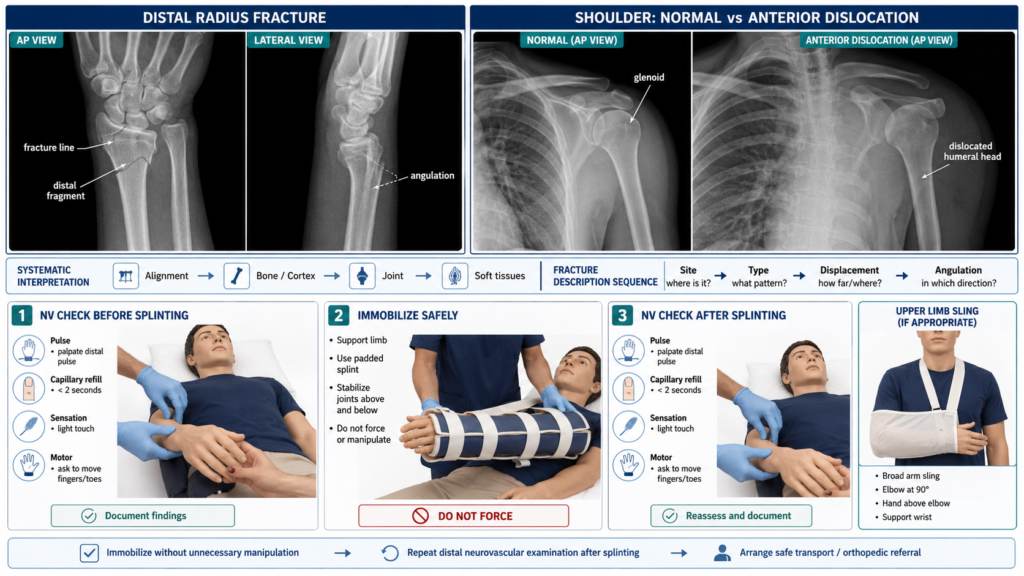
    2. Review systematically. Assess alignment and joints → bones and cortices → joint spaces → soft tissues without stopping after the first abnormality.
    3. Identify the injury. Decide whether the main abnormality represents a fracture, subluxation or complete dislocation.
    4. Describe a fracture. State the bone, side, proximal/middle/distal site, fracture pattern such as transverse, oblique, spiral or comminuted, and whether the articular surface is involved where visible.
    5. Describe displacement. Describe the position of the distal fragment, including translation and angulation; mention shortening or rotation only when reasonably assessable.
    6. Describe a dislocation. Name the affected joint and describe the direction of displacement of the distal component relative to the proximal component.
    7. Assess the injured limb clinically. Look for deformity, swelling or an open wound and assess distal pulse, capillary refill, sensation and relevant motor function before immobilization.
    8. Immobilize safely. Support the limb in a comfortable position using an appropriate padded splint or sling. For a long-bone injury, stabilize the fracture and adjacent joints where appropriate without forceful manipulation.
    9. Reassess and escalate. Repeat distal neurovascular examination after splinting, provide analgesia and arrange definitive orthopedic assessment; suspected open fractures or neurovascular compromise require urgent escalation.
Exam point: Never finish a fracture-management station without stating that distal neurovascular status should be checked before and after immobilization.
Safety: Do not repeatedly manipulate a painful or deformed limb. A dislocation or markedly displaced fracture requiring reduction should be managed by an appropriately trained clinician with suitable analgesia or sedation and monitoring.

🖼️ Visual Learning

VISUAL 1

Systematic X-Ray Recognition: Fracture vs Dislocation
🎥 Practical Video

🎥 Practical Demonstration
Musculoskeletal X-Ray Interpretation — Complete Step-by-Step Guide
Source: Learn Radiology with Dr. Ben

This demonstration reinforces a systematic approach to musculoskeletal radiographs and shows how fractures should be identified and described rather than relying on a quick visual impression.


▶ Watch Practical Video

📝 Important Viva Questions

Q1. What minimum information should you confirm before interpreting a limb X-ray?

Answer: Confirm the patient, anatomical region, side marker, date and available projections, and assess whether the radiograph is technically adequate.

Q2. Why are two radiographic views usually required when assessing a suspected fracture?

Answer: A fracture or displacement may be inapparent on one projection. Two approximately perpendicular views provide better assessment of the fracture and its alignment.

Q3. How should displacement of a fracture be described?

Answer: Describe the position of the distal fragment relative to the proximal fragment, including direction of translation and angulation.

Q4. What is the difference between a subluxation and a dislocation?

Answer: Subluxation is partial loss of normal joint congruity, whereas dislocation is complete loss of normal articulation between the joint surfaces.

Q5. What neurovascular findings should be assessed distal to an injured limb?

Answer: Assess distal pulses, capillary refill, skin colour or temperature, sensation and relevant motor function.

Q6. Why must neurovascular status be checked again after applying a splint?

Answer: Immobilization can reveal or worsen vascular or nerve compromise if the splint is too tight or limb alignment changes. Post-splint findings should therefore be documented.

Q7. What additional immediate concern should you look for in every fracture?

Answer: Determine whether the fracture is open by looking for a wound communicating with the fracture region, because this requires urgent specialist management.

Scroll to Top
💬 WhatsApp Support