
This chapter follows the supplied KMU outcomes and connects glomerular injury, renal edema, nephrotic syndrome, investigations and the major glomerular diseases into one clinical pathway. Understand the explanations first; use the AIM High-Yield Review at the end for revision.
Topic 1. Glomerular Disease, Facial Swelling & Nephrotic Syndrome Across the Lifespan
Renal Module — recognition of glomerular disease, interpretation of renal investigations, nephrotic and nephritic presentations, major glomerular disorders, management principles and pediatric counselling.
Topic Introduction
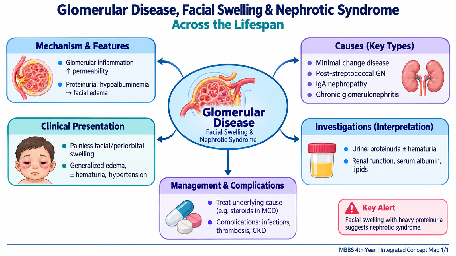
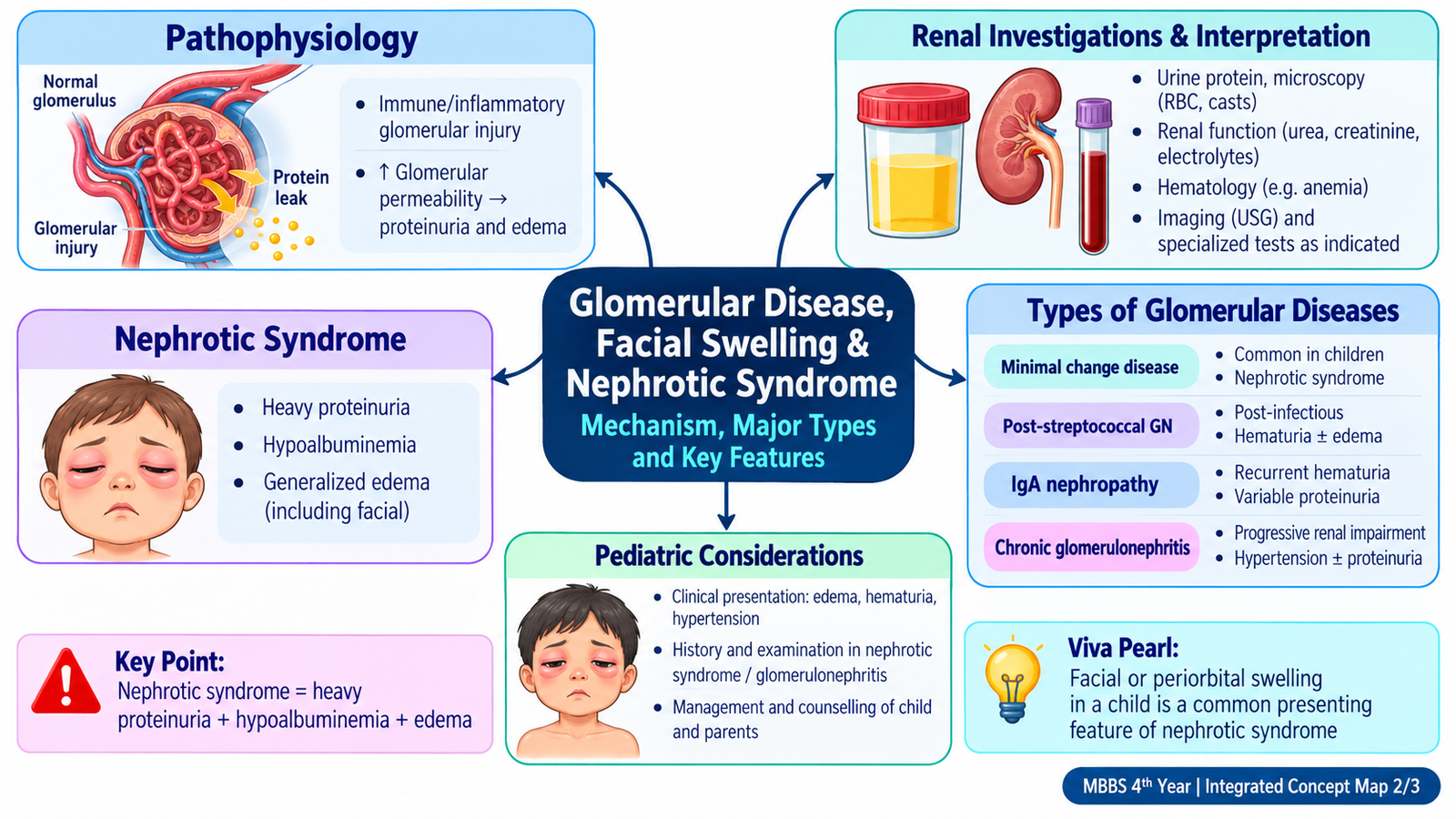
The glomerulus is the kidney’s filtration unit. When it is damaged, the patient may lose protein into the urine, leak blood into the urine, retain salt and water, develop hypertension, or gradually lose renal function. These changes create two important clinical patterns: a predominantly nephrotic pattern, in which heavy protein loss and edema dominate, and a predominantly nephritic pattern, in which glomerular inflammation produces hematuria, reduced filtration, edema and hypertension. This topic teaches how to recognize these patterns, assess facial swelling, interpret renal investigations and manage minimal change disease, post-streptococcal glomerulonephritis, IgA nephropathy and chronic glomerular disease in adults and children.
A. From Glomerular Injury to Renal Edema
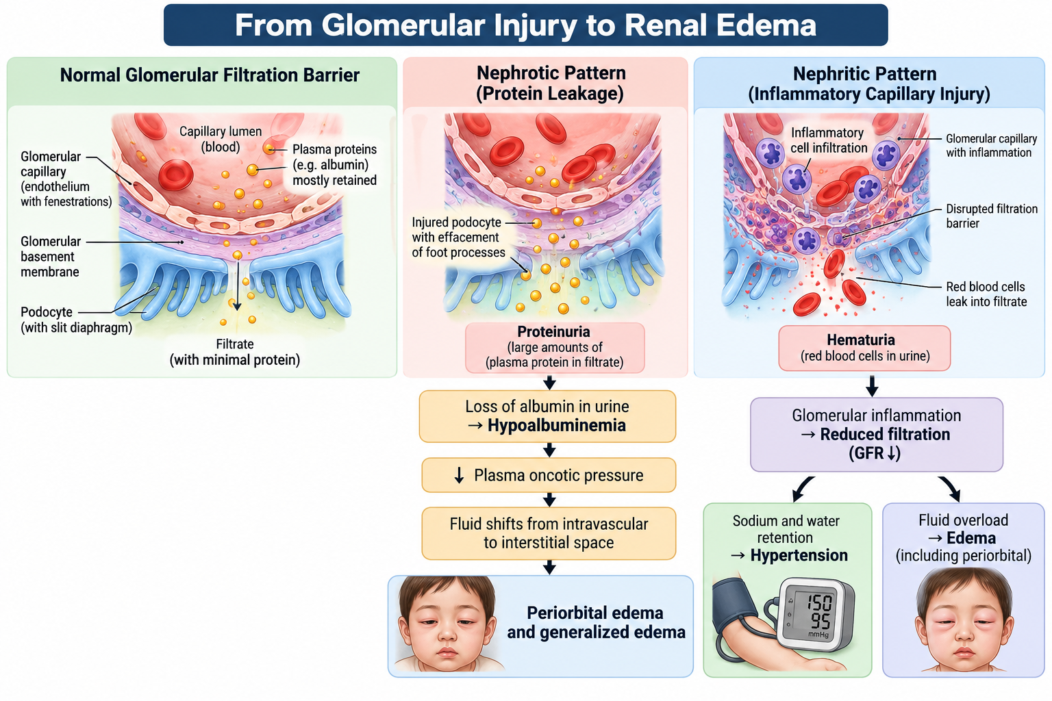
A healthy glomerular filtration barrier allows water and small solutes to pass while retaining blood cells and most plasma proteins. Glomerular disease disrupts this selectivity. The clinical picture depends on whether the dominant abnormality is protein leakage, inflammation with capillary injury, or progressive loss of functioning glomeruli.
Nephrotic pattern
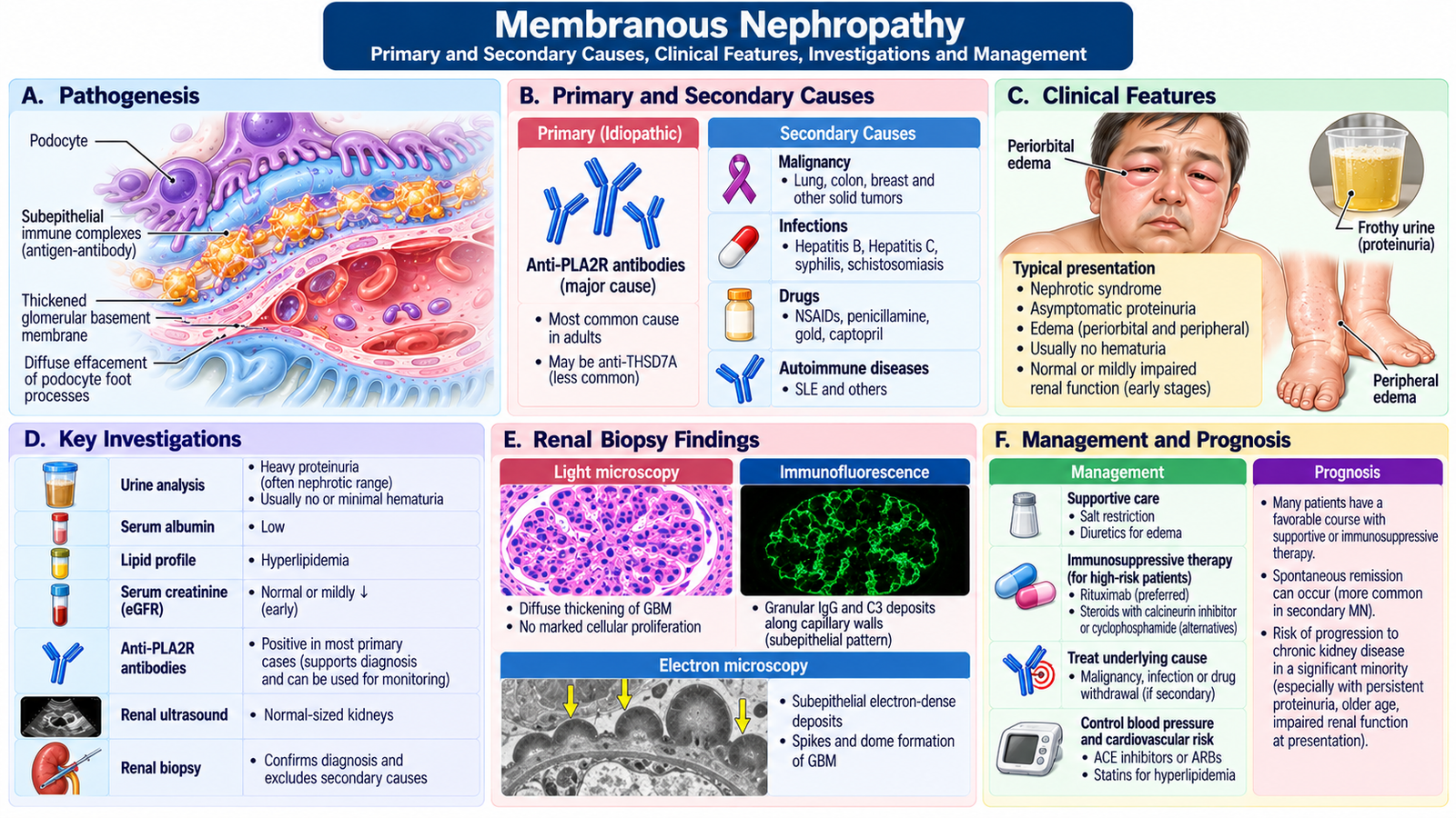
In nephrotic syndrome, increased glomerular permeability allows excessive albumin to escape into urine. Falling plasma albumin reduces plasma oncotic pressure, encouraging fluid to move from the intravascular compartment into the interstitial tissues. Renal sodium retention further amplifies edema.
The patient therefore develops proteinuria, hypoalbuminemia and edema. Hyperlipidemia may accompany the syndrome because the liver increases lipoprotein production in response to altered plasma protein balance.
Nephritic pattern
In nephritic disease, glomerular inflammation damages capillary walls. Red cells enter the filtrate, producing hematuria, while reduced filtration causes retention of salt, water and nitrogenous waste. The result may be hematuria, hypertension, edema, oliguria and impaired renal function.
Why is facial swelling prominent?
Renal edema is often first noticed around the eyes because loose periorbital tissue allows fluid to accumulate easily, particularly after lying flat overnight. With increasing fluid retention, edema may become dependent and generalized.
B. Clinical Assessment of Facial Swelling & Glomerular Disease
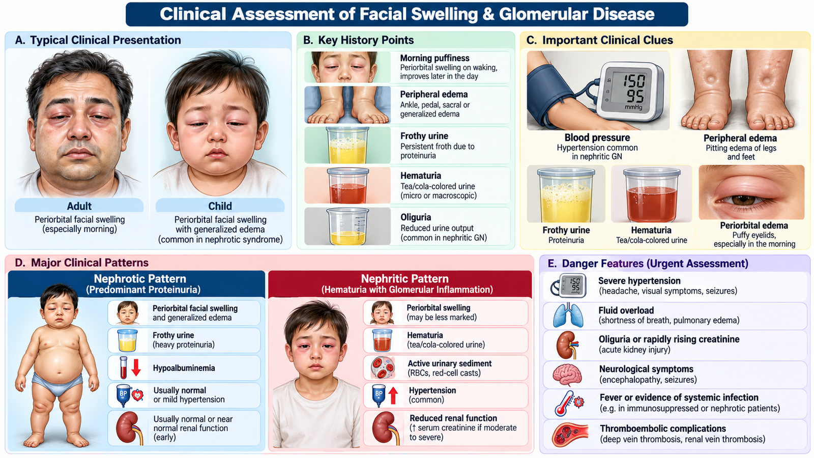
Facial swelling should not be labelled “renal” simply because it is around the eyes. The history and examination should determine whether the patient has a renal edema syndrome and whether the underlying pattern is predominantly nephrotic, nephritic or chronic renal disease.
Focused history
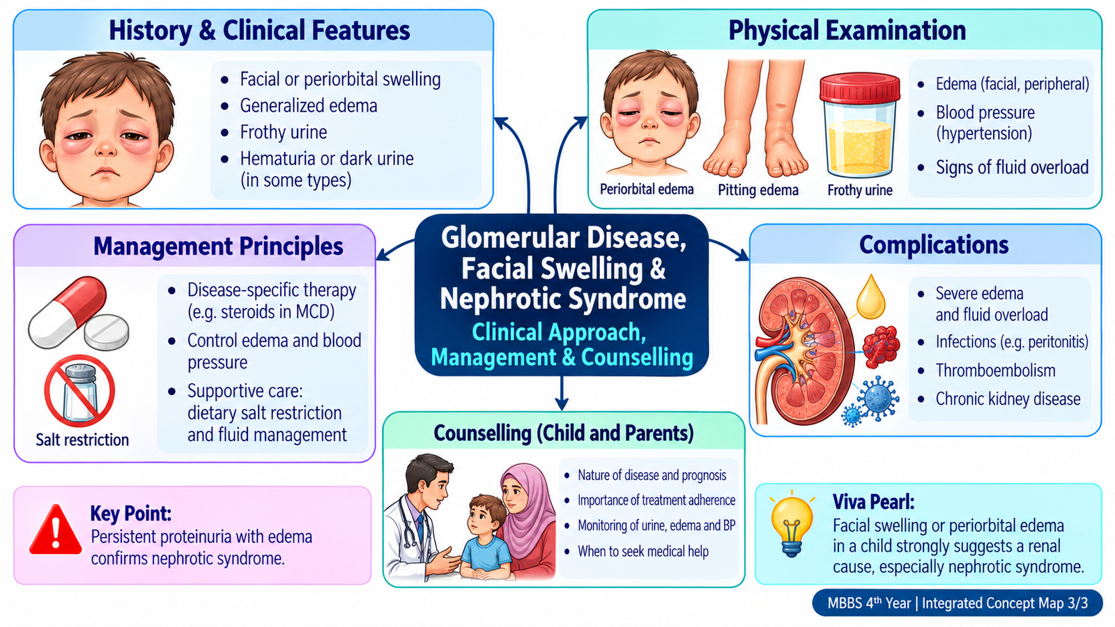
- Onset and progression: sudden edema suggests an acute process; persistent or progressive swelling raises concern for ongoing renal disease.
- Timing of facial swelling: periorbital puffiness noticed particularly in the morning supports renal edema.
- Urinary changes: frothy urine suggests significant proteinuria; red or cola-colored urine suggests glomerular hematuria; reduced urine output suggests impaired filtration.
- Recent infection: preceding sore throat or skin infection supports post-infectious glomerular disease; hematuria occurring around an upper respiratory illness suggests IgA nephropathy.
- Systemic symptoms: fever, rash, arthralgia or other multisystem features may suggest a secondary glomerular disorder and guide targeted investigations.
- Past renal disease: previous edema, hematuria, hypertension or abnormal renal function may indicate recurrent or chronic glomerular disease.
- Drug and family history: relevant nephrotoxic exposure and familial renal disease should be identified where clinically indicated.
Important examination findings
The examination is directed at confirming edema, assessing its severity and detecting evidence of glomerular inflammation or renal dysfunction.
- Periorbital edema: especially evident around the eyelids.
- Peripheral or generalized edema: supports significant sodium and fluid retention.
- Blood pressure: hypertension is particularly important in nephritic glomerular disease.
- Pulse, respiratory status and oxygenation: help identify significant fluid overload.
- Weight: serial increase may reflect fluid accumulation.
- Ascites or pleural fluid: may occur with severe nephrotic edema.
- Signs of chronic kidney disease: pallor, persistent hypertension or features of volume overload suggest more advanced disease.
Children: what changes?
In a child, edema may first be noticed by parents as swollen eyelids or increasing abdominal girth. History should also address urine appearance and output, recent infection, fever, previous episodes, treatment adherence and the child’s activity and appetite. Examination should include blood pressure, edema distribution, weight, evidence of ascites and features suggesting infection or significant intravascular depletion.
🩺 AIM Clinical Bridge
An 8-year-old child is brought because the parents noticed puffy eyelids on waking, followed by swelling of the legs and abdomen. The urine appears unusually frothy. Blood pressure is normal and there is no visible hematuria.
Think clinically:
- What renal syndrome is suggested by edema plus frothy urine?
- Which urine and blood investigations would confirm the pattern?
- Which common childhood glomerular disease should be considered?
C. Renal Investigations: What to Request and How to Interpret Them
Renal investigations should answer four practical questions: Is there glomerular injury? Is protein or blood being lost in urine? Is renal filtration impaired? Is there evidence pointing toward a particular glomerular disease?
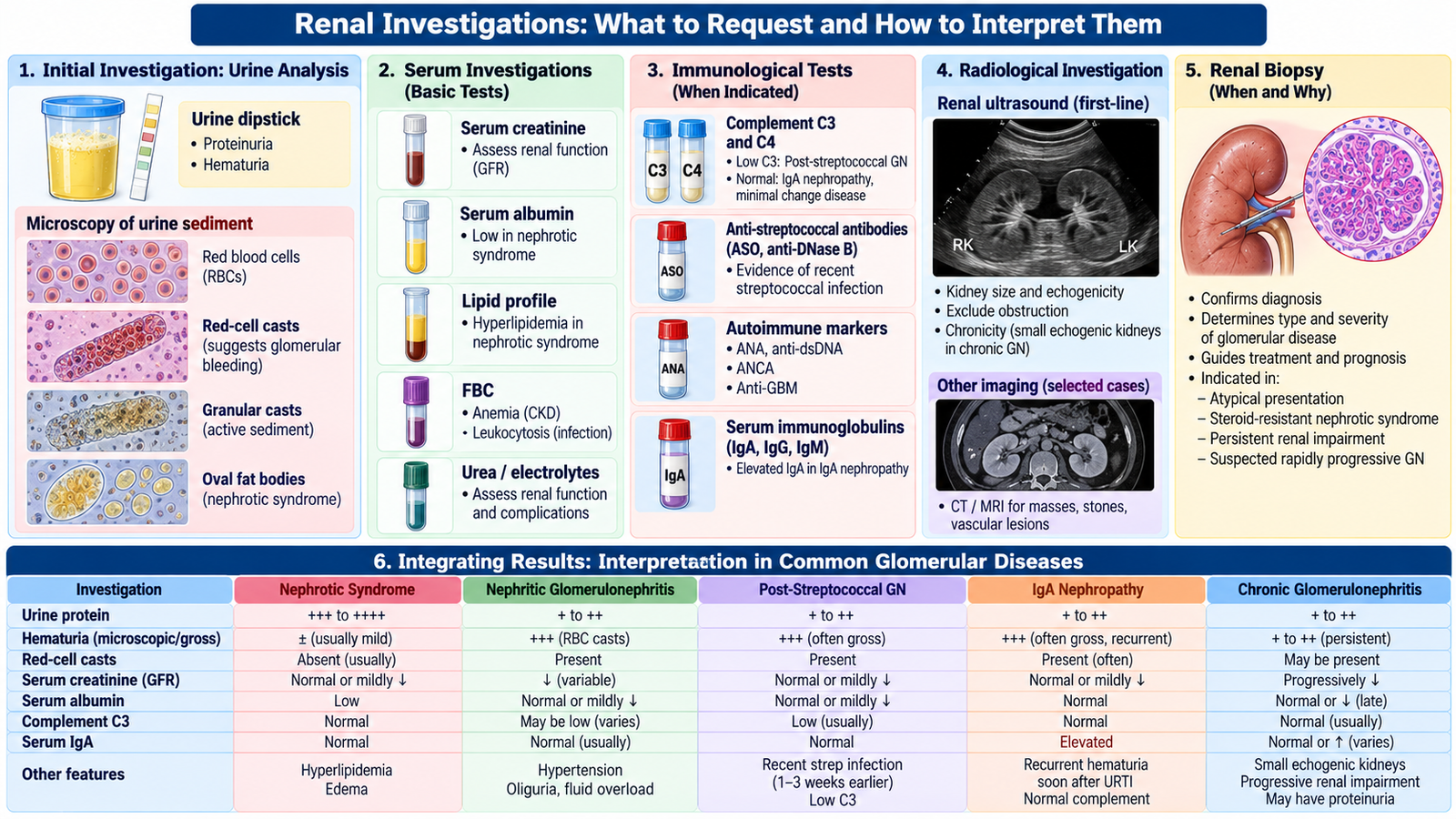
1. Urinalysis and urine microscopy
Protein on dipstick raises suspicion of a nephrotic process when substantial and persistent. Protein should then be quantified using an appropriate urine protein measurement.
Hematuria, particularly with dysmorphic red cells or red-cell casts, supports a glomerular source rather than bleeding from the lower urinary tract.
2. Serum renal profile
Serum creatinine and estimated glomerular filtration rate assess renal filtration. Rising creatinine suggests reduced filtration and changes the urgency of assessment, monitoring and referral. Urea and electrolytes help identify the biochemical consequences of renal dysfunction.
3. Serum proteins and lipid profile
Low serum albumin supports nephrotic syndrome when it occurs with significant proteinuria and edema. Hyperlipidemia commonly accompanies the nephrotic state.
4. Hematological investigations
A complete blood count may identify anemia in chronic renal disease or provide evidence of infection or systemic illness where clinically relevant.
5. Complement and disease-directed tests
Complement measurement helps distinguish some inflammatory glomerular disorders. Low complement supports post-infectious glomerulonephritis in the appropriate clinical setting, whereas complement is generally preserved in IgA nephropathy.
Evidence of recent streptococcal infection may be sought with appropriate streptococcal serology. Autoimmune testing is selected when the clinical picture suggests systemic autoimmune disease rather than being ordered indiscriminately.
6. Imaging
Renal ultrasonography evaluates kidney size, structure and obstruction. In chronic renal disease, small echogenic kidneys may support chronic irreversible parenchymal damage, although kidney size must always be interpreted in clinical context.
7. Renal biopsy
Biopsy provides histological and immunological information when the diagnosis is uncertain, when the clinical course is atypical, or when identifying the precise glomerular lesion would alter treatment. A typical child with uncomplicated steroid-responsive nephrotic syndrome does not automatically require biopsy at first presentation, while adults with nephrotic syndrome more commonly require histological diagnosis.
| Result / Pattern | What it means | Clinical implication |
|---|---|---|
| Marked proteinuria + low albumin + edema | Loss of glomerular protein selectivity | Nephrotic syndrome pattern |
| Hematuria + red-cell casts | Glomerular capillary inflammation | Nephritic/glomerulonephritic pattern |
| Rising creatinine | Reduced filtration | Assess severity, complications and need for escalation |
| Low complement after recent streptococcal infection | Complement consumption during immune glomerular injury | Supports post-streptococcal GN |
| Hematuria associated closely with respiratory infection + preserved complement | Pattern compatible with IgA-mediated glomerular disease | Consider IgA nephropathy |
D. Minimal Change Disease & Childhood Nephrotic Syndrome
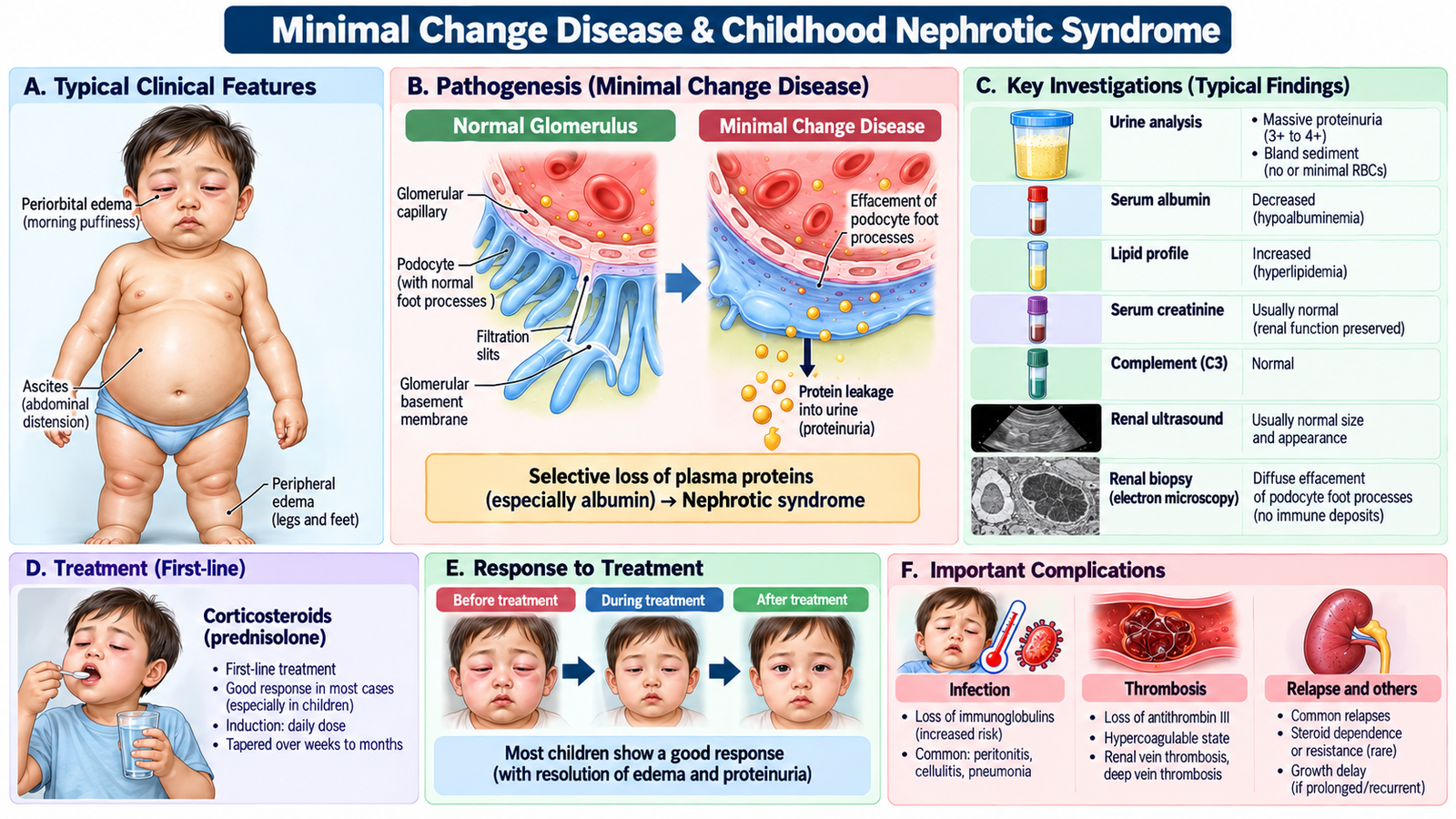
Minimal change disease is a major cause of nephrotic syndrome in children. The glomeruli may appear almost normal on routine light microscopy, but the filtration barrier becomes abnormally permeable to protein. The important clinical consequence is therefore not gross structural destruction but heavy protein loss with edema.
Clinical presentation
- Periorbital swelling progressing to dependent or generalized edema.
- Frothy urine due to significant proteinuria.
- Possible ascites when edema is severe.
- Blood pressure and renal function may remain relatively preserved in an uncomplicated presentation.
Diagnostic workup
The key pattern is significant urinary protein loss with low serum albumin and edema. Lipid levels may be elevated. Renal function and electrolytes are checked to identify complications or atypical disease. Urine microscopy is important because prominent hematuria or active urinary sediment may suggest another glomerular disorder.
A child with a typical presentation is commonly managed clinically without immediate renal biopsy. Biopsy becomes more relevant when features are atypical, renal dysfunction persists, or the clinical response is not as expected.
Management principles
Management aims to control the disease, manage edema safely and prevent complications. Corticosteroid therapy is the central treatment for typical childhood minimal change nephrotic syndrome. Salt intake is moderated when edema is significant, and fluid management is individualized according to volume status.
First-line treatment: Oral prednisolone is the standard initial treatment for childhood steroid-sensitive nephrotic syndrome, usually given over 8–12 weeks according to the prescribed regimen. Furosemide may be used cautiously for significant edema after assessing intravascular volume. Steroid-resistant, frequently relapsing or steroid-dependent disease requires pediatric nephrology referral.
Marked edema does not automatically mean that the circulation is overfilled. Some nephrotic patients may have reduced effective intravascular volume despite visible edema, so aggressive diuresis without clinical assessment can worsen hypovolemia or kidney injury.
Important complications
- Infection: nephrotic children have increased susceptibility to significant infection.
- Thrombosis: urinary loss of anticoagulant proteins and other changes in coagulation promote hypercoagulability.
- Hypovolemia: excessive movement of fluid out of the circulation may reduce effective circulating volume.
- Acute kidney injury: may occur with severe volume disturbance or other complications.
- Nutritional consequences: prolonged protein loss may affect nutritional status.
Counselling the child and parents
Parents should understand that nephrotic syndrome causes protein leakage through the kidneys and that swelling can recur. Explain the importance of taking prescribed treatment correctly, attending follow-up and having urine and clinical status monitored as advised.
They should seek medical assessment if the child develops fever or appears acutely unwell, develops breathing difficulty, marked abdominal pain, very low urine output, rapidly increasing edema or symptoms suggestive of thrombosis. Counselling should remain reassuring and family-centred because many children with typical minimal change disease respond well to treatment, although relapses may occur.
E. Post-Streptococcal Glomerulonephritis
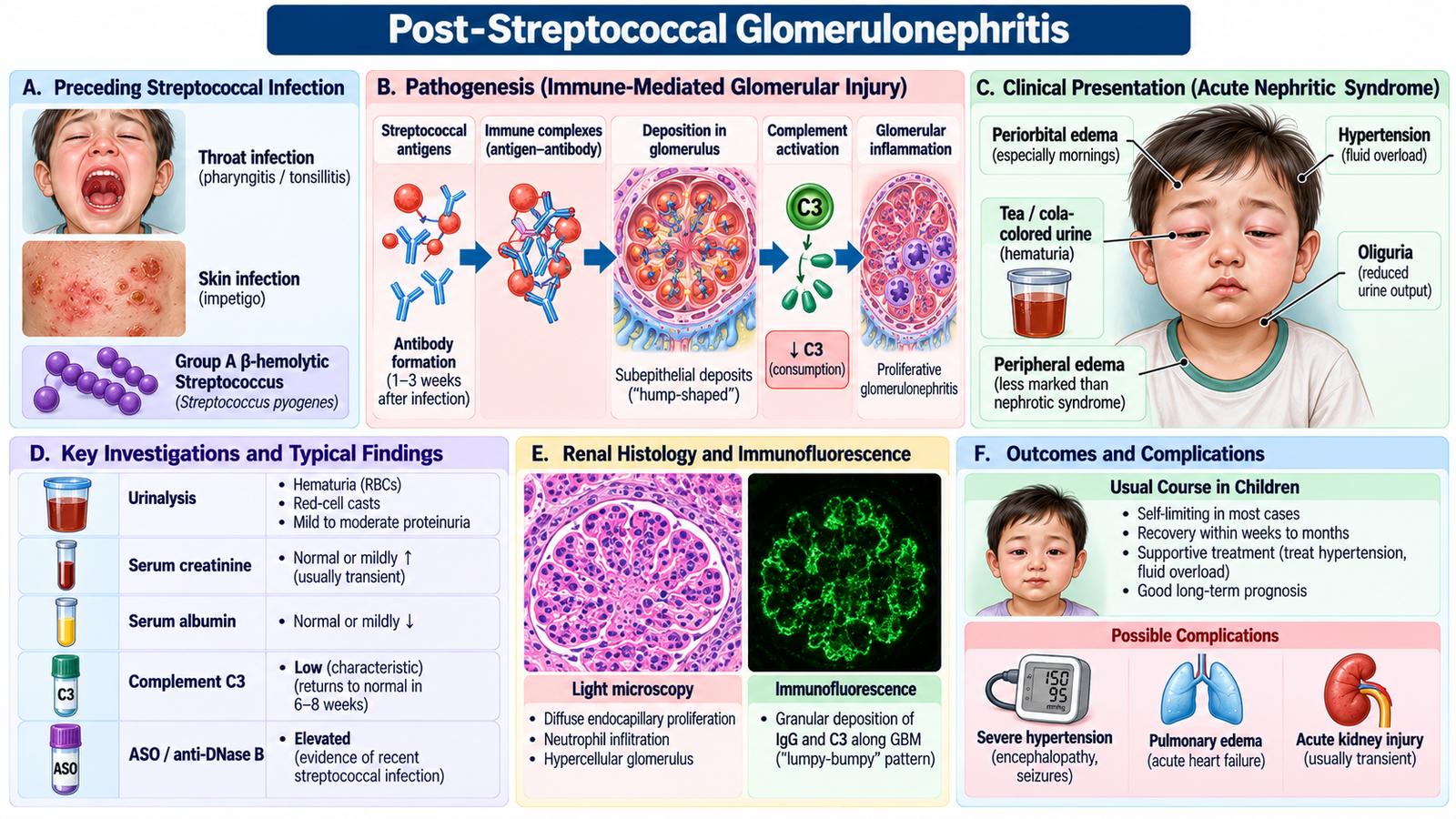
Post-streptococcal glomerulonephritis is an immune-mediated glomerular disease that develops after certain streptococcal throat or skin infections. The glomerular injury occurs because of an immune response rather than direct bacterial invasion of the kidney.
Clinical presentation
The patient typically develops features of an acute nephritic syndrome after a latent interval following infection. Important findings include:
- dark or cola-colored urine from glomerular hematuria;
- periorbital edema;
- hypertension;
- reduced urine output;
- variable renal impairment.
Diagnostic workup and interpretation
- Urinalysis: hematuria, proteinuria and an active glomerular sediment.
- Urine microscopy: red-cell casts strongly support glomerular inflammation.
- Serum creatinine: assesses reduction in filtration.
- Complement: low C3 supports post-infectious glomerular inflammation.
- Evidence of recent streptococcal infection: appropriate streptococcal antibodies may support the preceding infection when interpreted with the clinical picture.
Management
Management is predominantly supportive. Salt and fluid management, control of hypertension and treatment of significant edema are central. Renal function and electrolytes are monitored. Antibiotic therapy may be used to eradicate persistent streptococcal infection when present, but it does not reverse immune glomerular injury that has already developed.
Specific treatment: Furosemide is used for significant fluid overload, while antihypertensive agents such as amlodipine may be required for persistent hypertension. Penicillin is given to eradicate residual streptococcal infection. Severe pulmonary edema, hypertensive emergency or acute kidney injury requires urgent hospital management.
F. IgA Nephropathy
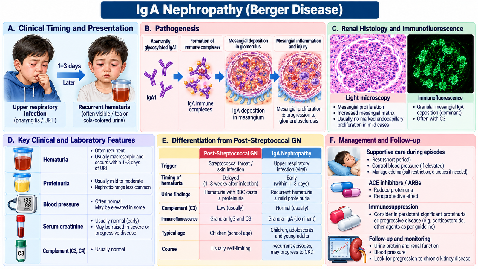
IgA nephropathy is a glomerular disorder in which IgA-containing immune deposits accumulate mainly within the mesangium. The disease is important because it often presents with recurrent glomerular hematuria and may gradually progress in some patients.
Recognizing the clinical pattern
A classic clue is visible hematuria occurring during or very soon after an upper respiratory infection. This timing differs from post-streptococcal glomerulonephritis, which characteristically follows the infection after a latent interval.
Some patients are identified through persistent microscopic hematuria or proteinuria rather than dramatic visible hematuria. Blood pressure and renal function must therefore be assessed even when symptoms seem mild.
Investigations
- Urinalysis: hematuria with variable proteinuria.
- Urine microscopy: glomerular red-cell morphology or red-cell casts may be present.
- Renal function: establishes baseline filtration and identifies progression.
- Complement: typically remains normal, helping distinguish the pattern from post-infectious GN.
- Renal biopsy: when clinically indicated, confirms mesangial IgA deposition and defines the glomerular lesion.
Management
Management depends on the severity of proteinuria, blood pressure and renal impairment. General renal-protective measures include good blood-pressure control and reduction of persistent proteinuria. Patients with more significant or progressive disease require nephrology assessment, and immunosuppressive treatment is reserved for selected circumstances rather than being routine for every patient.
First-line renal protection: ACE inhibitors such as ramipril or ARBs such as losartan are important treatments for persistent proteinuria and blood-pressure control, with monitoring of creatinine and potassium. Patients with persistent significant proteinuria or declining kidney function require nephrology referral for further disease-specific treatment.
Complications
The main long-term concern is progressive loss of renal function leading to chronic kidney disease in a proportion of patients. Persistent proteinuria, hypertension and worsening renal function therefore require continued monitoring.
G. Chronic Glomerulonephritis: Recognizing Progressive Glomerular Damage
Chronic glomerulonephritis describes progressive glomerular injury that eventually reduces the number of functioning nephrons and leads to chronic kidney disease. By the time the patient becomes symptomatic, the original glomerular disorder may be less obvious than the consequences of chronic renal dysfunction.
Clinical clues
- Persistent proteinuria or hematuria.
- Progressive hypertension.
- Gradually rising serum creatinine and falling filtration.
- Edema or fluid overload.
- Pallor due to anemia in established chronic kidney disease.
- Later manifestations of uremia if renal dysfunction becomes advanced.
Diagnostic workup
Assessment includes urinalysis, quantification of protein loss, renal function, electrolytes, complete blood count and renal imaging. Ultrasound may demonstrate chronic parenchymal change. Previous renal results are particularly valuable because chronicity is established by persistence or progression rather than by a single abnormal creatinine value.
Management principles
Management aims to preserve remaining renal function and address the underlying glomerular disease when it remains treatable. Blood pressure and proteinuria are controlled, nephrotoxic exposures are avoided where possible, and renal function and complications are monitored. Progressive renal impairment requires nephrology involvement and broader chronic kidney disease management.
Renoprotective treatment: ACE inhibitors or ARBs are preferred when indicated for proteinuric chronic kidney disease, with monitoring of renal function and potassium. Treat CKD complications such as anemia, fluid overload and electrolyte disturbances; advanced kidney failure may require dialysis or kidney transplantation.
Patients should understand that monitoring is not merely “repeat testing.” Serial blood pressure, urine protein and renal function show whether glomerular damage is stable, improving or progressing.
Important Comparison — Nephrotic vs Nephritic Pattern
| Feature | Nephrotic Pattern | Nephritic Pattern |
|---|---|---|
| Main glomerular problem | Increased protein permeability | Inflammatory capillary injury |
| Dominant urine finding | Heavy proteinuria | Hematuria with active sediment |
| Serum albumin | Markedly reduced pattern | Not the dominant abnormality |
| Edema mechanism | Protein loss, low oncotic pressure and sodium retention | Reduced filtration with salt and water retention |
| Blood pressure | May be normal or elevated | Hypertension is an important feature |
| Typical examples in this topic | Minimal change disease | Post-streptococcal GN, IgA nephropathy |
Integrated Mechanism Flow
Immune injury or permeability disturbance
Protein and/or blood enters urine
Protein loss or reduced filtration
Edema, hematuria, hypertension or oliguria
Urine + renal profile + disease-directed tests
Treat disease, control fluid/BP and monitor renal function
AIM High-Yield Review
- ⭐ Nephrotic syndrome is dominated by significant proteinuria, hypoalbuminemia and edema; minimal change disease is an important childhood cause.
- Renal facial edema is commonly periorbital and often most noticeable after lying flat overnight.
- ⭐ Hematuria with red-cell casts strongly supports a glomerular inflammatory source.
- Rising serum creatinine means filtration is impaired and should prompt assessment of severity and complications.
- Minimal change disease often presents in children with edema and heavy proteinuria and commonly responds to corticosteroid therapy.
- Nephrotic complications include significant infection, thrombosis, hypovolemia and acute kidney injury.
- ⭐ Post-streptococcal GN produces a nephritic picture after a preceding streptococcal infection and is supported by low complement in the appropriate clinical context.
- PSGN treatment is mainly supportive: control fluid overload and hypertension while monitoring renal function and electrolytes.
- ⭐ IgA nephropathy classically causes hematuria during or soon after an upper respiratory illness; complement is generally preserved.
- Renal biopsy is used when histological diagnosis is needed to clarify an atypical or significant glomerular disorder; it is not automatically required in every typical child with nephrotic syndrome.
- Chronic glomerular injury may progress through persistent proteinuria, hypertension and nephron loss to chronic kidney disease.
- Pediatric counselling should cover treatment adherence, recurrence, follow-up and warning symptoms such as fever, respiratory difficulty, severe edema or markedly reduced urine output.
Glomerular Disease, Facial Swelling & Nephrotic Syndrome Across the Lifespan
Use this video to reinforce glomerular disease patterns, nephrotic syndrome, minimal change disease, post-streptococcal glomerulonephritis, IgA nephropathy and renal edema.