
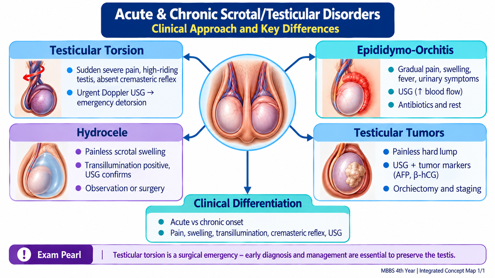
In any painful or swollen scrotum, first decide whether the problem is acute or chronic and whether testicular torsion must be excluded urgently. Then use age, onset, pain, examination findings and imaging to distinguish torsion, epididymo-orchitis, hydrocele and testicular tumor.
Topic 6. Acute & Chronic Scrotal/Testicular Disorders
Renal Module • Surgical approach to testicular torsion, epididymo-orchitis, hydrocele and testicular tumors
Topic Introduction
Scrotal pain and swelling range from benign fluid collections to conditions threatening the testis or indicating malignancy. The key clinical task is to recognize the pattern quickly. Sudden severe pain raises concern for testicular torsion; inflammatory pain with urinary or infective features suggests epididymo-orchitis; a painless cystic swelling may represent hydrocele; and a persistent intratesticular mass must raise suspicion of testicular tumor. The approach therefore depends on careful clinical differentiation followed by appropriately selected investigations and management.
1. The Acute Scrotum: Recognize the Emergency First
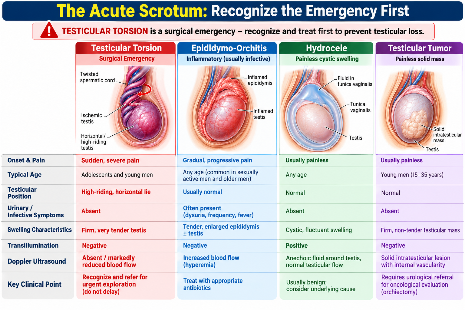
The acute scrotum means acute pain, swelling or tenderness involving the scrotal contents. Several disorders can produce this presentation, but the first priority is to identify testicular torsion because prolonged interruption of testicular blood flow can cause irreversible ischemic injury.
A useful first distinction is:
Think first about testicular torsion, particularly when onset is abrupt and accompanied by nausea or vomiting.
Epididymo-orchitis becomes more likely when pain develops more gradually with fever, urinary symptoms or evidence of infection.
Focused clinical assessment
History should establish the time and speed of onset, severity and location of pain, associated swelling, nausea or vomiting, fever, dysuria or urethral symptoms, previous similar episodes, trauma and relevant sexual or urinary history.
Examination should identify whether the abnormality is primarily testicular, epididymal or surrounding the testis. Important findings include tenderness, swelling, testicular position, scrotal skin changes and whether a swelling is cystic or solid. These findings guide the differential diagnosis, but when torsion is strongly suspected they must not create unnecessary delay.
2. Testicular Torsion: Diagnosis & Emergency Management
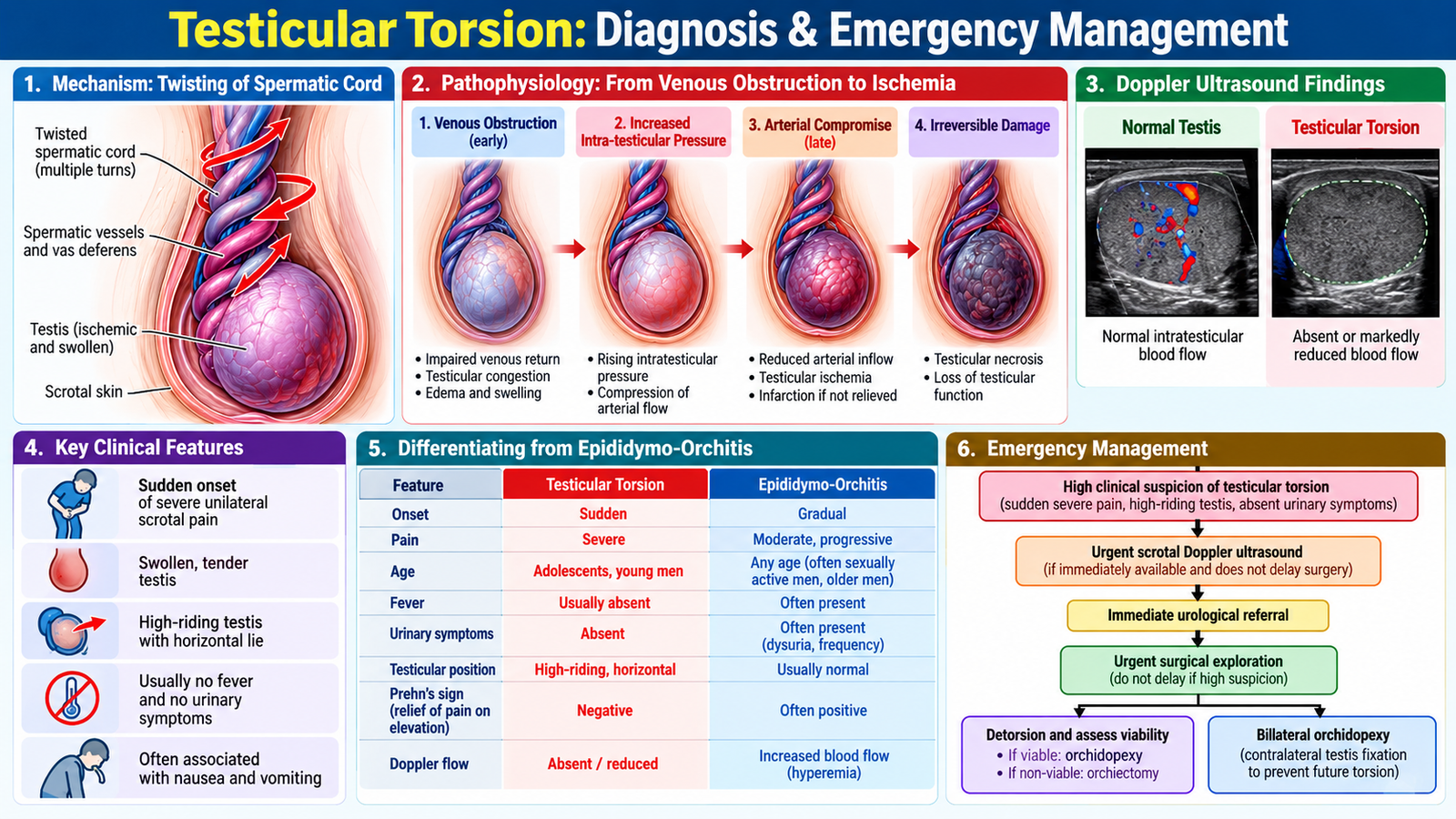
Testicular torsion occurs when the spermatic cord twists, compromising testicular vascular supply. Venous outflow is impaired first, producing congestion and swelling; continuing torsion then reduces arterial inflow and causes ischemia. This explains why torsion is a time-critical surgical emergency.
Clinical clues
The classic presentation is sudden, severe unilateral scrotal pain, often associated with nausea or vomiting. Examination may show a tender swollen testis lying higher than the opposite side or in an abnormal orientation. The cremasteric reflex may be absent on the affected side.
No individual sign should be considered perfect. The important clinical principle is the overall probability of torsion in a patient with an acute painful testis.
Diagnostic workup
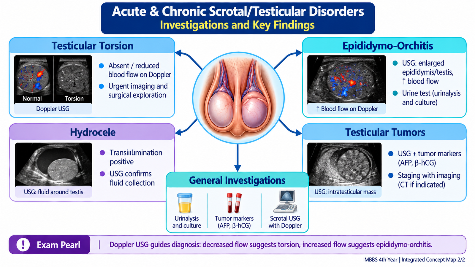
When the clinical picture is strongly suggestive, urgent surgical assessment is required. Color Doppler ultrasonography is useful when the diagnosis is uncertain because it assesses testicular perfusion. Reduced or absent blood flow supports torsion, whereas increased inflammatory flow is more compatible with epididymo-orchitis.
Acute pain + examination strongly suggesting torsion → urgent surgical action. Imaging is helpful when uncertainty exists, but investigation should not cause harmful delay when suspicion is high.
Emergency management
The definitive principle is urgent scrotal exploration and detorsion, followed by assessment of testicular viability. A viable testis is fixed to prevent recurrent torsion. The contralateral testis is also usually fixed because the anatomical predisposition may be bilateral. If the affected testis is clearly non-viable, removal may be required.
Manual detorsion may occasionally be attempted as a temporary emergency measure where appropriate, but successful pain relief does not replace definitive surgical exploration and fixation.
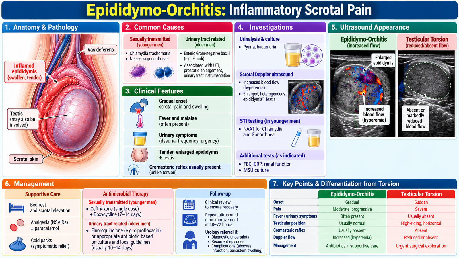
3. Epididymo-Orchitis: Inflammatory Scrotal Pain
Epididymitis is inflammation of the epididymis; when inflammation also involves the testis it is termed epididymo-orchitis. Infection may reach the epididymis through the genitourinary tract. The likely infectious source depends on age, sexual history and underlying urinary-tract factors.
How it presents
Pain and swelling usually develop more gradually than in torsion. Epididymal tenderness may progress to generalized scrotal tenderness and swelling. Fever, dysuria, urinary frequency or urethral symptoms support an infective process.
The most important diagnostic challenge is not simply proving infection—it is ensuring that torsion has not been missed.
Investigation and interpretation
- Urinalysis may demonstrate evidence of urinary inflammation or infection.
- Urine culture helps identify a urinary pathogen when bacterial urinary infection is suspected.
- Appropriate testing for a sexually transmitted infection is considered when supported by the sexual history and clinical presentation.
- Color Doppler ultrasound is particularly useful when torsion remains in the differential. Increased vascularity supports inflammation, whereas reduced testicular perfusion raises concern for torsion.
Management principles
Treatment addresses the suspected infectious cause and provides symptomatic support with analgesia, rest and scrotal support where appropriate. Antimicrobial choice should be directed by the likely source of infection and relevant microbiological information rather than treating every patient identically.In sexually transmitted epididymo-orchitis, first-line treatment is intramuscular ceftriaxone plus oral doxycycline for 10 days. When enteric organisms are responsible, oral levofloxacin may be used if susceptibility and safety considerations permit and gonorrhea has been excluded. Sexual partners should be assessed and treated when an STI is identified.
Failure to improve, severe systemic illness, marked swelling or diagnostic uncertainty requires reassessment for complications or an alternative diagnosis.
4. Hydrocele: Recognizing & Managing a Cystic Scrotal Swelling
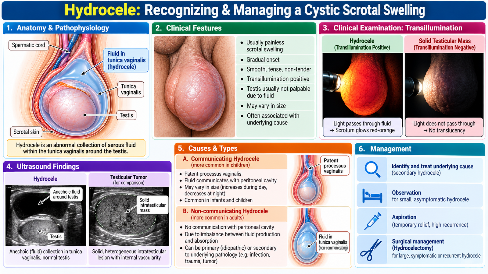
A hydrocele is an abnormal collection of serous fluid within the tunica vaginalis surrounding the testis. It commonly presents as a painless scrotal swelling, although a large hydrocele may produce heaviness or discomfort.
Clinical recognition
The swelling is usually smooth and fluctuant. Because fluid surrounds the testis, the testis may be difficult to palpate separately when the hydrocele is large. A typical hydrocele transilluminates, reflecting transmission of light through clear fluid.
Diagnostic workup
Clinical examination may establish a straightforward hydrocele. Scrotal ultrasonography becomes particularly valuable when the testis cannot be adequately examined, the swelling is atypical, or an underlying testicular lesion needs to be excluded. Ultrasound demonstrates the fluid collection and allows assessment of the testis itself.
Management
Management depends on symptoms, size and the underlying cause. Small uncomplicated hydroceles may not require immediate intervention. A persistent, large or troublesome hydrocele may require definitive surgical treatment. In adults, a newly developed hydrocele should prompt attention to possible underlying testicular or epididymal disease rather than assuming that the fluid collection is the entire diagnosis. Hydrocelectomy is the definitive treatment for a large, persistent or symptomatic adult hydrocele. In infants, uncomplicated communicating hydroceles may resolve spontaneously, while persistent cases or associated inguinal hernia require pediatric surgical assessment.
5. Testicular Tumors: From Scrotal Mass to Diagnosis & Management
A testicular tumor should be considered whenever a patient has a persistent solid intratesticular mass. Many tumors present as a painless testicular lump or enlargement, although some patients report discomfort or heaviness. The essential clinical principle is that a suspicious intratesticular mass requires prompt evaluation.
Clinical assessment
History should establish the duration and progression of swelling, pain or heaviness and relevant previous testicular problems. Examination aims to determine whether the lesion is intratesticular or extratesticular. A firm testicular mass is more concerning than a clearly cystic extratesticular swelling.
Diagnostic workup
Scrotal ultrasound is the key initial imaging investigation because it can establish whether a lesion is solid and intratesticular. When malignancy is suspected, serum tumor markers such as alpha-fetoprotein (AFP), beta-human chorionic gonadotropin (β-hCG) and lactate dehydrogenase (LDH) contribute to diagnostic assessment, classification and subsequent monitoring.
Tumor markers must be interpreted in context. A normal marker result does not exclude testicular malignancy because not every tumor produces measurable markers.
Management principle
A suspicious testicular malignancy requires specialist urological management. The usual diagnostic and therapeutic approach to a malignant-appearing intratesticular lesion is radical inguinal orchiectomy, which provides tissue diagnosis while respecting the lymphatic drainage of the testis. Subsequent management depends on histology, tumor markers and disease extent.
Following diagnosis, contrast-enhanced CT of the chest, abdomen and pelvis is used for staging. Depending on histology and stage, subsequent treatment may involve surveillance, chemotherapy or radiotherapy. Sperm banking should be discussed before treatment because fertility may be affected.
Important complications
Untreated malignant tumors may progress locally or metastasize. Lymphatic spread classically follows the testicular lymphatic drainage toward retroperitoneal lymph nodes; advanced disease may spread more widely. Treatment may also have implications for fertility and endocrine function, making appropriate counselling part of patient-centred management.
6. Putting Scrotal Pain & Swelling Together: Clinical Differentiation
The four conditions become easier to separate when the student combines time course, pain, systemic or urinary features, examination and ultrasound findings rather than relying on a single sign.
| Feature | Testicular torsion | Epididymo-orchitis | Hydrocele | Testicular tumor |
|---|---|---|---|---|
| Typical onset | Sudden | Usually more gradual | Gradual | Usually gradual |
| Pain | Severe acute pain | Tender inflammatory pain | Usually painless/heaviness | Often painless lump |
| Associated clues | Nausea/vomiting may occur | Fever or urinary/urethral symptoms may occur | Fluid-filled swelling | Persistent firm intratesticular lesion |
| Key examination clue | Tender, possibly high-riding testis | Tender epididymal/testicular swelling | Cystic, usually transilluminant | Solid testicular mass |
| Doppler / ultrasound logic | Reduced/absent perfusion supports torsion | Increased inflammatory vascularity | Fluid around testis | Solid intratesticular lesion |
| Management priority | Emergency surgical assessment | Treat infection and exclude torsion | Treat if symptomatic/persistent or underlying cause identified | Urgent urological evaluation |
A young man develops sudden severe unilateral scrotal pain with nausea. Examination shows a very tender testis in an abnormal high position. There are no convincing urinary symptoms.
Think clinically:
- What diagnosis must be prioritized?
- Would waiting for routine investigations be safe if clinical suspicion is high?
- What is the definitive management principle?
AIM High-Yield Review
- Sudden severe unilateral scrotal pain should immediately raise concern for testicular torsion.
- Torsion causes vascular compromise → ischemia → potential testicular infarction, making rapid treatment essential.
- Color Doppler ultrasound helps distinguish reduced perfusion in torsion from increased inflammatory flow in epididymo-orchitis when diagnostic uncertainty exists.
- A strong clinical suspicion of torsion requires urgent surgical assessment rather than harmful diagnostic delay.
- Epididymo-orchitis is favored by a more gradual painful swelling with infective, urinary or urethral features.
- A hydrocele is a fluid collection around the testis; ultrasound is especially useful when the testis cannot be adequately assessed clinically.
- A solid intratesticular mass should be regarded as suspicious for testicular malignancy until properly evaluated.
- AFP, β-hCG and LDH assist assessment of suspected testicular tumors, but normal markers do not exclude malignancy.
- Suspected testicular cancer is managed through an inguinal oncological approach, not routine trans-scrotal biopsy.
Acute & Chronic Scrotal/Testicular Disorders
Testicular torsion • Epididymo-orchitis • Hydrocele • Testicular tumor • Clinical examination