
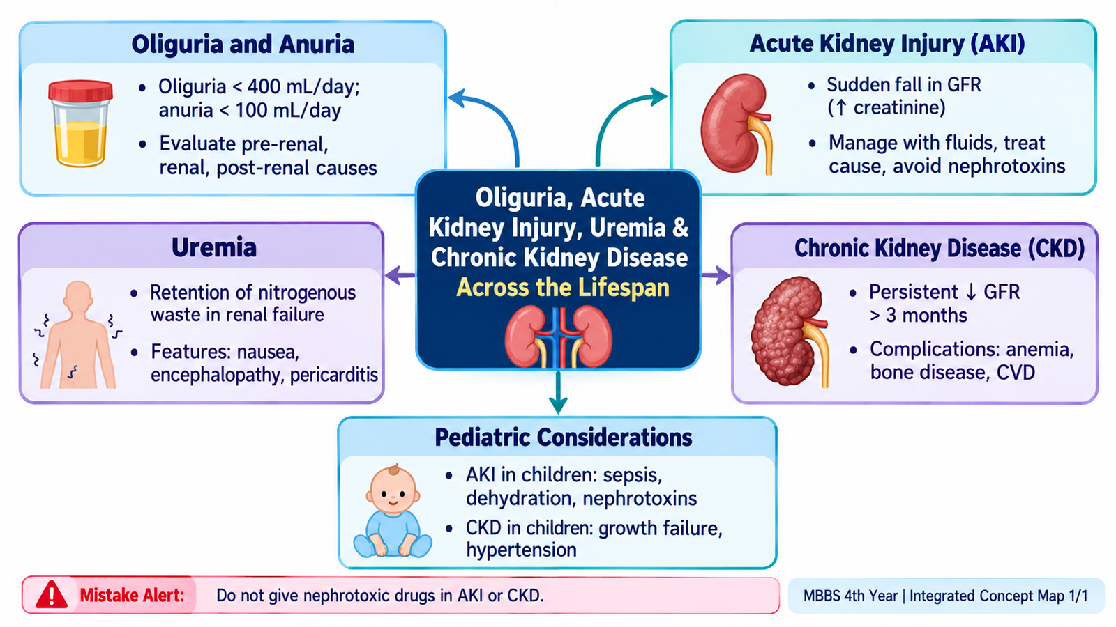
This chapter follows the KMU outcomes for oliguria, acute kidney injury, uremia and chronic kidney disease in adults and children. First understand the sequence from reduced kidney function → clinical consequences → investigation → management; use the final High-Yield Review for revision.
Topic 3. Oliguria, Acute Kidney Injury, Uremia & Chronic Kidney Disease Across the Lifespan
Renal Module — a clinically connected approach to reduced urine output, acute and chronic loss of kidney function, uremic manifestations, investigation and management in adults and children.
Topic Introduction
Kidney dysfunction may present suddenly with oliguria or anuria, develop as acute kidney injury (AKI), or progress over months to years as chronic kidney disease (CKD). As filtration and regulatory functions fail, water, electrolytes, acids and metabolic waste products accumulate. Severe retention of these substances produces the clinical syndrome of uremia. The practical approach is therefore to recognize reduced urine output, determine whether the problem is acute or chronic, identify reversible causes, assess complications and restore or support kidney function. The same principles apply in children, but causes, fluid assessment, growth and drug/fluid calculations require pediatric attention.
A. Oliguria & Anuria: Recognizing the Clinical Problem
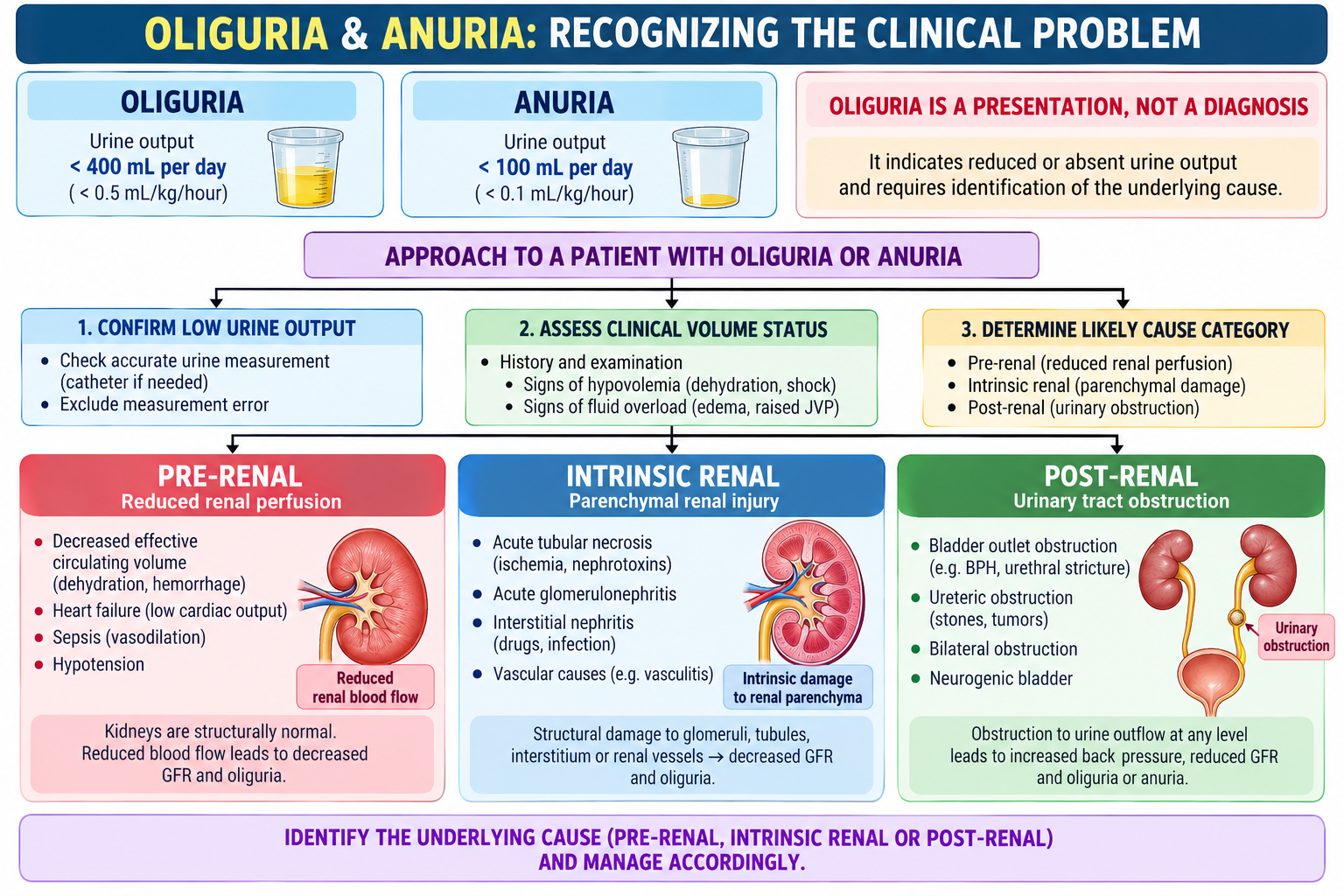
Oliguria means abnormally reduced urine output, while anuria represents extremely little or virtually absent urine production. They are clinical signs, not diagnoses. The first task is to confirm that urine output is genuinely reduced and then identify whether renal perfusion, renal tissue or urinary outflow is responsible.
Think in three compartments
Reduced renal perfusion without primary structural kidney damage, such as volume depletion or reduced effective circulation.
Damage within the kidney itself involving glomeruli, tubules, interstitium or renal vessels.
Obstruction to urinary drainage causing pressure to build upstream and impair filtration.
Focused assessment
Ask about vomiting, diarrhea, bleeding, fever, poor intake, recent illness, nephrotoxic medicines, urinary symptoms and difficulty passing urine. Look for dehydration, hypotension, edema, signs of systemic illness and bladder distension. The clinical question is: Is renal blood flow reduced, is the kidney damaged, or is urine unable to leave?
B. Acute Kidney Injury: Mechanism, Recognition & Clinical Presentation
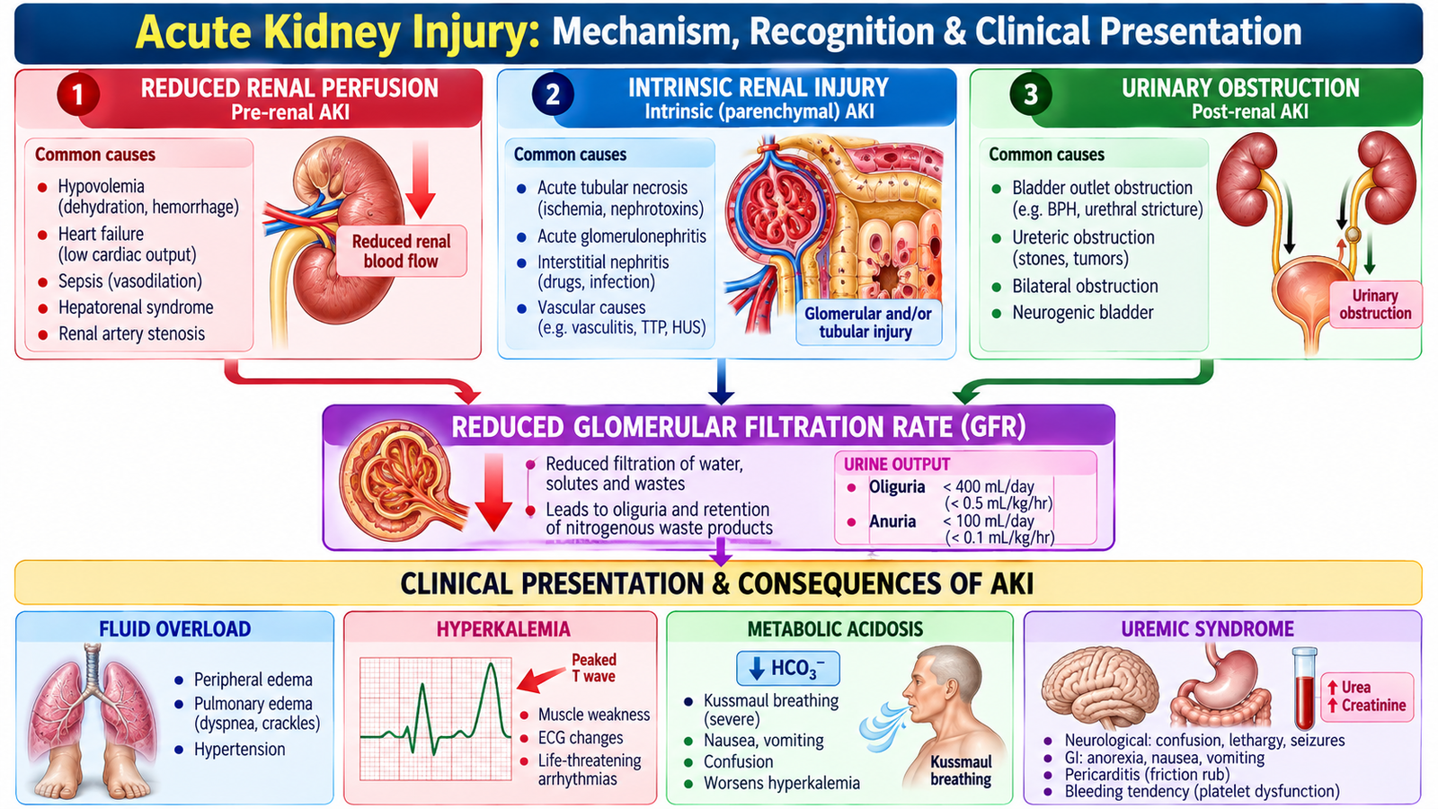
Acute kidney injury is an abrupt deterioration in kidney function. The fall in filtration causes retention of nitrogenous waste products and may disturb fluid, electrolyte and acid–base balance. AKI can occur with or without obvious oliguria, so urine output and renal-function tests must be interpreted together.
How AKI develops
Pre-renal AKI begins with inadequate renal perfusion. If perfusion is restored early, kidney function may recover rapidly. Persistent ischemia, however, may progress to intrinsic tubular injury.
Intrinsic AKI results from direct damage to renal structures. Common mechanisms include tubular injury, glomerular disease, interstitial inflammation and vascular injury.
Post-renal AKI results from urinary tract obstruction. Relief of significant obstruction can restore filtration, making early recognition particularly important.
Clinical manifestations
- Reduced urine output may occur, although some patients remain non-oliguric.
- Fluid retention may cause peripheral edema, hypertension or pulmonary congestion.
- Hyperkalemia may develop because potassium excretion falls.
- Metabolic acidosis may occur because acid excretion is impaired.
- Progressive nitrogenous waste retention increases serum urea and creatinine.
- Severe kidney dysfunction may progress to symptomatic uremia.
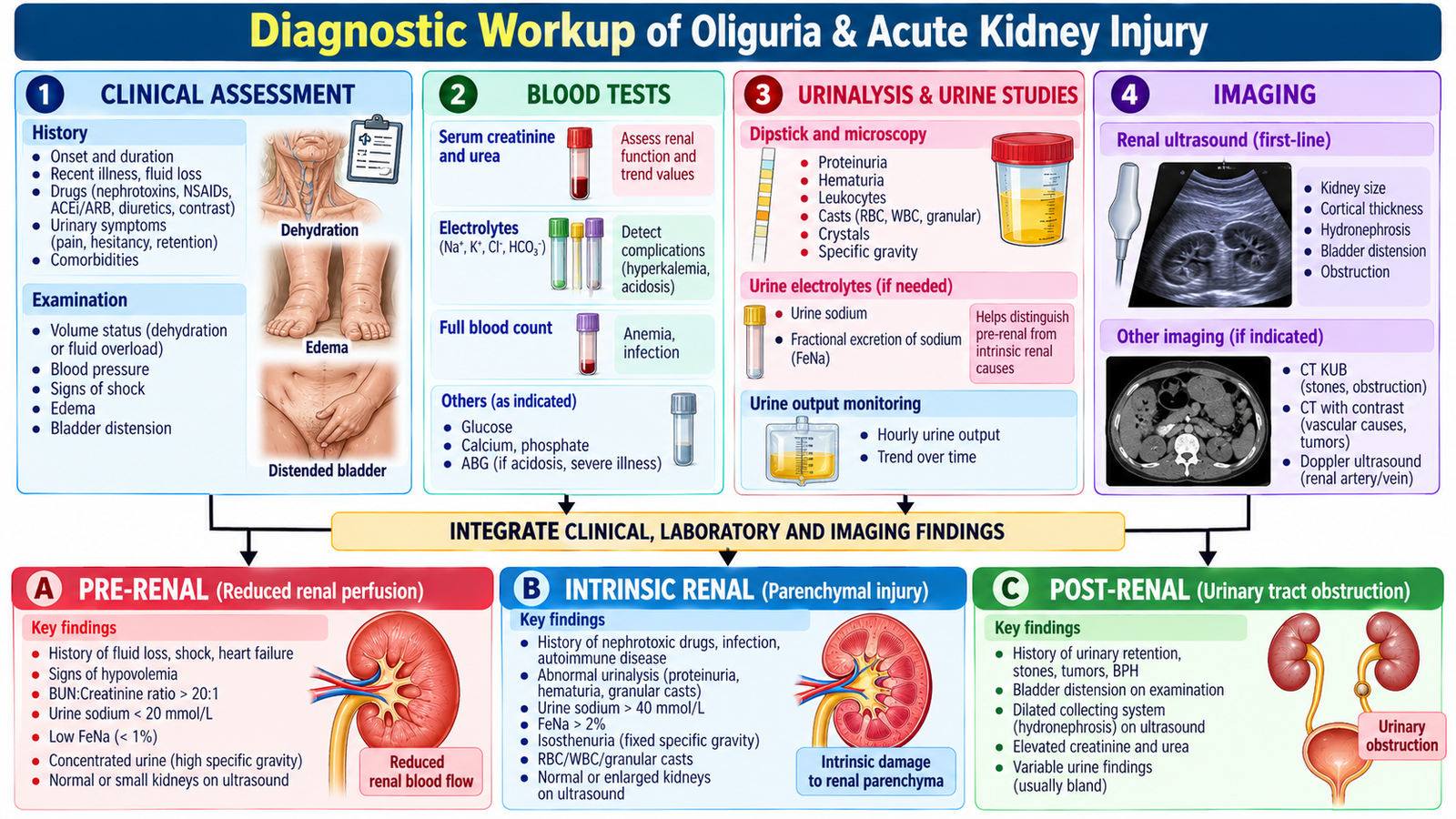
C. Diagnostic Workup of Oliguria & Acute Kidney Injury
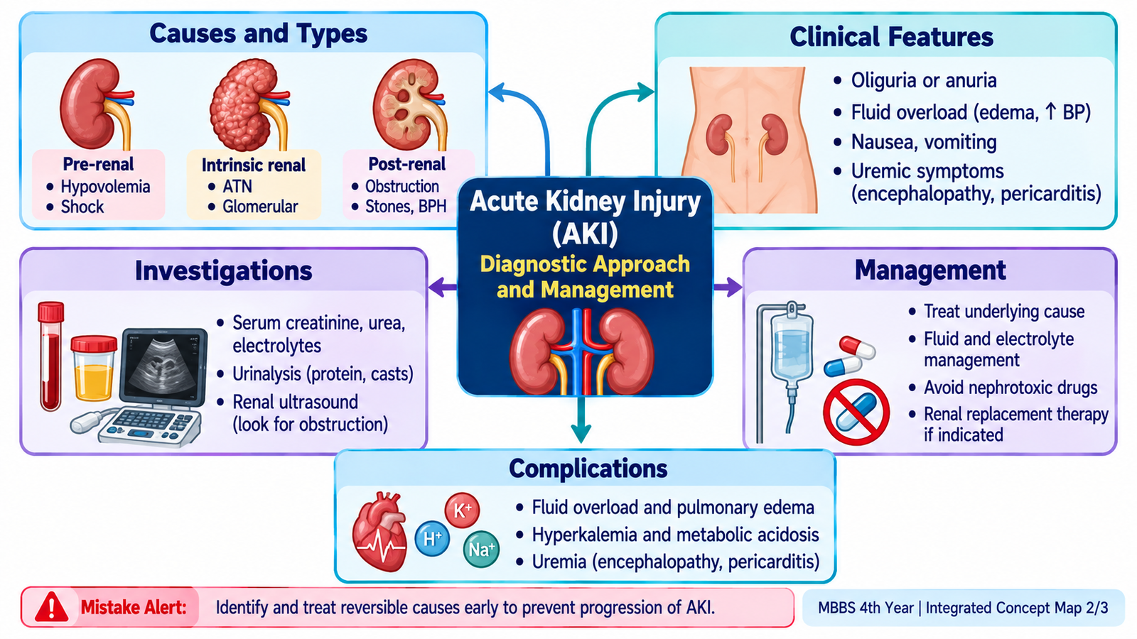
Investigation should answer four questions: Is kidney function impaired? How severe is the disturbance? What is causing it? Are dangerous complications already present? Laboratory results should never be interpreted without the clinical volume status, urine output and possibility of obstruction.
1. Blood investigations
- Serum creatinine and urea: indicate accumulation of substances normally cleared by the kidneys and are followed serially.
- Electrolytes: particularly potassium, because significant hyperkalemia may be immediately dangerous.
- Bicarbonate / acid–base assessment: identifies metabolic acidosis.
- Complete blood count: may provide clues to infection, anemia or an underlying systemic process.
2. Urine assessment
Urinalysis can provide important etiological clues. Blood, protein, leukocytes and sediment findings may direct attention toward glomerular, interstitial or infective processes. Urine volume must be monitored because its trend helps assess progression and response to treatment.
3. Imaging
Renal and urinary tract ultrasonography is particularly useful when obstruction is suspected. It may demonstrate hydronephrosis and can also provide information about kidney size and structure.
4. Search for the cause
Combine history, examination, laboratory findings and imaging. A dehydrated patient after gastrointestinal fluid loss suggests a pre-renal mechanism; active urinary sediment suggests intrinsic renal disease; bladder distension or hydronephrosis points toward obstruction.
An older patient develops very low urine output after several days of vomiting. The patient is clinically dehydrated and serum creatinine has risen.
D. Management of Acute Kidney Injury & Oliguria
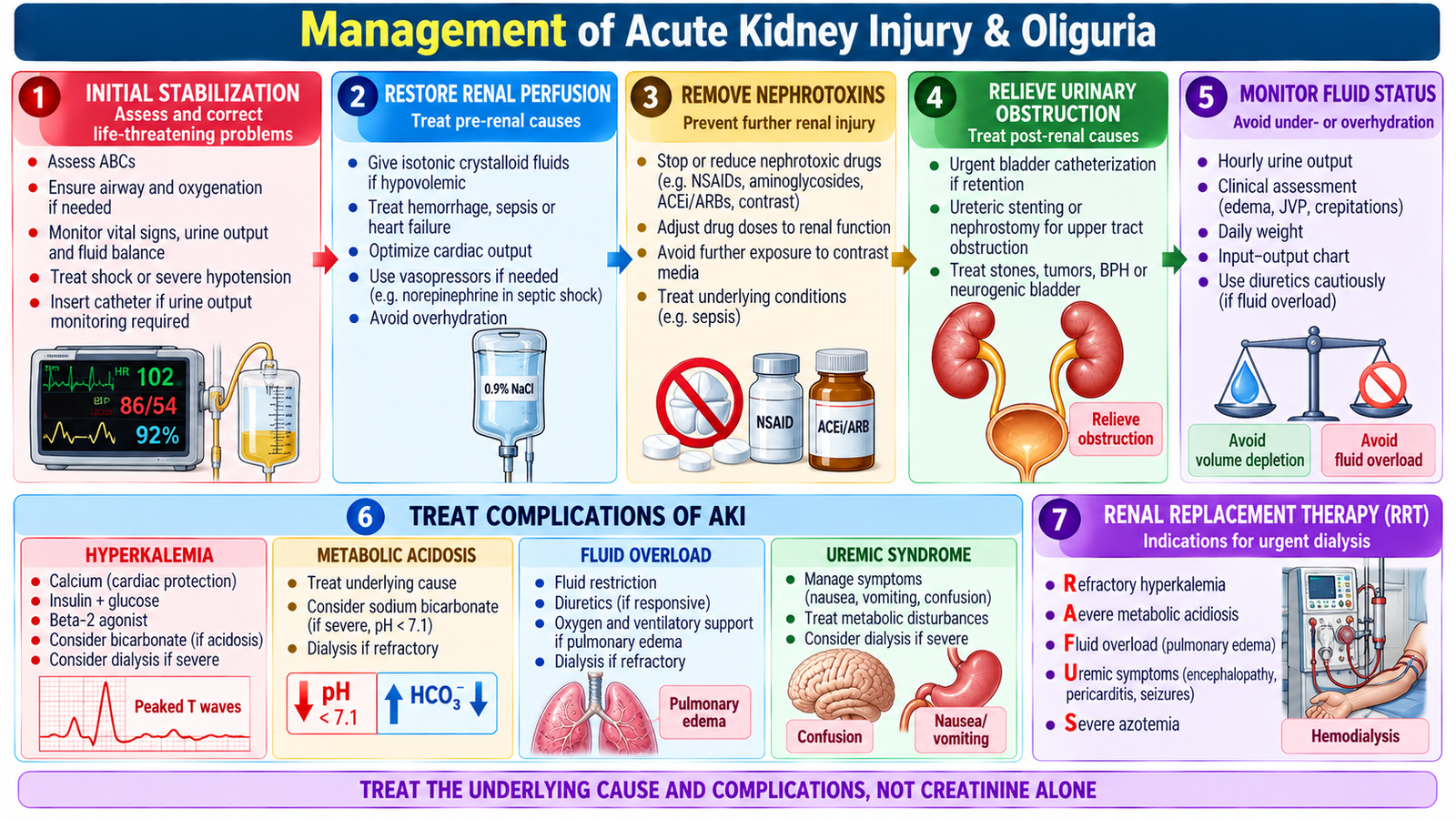
AKI management is based on treating the cause, protecting remaining kidney function and preventing or treating complications. Management must be individualized because the correct action for a volume-depleted patient may be harmful in a patient already overloaded with fluid.
Correct reversible causes
- Hypovolemia: restore circulating volume appropriately while monitoring response.
- Sepsis or systemic illness: treat the underlying cause and maintain adequate perfusion.
- Nephrotoxic exposure: review and stop avoidable nephrotoxic agents where appropriate.
- Urinary obstruction: identify and relieve obstruction promptly.
Support kidney function
Monitor fluid balance, urine output, body weight, renal-function tests and electrolytes. Drug prescriptions should be reviewed because reduced renal clearance can lead to accumulation of renally eliminated medicines.
Treat complications
Hyperkalemia, pulmonary edema, significant metabolic acidosis and uremic complications require active treatment. When these problems cannot be adequately controlled medically, renal replacement therapy may be required.
Specific treatment: Give IV isotonic crystalloid for hypovolemia with reassessment of fluid status. Relieve urinary retention by catheterization and arrange urgent decompression for significant upper urinary tract obstruction. Severe hyperkalemia with ECG changes requires IV calcium gluconate for cardiac protection and insulin with glucose to lower serum potassium. Furosemide may treat fluid overload but should not be used routinely to reverse AKI. Refractory hyperkalemia, acidosis, pulmonary edema or uremic complications require urgent consideration of dialysis.
E. Uremia: From Reduced Filtration to Multisystem Disease
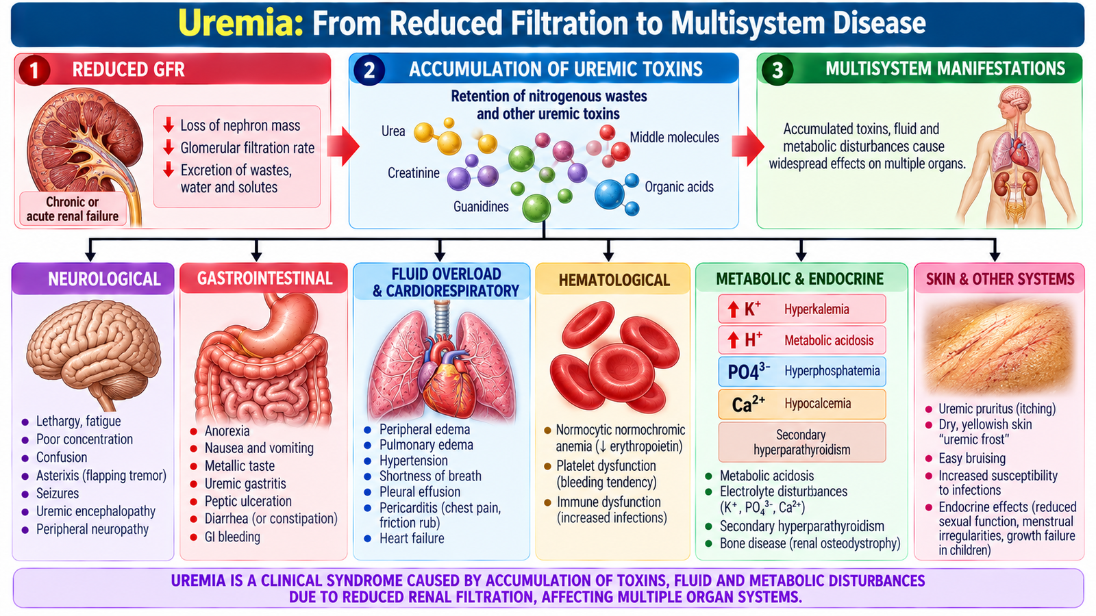
Uremia is the clinical syndrome produced by advanced loss of kidney function with accumulation of metabolic waste products together with fluid, electrolyte, endocrine and acid–base disturbances. It is therefore more than an elevated serum urea concentration: it represents systemic illness caused by severe renal dysfunction.
Why symptoms occur
- General/GI: fatigue, anorexia, nausea and vomiting may occur.
- Neurological: impaired concentration, drowsiness, confusion and severe encephalopathic manifestations can develop.
- Cardiovascular: sodium and water retention contribute to hypertension, edema and pulmonary congestion; advanced uremia may cause pericardial involvement.
- Hematological: reduced erythropoietin production contributes to anemia in chronic kidney dysfunction; platelet dysfunction may increase bleeding tendency in advanced uremia.
- Metabolic: hyperkalemia, metabolic acidosis and mineral-bone disturbances may accompany advanced renal failure.
Management principle
Treat reversible contributors, control fluid and metabolic disturbances, review medications and manage complications. Severe symptomatic uremia is an important reason to consider renal replacement therapy rather than relying on a single biochemical value.
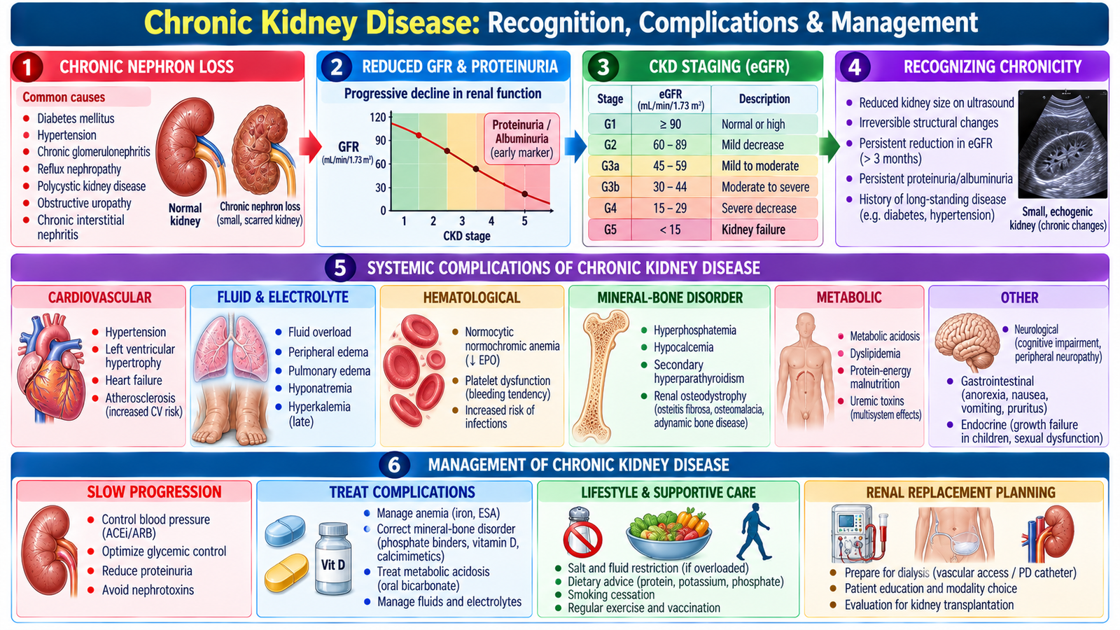
F. Chronic Kidney Disease: Recognition, Complications & Management
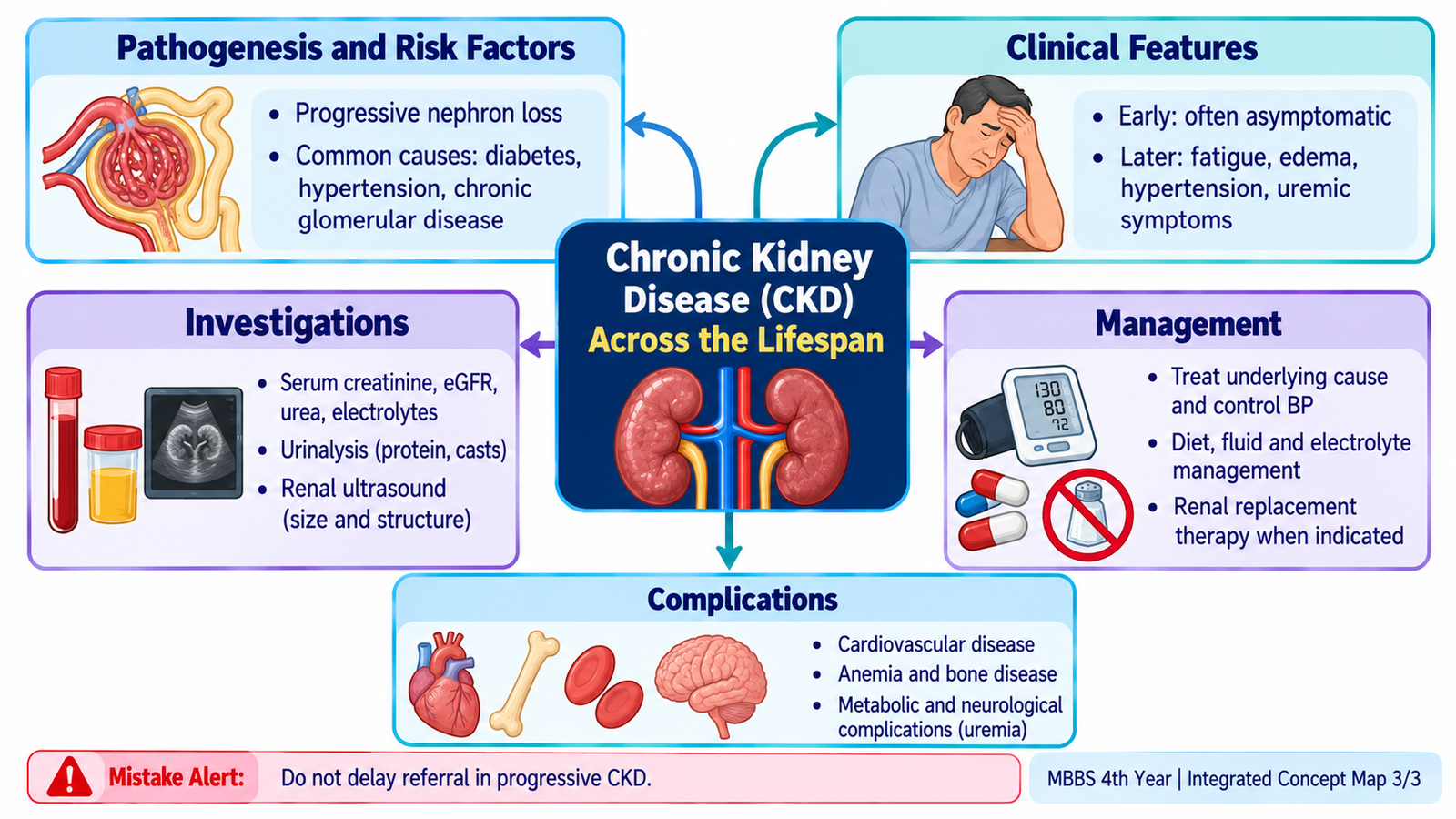
Chronic kidney disease represents persistent structural or functional kidney abnormality. Unlike AKI, the process is prolonged and often progressive. The major clinical task is to establish chronicity, identify the cause, assess the degree of kidney dysfunction and kidney damage, recognize complications and slow further progression.
Clinical presentation
Early CKD may be clinically silent and detected through abnormal renal function or urine testing. As function declines, patients may develop fatigue, anorexia, nausea, pruritus, edema, hypertension, reduced exercise tolerance and eventually manifestations of uremia.
Diagnostic workup
- Renal function: serum creatinine with estimated glomerular filtration provides an assessment of filtration.
- Urine: urinalysis and assessment of urinary protein/albumin help identify kidney damage and underlying disease.
- Blood tests: evaluate potassium, bicarbonate, hemoglobin and abnormalities related to mineral and bone metabolism.
- Ultrasound: evaluates kidney structure and obstruction; chronically diseased kidneys may become small and echogenic, although kidney size depends on the underlying disorder.
- Etiological assessment: history, examination and selected investigations are directed by the suspected underlying renal or systemic disease.
Major complications
Management principles
Management aims to treat the underlying cause where possible, slow progression, control blood pressure and fluid status, reduce further renal injury, manage anemia and mineral-bone abnormalities, correct important electrolyte or acid–base disturbances, adjust medications for renal function and reduce cardiovascular risk. Advanced disease requires preparation for renal replacement therapy when indicated.
Specific treatment: ACE inhibitors such as ramipril or ARBs such as losartan are used for indicated proteinuric CKD, with monitoring of creatinine and potassium. SGLT2 inhibitors such as dapagliflozin provide additional renal protection in eligible adults. Furosemide may control fluid overload; iron replacement and erythropoiesis-stimulating agents treat selected patients with CKD-related anemia. Advanced CKD requires nephrology assessment for dialysis or kidney transplantation when indicated.
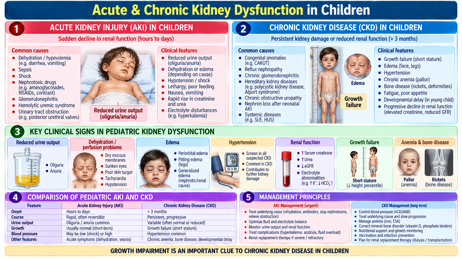
G. Acute & Chronic Kidney Dysfunction in Children
Children develop the same fundamental consequences of impaired renal function as adults, but the clinical context differs. Fluid status must be assessed carefully, urine output is interpreted relative to body size, and chronic renal disease may affect growth, nutrition and development. Management therefore combines renal stabilization with pediatric supportive care.
Pediatric acute kidney injury
AKI may present with reduced urine output, edema, hypertension, electrolyte abnormalities or rising renal-function markers. History should look for fluid losses, severe infection, recent illness, nephrotoxic exposure and urinary obstruction. Examination emphasizes hydration, perfusion, edema, blood pressure and evidence of systemic disease.
Investigation follows the same clinical logic as in adults: assess renal function and electrolytes, examine urine, determine fluid status and use imaging when structural disease or obstruction is suspected. Management corrects the underlying cause, carefully manages fluid and electrolytes, avoids further nephrotoxicity and treats complications. Fluid and medication decisions must be appropriate for the child’s weight and clinical state.
Pediatric chronic kidney disease
Children with CKD may show fatigue, pallor, poor appetite, polyuria or reduced urine output depending on the stage and cause, edema, hypertension and impaired growth. Investigation assesses renal function, urine abnormalities, electrolytes, anemia, mineral-bone disturbances and renal structure.
Management includes treatment of the underlying renal disorder where possible, control of blood pressure and fluid status, nutritional support, correction of anemia and mineral-bone abnormalities, monitoring of growth and development, and renal replacement therapy in advanced disease when required.
Integrated Mechanism Flow
Hypoperfusion, intrinsic injury or obstruction
Reduced filtration
Water, K⁺, acid, wastes
Edema, acidosis, hyperkalemia, uremia
Correct cause + support renal function
Important Comparison — AKI vs CKD
| Feature | Acute Kidney Injury | Chronic Kidney Disease |
|---|---|---|
| Course | Abrupt deterioration | Persistent, long-term abnormality |
| Potential reversibility | Often substantially reversible if cause corrected | Often progressive; focus on slowing progression |
| Anemia / bone disease | Usually not caused by a brief AKI episode | Support chronicity when present in the appropriate context |
| Ultrasound | Kidney size may remain preserved | May show small echogenic kidneys in established disease |
| Main priority | Identify reversible cause and dangerous complications | Define cause, complications and progression |
AIM High-Yield Review
- Oliguria and anuria are presentations; always search for pre-renal, intrinsic renal and post-renal causes.
- ⭐ A reversible fall in renal perfusion can cause pre-renal AKI; prolonged hypoperfusion may progress to intrinsic renal injury.
- AKI may occur without oliguria, so urine output alone cannot exclude it.
- ⭐ In AKI, immediately look for dangerous hyperkalemia, pulmonary edema, metabolic acidosis and uremic complications.
- Urinalysis helps localize renal pathology, while ultrasound is particularly important when urinary obstruction is suspected.
- Management of AKI begins with identifying and correcting the cause rather than simply trying to increase urine output.
- ⭐ Uremia is a clinical syndrome of advanced renal dysfunction, not simply a raised blood urea concentration.
- CKD produces systemic consequences including fluid overload, hypertension, anemia, metabolic acidosis and mineral-bone abnormalities.
- Evidence of long-standing anemia, mineral-bone disturbance or chronically abnormal renal structure can support chronic rather than purely acute kidney disease.
- Renal replacement therapy is considered when severe complications or symptomatic uremia cannot be adequately controlled by conservative treatment.
- ⭐ In children, renal disease can affect growth and development in addition to producing the usual renal and metabolic manifestations.
- In both adults and children, successful renal assessment connects clinical state → urine output → renal function → electrolytes → urine findings → imaging → cause → management.
Oliguria, Acute Kidney Injury, Uremia & Chronic Kidney Disease
Watch these videos after completing the AIM Learning Material to reinforce the clinical approach to acute and chronic kidney dysfunction.
Video 1 — Acute Kidney Injury: Clinical Approach
Focus on pre-renal, intrinsic and post-renal AKI, complications, diagnostic approach and management.
Video 2 — Chronic Kidney Disease
Reinforces CKD causes, clinical manifestations, diagnosis, complications and treatment principles.