
This chapter follows the KMU outcomes for nephrolithiasis. First understand how stones form, how renal colic presents, and how investigations guide treatment; then use the final AIM High-Yield Review for rapid revision.
TOPIC 5. Nephrolithiasis: Acute Flank Pain, Evaluation, Treatment & Prevention
A clinically focused approach to renal calculi—from stone formation and acute renal colic to diagnostic evaluation, treatment selection, recurrence prevention and patient counselling.
Topic Introduction
Nephrolithiasis means formation of calculi within the urinary tract, most commonly within the kidney, with symptoms often appearing when a stone moves into the ureter and obstructs urine flow. The result may be sudden severe flank pain, hematuria, nausea or urinary symptoms. For a Final Year student, the important task is not simply to memorize stone types: it is to connect risk factors with stone formation, recognize renal colic, select and interpret appropriate investigations, identify patients requiring urgent intervention, understand treatment options and prevent recurrence.
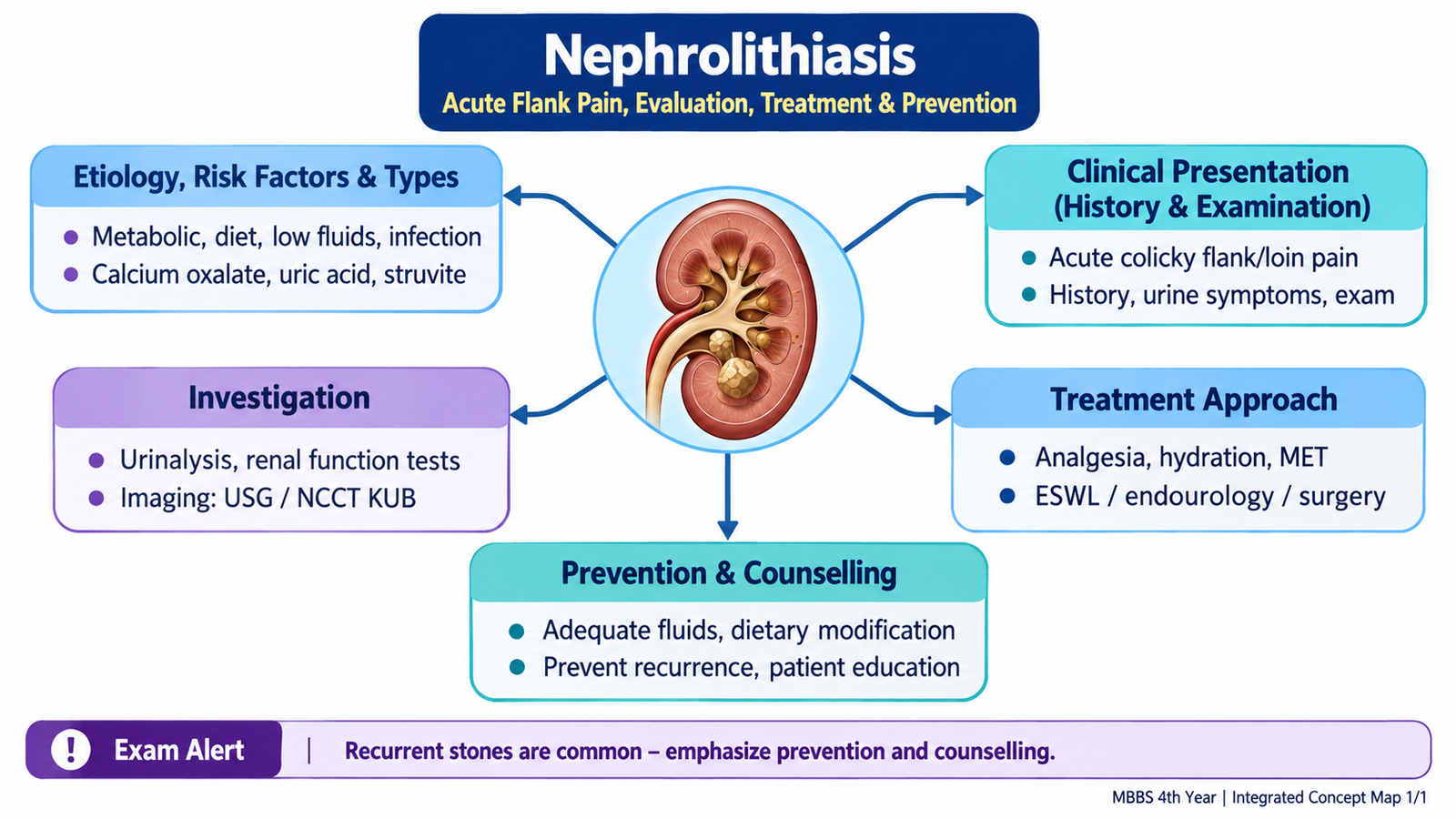
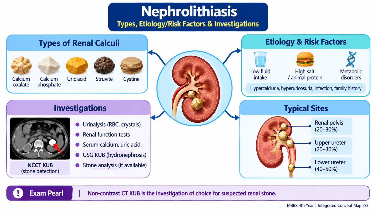
A. Understanding Renal Calculi: Formation, Etiology & Risk Factors
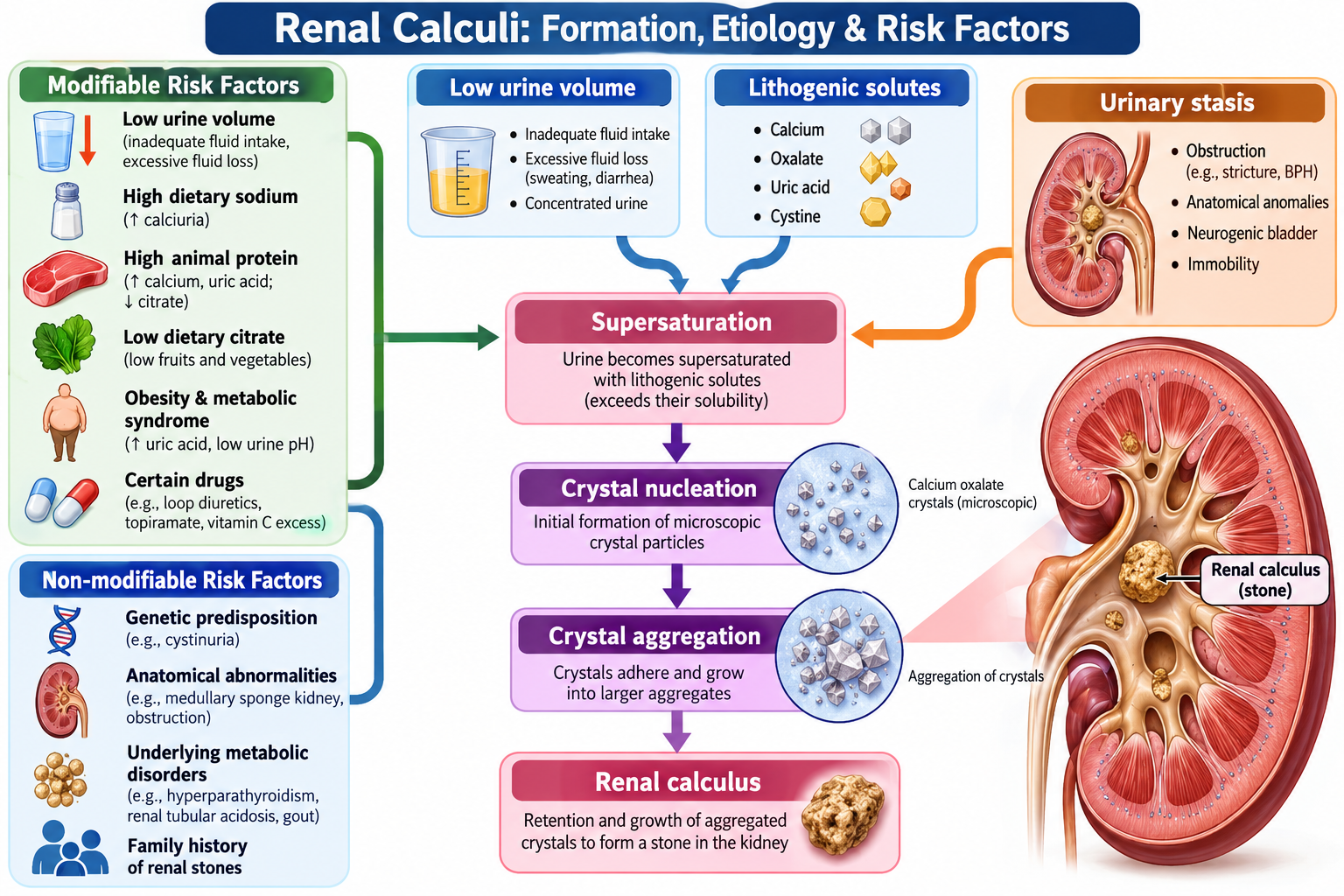
Urine normally contains dissolved salts and minerals. A stone develops when the balance between stone-forming substances, urinary concentration and natural inhibitors of crystallization is disturbed. Supersaturation permits crystals to form, enlarge and aggregate. Low urine volume is therefore an important common pathway because it increases the concentration of lithogenic substances.
How a stone forms
Urinary supersaturation → crystal nucleation → crystal growth and aggregation → retention within the urinary tract → calculus formation.
Important risk factors
- Low urine volume: inadequate fluid intake or increased fluid loss concentrates stone-forming solutes.
- Dietary factors: excessive sodium and certain dietary patterns can increase urinary stone risk in susceptible patients.
- Metabolic abnormalities: increased urinary calcium, oxalate or uric acid can promote crystallization.
- Urinary infection: infection with urease-producing organisms can promote infection-related stones.
- Urinary stasis or obstruction: impaired drainage favors crystal retention and stone growth.
- Previous or family history: recurrent stone disease suggests an underlying metabolic, anatomical or behavioral predisposition.
B. Types of Renal Calculi & Their Clinical Meaning
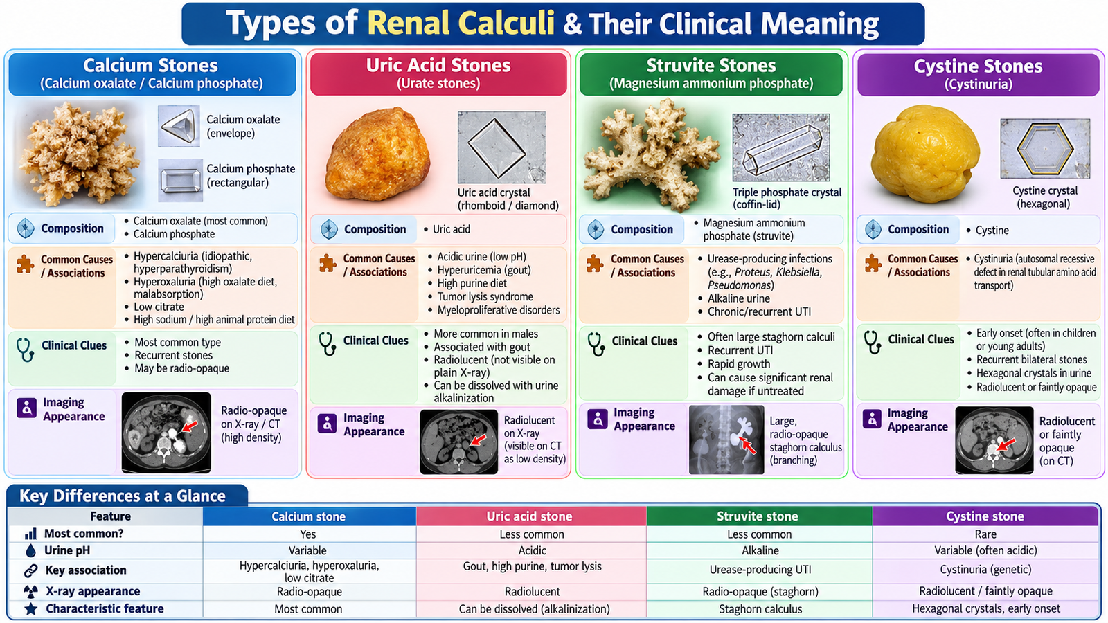
Stone composition matters because it gives clues to the underlying cause and helps guide prevention. The major stone types can be understood by linking each composition to the urinary environment in which it forms.
| Stone type | Important association | Useful clue |
|---|---|---|
| Calcium stones | Most common group; usually calcium oxalate, sometimes calcium phosphate | Generally radiopaque |
| Uric acid stones | Hyperuricosuria and persistently acidic urine | May not be visible on a plain X-ray but are detected by non-contrast CT |
| Struvite stones | Urease-producing urinary infection → alkaline urine | May form large branching staghorn calculi |
| Cystine stones | Inherited cystinuria | Consider particularly in young patients with recurrent stones |
C. Acute Flank Pain: Clinical Presentation, History & Examination
A renal calculus may remain asymptomatic while confined to the kidney. The classic acute presentation occurs when a stone enters the ureter and causes obstruction, ureteric distension and spasm. This produces renal colic—severe pain that typically begins in the flank and may radiate toward the groin as the stone moves distally.
Typical presentation
- Sudden, severe unilateral flank or loin pain, often occurring in waves.
- Radiation toward the lower abdomen, groin, testis or labium depending on stone position.
- Nausea and vomiting due to intense visceral pain.
- Microscopic or visible hematuria may occur from urothelial irritation.
- Frequency, urgency or dysuria may become prominent as a stone approaches the vesicoureteric junction.
- Patients with renal colic are often restless and unable to find a comfortable position.
Focused history
Clarify the onset, site, severity, radiation and character of pain. Ask about hematuria, urinary frequency, dysuria, fever, rigors, nausea and vomiting. Previous stones, previous procedures, urinary infection, reduced fluid intake, dietary habits, family history and relevant metabolic disease help estimate cause and recurrence risk.
Always establish whether the patient has a solitary functioning kidney or known renal impairment, because obstruction in such a patient carries greater risk.
Physical examination
Assess temperature, pulse, blood pressure, hydration and overall clinical stability. Abdominal and flank examination may show loin or costovertebral-angle tenderness. The examination is also important for detecting alternative causes of acute abdominal or flank pain and for identifying systemic infection.
Fever or systemic illness in a patient with an obstructed urinary tract raises concern for an infected obstructed system. This is a urological emergency because infection cannot be safely controlled if the obstructed collecting system is not adequately drained.

🩺 AIM Clinical Bridge
A 34-year-old man develops sudden severe right-sided flank pain radiating toward the groin. He is restless and nauseated and reports reddish urine. He has had a similar episode previously. He is afebrile and hemodynamically stable.
Think clinically:
- The pain pattern strongly suggests ureteric colic.
- Hematuria supports urinary tract irritation but does not by itself establish the diagnosis.
- The next step is to confirm the stone, define its location and obstruction, and assess renal function and infection.
D. Diagnostic Evaluation: Confirm the Stone, Obstruction & Complications
Investigation should answer several practical questions: Is there a stone? Where is it? How large is it? Is it causing obstruction? Is renal function affected? Is infection present? These answers determine whether conservative management is reasonable or intervention is needed.
Urinalysis
Urinalysis may demonstrate blood. Leukocytes, nitrites or other evidence of infection should increase concern for associated urinary infection. Urinary pH may also provide a clue to stone composition.
Blood investigations
- Renal function: assesses the effect of obstruction and establishes a baseline before treatment.
- Full blood count and inflammatory assessment: useful when infection or systemic inflammation is suspected.
- Relevant metabolic tests: selected according to the clinical setting and recurrent-stone risk.
Imaging
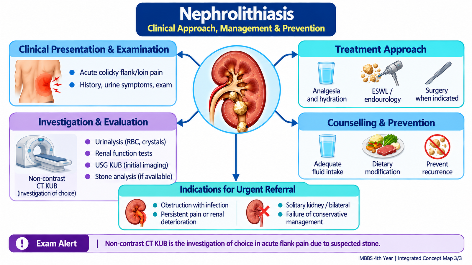
Non-contrast CT of the urinary tract is highly useful in adults with suspected renal/ureteric calculi because it can identify stone location and size and demonstrate obstruction. It also helps identify alternative pathology when the diagnosis is uncertain.
Ultrasonography can demonstrate renal stones and hydronephrosis without ionizing radiation and is particularly valuable when radiation avoidance is important. Its sensitivity for ureteric stones is lower than CT.
A plain radiograph may demonstrate radiopaque stones and can have a role in follow-up of suitable known calculi, but it does not reliably exclude stone disease.

E. Treatment: From Acute Renal Colic to Definitive Stone Management
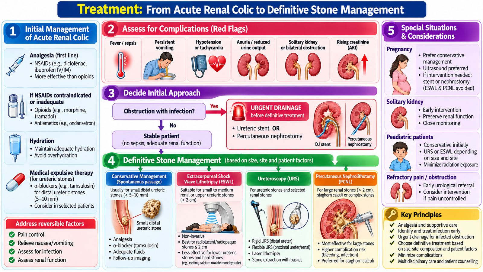
Treatment depends on the patient’s clinical stability, severity of pain, presence of infection or obstruction, renal function, and the stone’s size and location. The key distinction is between a patient suitable for conservative passage and one requiring urgent or definitive urological intervention.
Initial management
- Analgesia: adequate pain control is an immediate priority; NSAIDs are commonly effective in renal colic when not contraindicated.
- Antiemetic treatment: may be needed for significant nausea or vomiting.
- Hydration: correct dehydration and maintain appropriate fluid intake, but forced excessive fluid administration does not mechanically “flush out” an obstructing stone.
- Assess infection and renal function: these findings can change the urgency of management.
First-line analgesia: NSAIDs such as diclofenac or ketorolac are preferred for acute renal colic when renal function and other contraindications permit. IV paracetamol is an alternative when NSAIDs are unsuitable or insufficient; opioids may be required for refractory pain.
Conservative management
A clinically stable patient with a stone likely to pass spontaneously, controlled symptoms, no significant infection and no threatened renal function may be managed expectantly with analgesia, appropriate hydration and follow-up. Selected patients may receive medical expulsive therapy according to clinical circumstances.
Medical expulsive therapy: Tamsulosin (an alpha-1 blocker) may facilitate passage of distal ureteric stones, particularly those measuring 5–10 mm. This is an off-label option for selected stable patients without infection, uncontrolled pain or deteriorating renal function.
When intervention becomes important
Urological assessment is required when spontaneous passage is unlikely or unsafe—for example with persistent obstruction, uncontrolled symptoms, impaired renal function, an infected obstructed system or other high-risk circumstances.
Definitive treatment principles
- Extracorporeal shock-wave lithotripsy (ESWL): fragments selected stones so fragments can subsequently pass.
- Ureteroscopy: permits endoscopic access to ureteric/selected renal stones for extraction or fragmentation.
- Percutaneous nephrolithotomy (PCNL): provides percutaneous access for removal of larger or complex renal stone burdens.
An infected obstructed collecting system requires urgent drainage of the urinary tract together with appropriate treatment of infection. Definitive stone treatment is generally addressed after the acute septic risk has been controlled.
Emergency decompression: An infected obstructed kidney requires immediate IV antibiotics and urgent urinary drainage by ureteric double-J stenting or percutaneous nephrostomy. Definitive stone removal is delayed until sepsis has resolved.
F. Recurrence Prevention & Patient Counselling
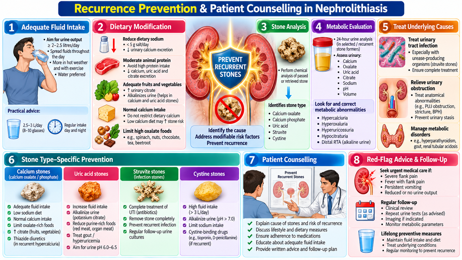
Removing or passing a stone does not remove the patient’s tendency to form another one. Prevention therefore begins by identifying modifiable risks and, particularly in recurrent or high-risk disease, determining whether a specific metabolic or stone-related abnormality is present.
General prevention principles
- Maintain adequate fluid intake: the central aim is to avoid persistently concentrated urine.
- Reduce excessive dietary salt: high sodium intake can promote urinary calcium loss and recurrent calcium stone formation.
- Avoid unnecessary dietary extremes: prevention should be individualized rather than based on indiscriminate restriction.
- Address the underlying cause: infection, metabolic abnormalities or urinary obstruction should be treated when identified.
- Stone analysis: when a stone is available, determining its composition can guide more specific preventive measures.
- Further metabolic evaluation: is particularly useful in recurrent or otherwise high-risk stone formers.
How to counsel the patient
Explain that recurrence can often be reduced by consistent preventive behavior. Encourage regular fluid intake across the day, with extra attention during hot weather or heavy activity. Dietary advice should be appropriate to the patient’s stone type and metabolic findings rather than simply telling every patient to eliminate calcium-containing foods.
Patients should understand when to seek urgent care. Fever or rigors with flank pain, inability to maintain oral intake, worsening uncontrolled pain, markedly reduced urine output or significant systemic illness require prompt reassessment.
Integrated Mechanism Flow
AIM High-Yield Review
- ⭐ Renal colic classically causes severe flank pain radiating toward the groin; the patient is often restless.
- Stone formation follows urinary supersaturation → crystallization → aggregation → calculus formation.
- Low urine volume is an important modifiable contributor to stone formation and recurrence.
- Calcium-containing calculi form the commonest stone group; uric acid, struvite and cystine stones have distinctive etiological associations.
- ⭐ Non-contrast CT is highly useful for defining stone site, size and obstruction in adults with suspected renal/ureteric calculi.
- Urinalysis helps identify hematuria, infection clues and urinary pH; renal function testing assesses the physiological impact of obstruction.
- A small uncomplicated stone in a stable patient may pass spontaneously, so management can include analgesia, appropriate hydration and observation with follow-up.
- ⭐ Obstruction plus infection is a urological emergency and requires urgent drainage together with treatment of infection.
- ESWL fragments selected stones, ureteroscopy provides endoscopic treatment, and PCNL is particularly useful for larger or complex renal stone burdens.
- A staghorn calculus should raise consideration of an infection-related struvite stone.
- Uric acid stones may be radiolucent on plain radiography but remain detectable on non-contrast CT.
- ⭐ Long-term management is incomplete without recurrence prevention: adequate fluid intake, appropriate dietary measures, cause-directed management and targeted evaluation in recurrent/high-risk patients.
- Patient counselling must include red flags such as fever with flank pain, reduced urine output, persistent vomiting, systemic illness or uncontrolled pain.
Nephrolithiasis — Types, Clinical Features, Diagnosis & Treatment
Watch this video to reinforce renal stone types, acute renal colic, diagnostic evaluation, treatment options and prevention of recurrence.