
This chapter follows the supplied KMU outcomes and connects the presenting urinary symptoms with focused assessment, investigation and management. First understand the clinical logic; use the AIM High-Yield Review at the end for revision.
TOPIC 4. Hematuria, Dysuria, Loin Pain & Urinary Tract/Prostatic Infection
Renal Module — a patient-centred approach to common urinary presentations, acute pyelonephritis and prostatitis, integrating Medicine/Nephrology with Surgery.
Topic Introduction
Blood in urine, painful urination and loin pain are common presentations, but the underlying problem may arise anywhere from the kidney to the urethra or prostate. The important Final-Year skill is therefore not simply to memorize causes, but to recognize the symptom pattern, identify clues that localize the disease, select appropriate investigations and understand the first management priorities. In this topic, you will learn how to approach hematuria systematically, evaluate dysuria and loin pain, recognize and manage acute pyelonephritis, and distinguish acute from chronic prostatitis. These are the exact clinical areas emphasized in the supplied Renal Module curriculum.
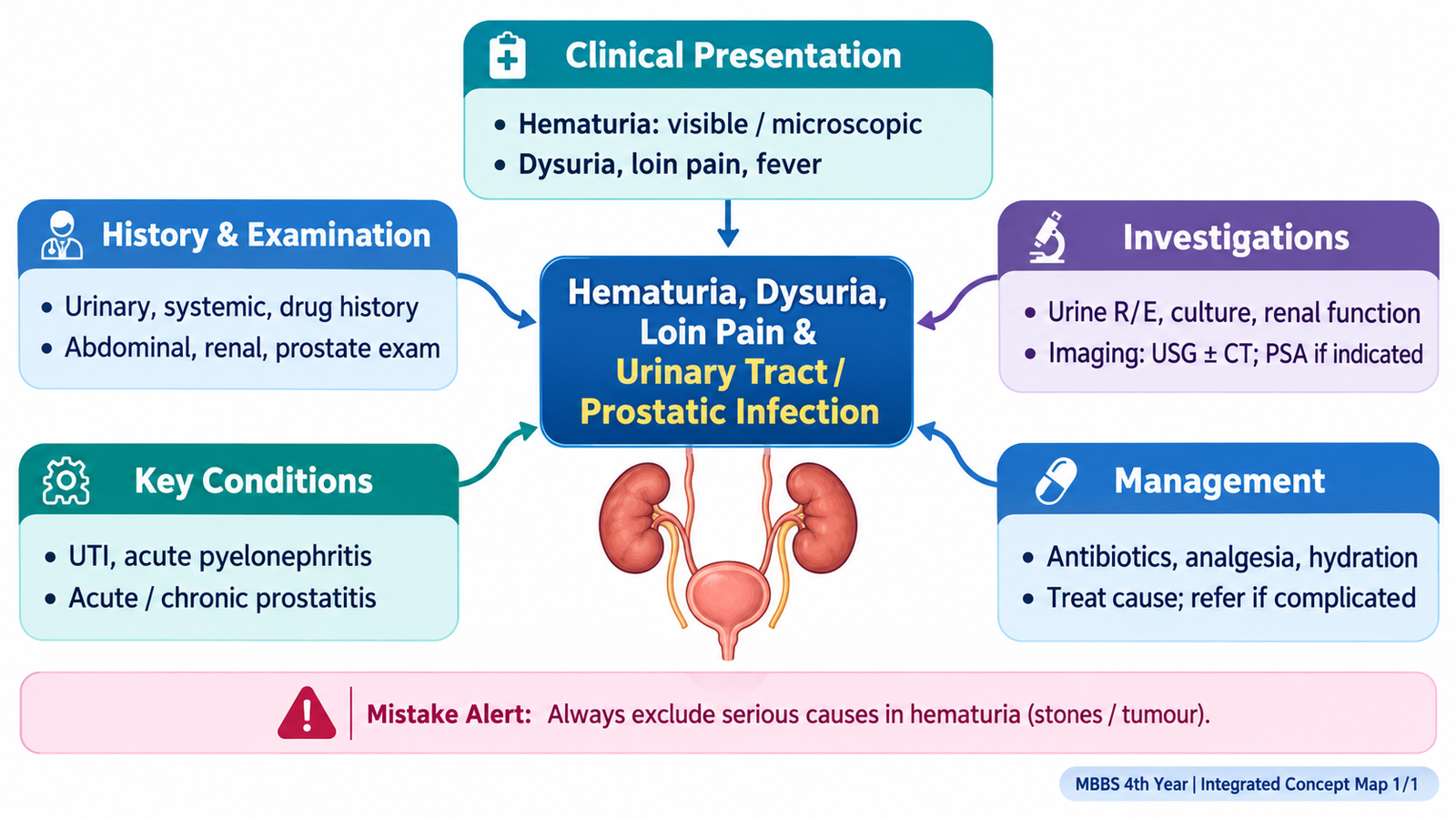
A. Recognizing and Assessing Hematuria
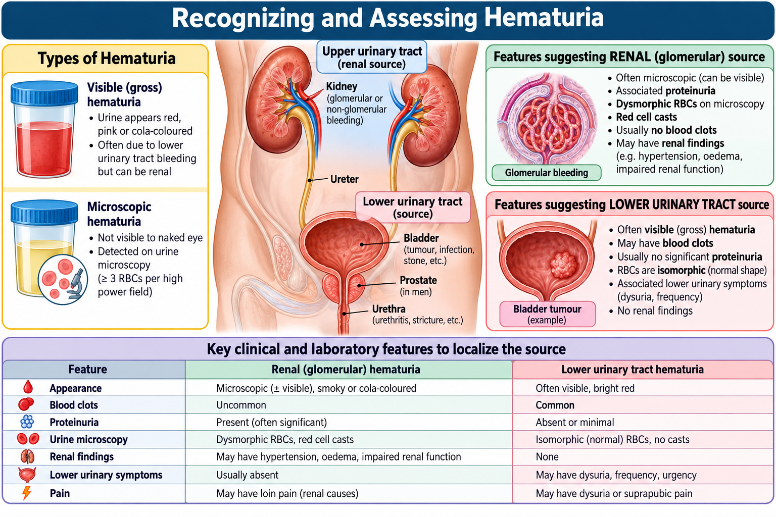
Hematuria means the presence of red blood cells in urine. It may be visible to the patient or detected only on urine testing. The key clinical task is to decide whether the bleeding is likely to arise from the renal parenchyma or from the urinary collecting system, bladder, prostate or urethra. A careful history and focused examination often provide the first important localization clues.
Focused history
Begin by establishing that the discoloration is likely to represent blood and determine how it relates to urination. The pattern matters because different parts of the urinary tract produce different associated symptoms.
- Visible or non-visible hematuria: ask whether the patient actually saw red or brown urine or whether blood was detected on testing.
- Timing and duration: establish sudden versus persistent or recurrent bleeding.
- Pain: hematuria with severe colicky loin pain suggests urinary tract obstruction such as a calculus, whereas painless visible hematuria requires careful evaluation for significant urinary tract pathology.
- Dysuria, frequency and urgency: support lower urinary tract inflammation or infection.
- Fever with loin pain: raises concern for upper urinary tract infection.
- Clots: favor a nonglomerular source because glomerular bleeding is usually mixed with urine before clot formation.
- Recent urinary instrumentation, trauma or surgery: may explain urinary tract bleeding.
- Drug history: anticoagulant or antiplatelet use may increase bleeding, but does not remove the need to consider underlying urinary pathology.
- Previous stones, urinary infection or urological disease: help prioritize the differential diagnosis.
- Smoking and occupational exposure: are clinically important when evaluating possible urothelial pathology.
Focused examination
Examination looks for evidence of infection, renal disease, obstruction or a urological source rather than attempting to reproduce a complete general examination.
- Vital signs: fever supports infection; hypotension or tachycardia may indicate significant illness or sepsis.
- General examination: pallor may accompany prolonged blood loss; edema or hypertension suggests possible renal parenchymal disease.
- Abdominal examination: assess for tenderness, masses and bladder distension.
- Renal angle tenderness: supports inflammation involving the upper urinary tract when accompanied by fever and urinary symptoms.
- Prostate assessment where clinically indicated: may identify tenderness or enlargement associated with prostatic disease.
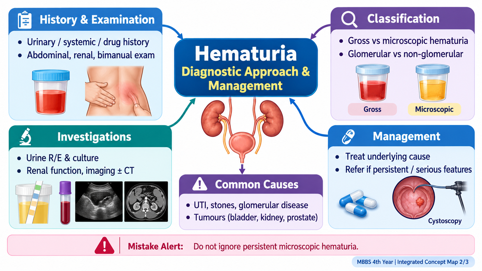
B. Diagnostic Workup and Management Approach to Hematuria
Once hematuria has been recognized clinically, investigation should answer three practical questions: Is blood truly present? Does the pattern suggest renal or urological disease? and is there an important structural cause that requires urological evaluation? The investigation pathway therefore moves from urine assessment to renal function, imaging and selected specialist evaluation.
Urinalysis and microscopy
Urine dipstick is a useful initial test because it detects heme. A positive result should be interpreted in context because dipstick positivity does not by itself identify the anatomical source.
- Red blood cells on microscopy confirm true urinary red-cell loss.
- Proteinuria accompanying hematuria raises suspicion of renal parenchymal, particularly glomerular, disease.
- White cells, leukocyte esterase or nitrites support urinary infection in the appropriate clinical setting.
- Urine culture is particularly useful when infection is suspected and guides antimicrobial therapy.
Hematuria + significant proteinuria or other renal features → consider a renal parenchymal source. Hematuria + dysuria/pyuria → infection becomes more likely. Visible hematuria without infection still requires appropriate structural evaluation.
Blood tests
Renal function testing helps determine whether the urinary presentation is associated with impaired kidney function. A full blood count may identify anemia from significant bleeding or leukocytosis in infection. Further tests should be selected according to the suspected cause rather than ordered indiscriminately.
Imaging and urological assessment
Imaging is chosen to look for structural urinary tract disease such as obstruction, calculi or a mass. Ultrasound may demonstrate hydronephrosis, bladder abnormalities or other structural changes. Cross-sectional urinary tract imaging may be required when a significant urological cause is suspected. Lower urinary tract evaluation, including cystoscopic assessment, is considered when the clinical pattern warrants direct assessment of the bladder and urethral outlet.
Management principle
Hematuria itself is a sign rather than a final diagnosis. Management therefore targets the identified cause. Infection is treated as infection; obstruction requires relief where clinically necessary; renal parenchymal disease follows an appropriate nephrological pathway; and suspected structural or malignant disease requires urological evaluation.
Severe hematuria with clot retention requires urgent urological assessment, insertion of a large-bore three-way urinary catheter and bladder irrigation to relieve obstruction. Persistent unexplained visible hematuria requires urological evaluation, including cystoscopy when indicated, to exclude malignancy.
- Hemodynamic instability or heavy ongoing bleeding.
- Clot retention with inability to pass urine.
- Hematuria associated with acute renal deterioration.
- Visible hematuria with features suspicious for significant structural urinary disease.

C. Dysuria and Loin Pain: Clinical Assessment, Investigation and Management
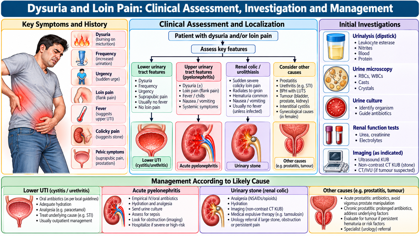
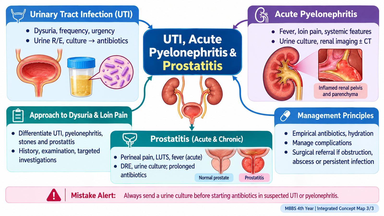
Dysuria is pain or burning associated with urination. Loin pain arises from the flank or renal region and may result from infection, obstruction or another urinary tract process. When they occur together, the pattern of associated symptoms helps localize the problem. The crucial distinction is whether the presentation is mainly lower urinary tract disease or whether the kidneys and upper urinary tract may be involved.
Understanding the symptom pattern
- Dysuria + frequency + urgency: points toward lower urinary tract irritation or infection.
- Dysuria + fever + loin pain: raises concern for upper urinary tract infection.
- Severe colicky loin-to-groin pain: suggests ureteric obstruction, particularly calculus disease.
- Loin pain with systemic illness: requires assessment for significant infection and complications.
- Male urinary symptoms with pelvic/perineal discomfort: should prompt consideration of prostatic infection.
Focused history
The history should define the urinary symptom complex and identify features suggesting upper tract disease, obstruction or prostatic involvement.
- Onset, duration and severity of dysuria or pain.
- Frequency, urgency and nocturia.
- Visible hematuria.
- Fever, chills or rigors.
- Loin, suprapubic, pelvic or perineal pain.
- Nausea and vomiting in a patient with upper tract disease.
- Previous urinary infections or stone disease.
- Urinary obstruction symptoms where relevant.
- Recent catheterization or instrumentation.
Focused examination
Assess temperature, pulse, blood pressure and overall clinical condition first. Abdominal and renal-angle examination may identify suprapubic tenderness, bladder distension or renal-angle tenderness. In men with symptoms suggesting prostatic disease, the prostate may require careful clinical assessment.
Diagnostic workup
Urinalysis is the main initial investigation because it can demonstrate blood and inflammatory changes. Urine culture is important when bacterial infection is suspected, particularly in clinically significant or upper tract infection. Renal function tests help identify associated renal dysfunction. Imaging is selected when obstruction, calculus, structural disease or a complicated urinary infection is suspected.
Management approach
The management of dysuria depends on the cause rather than the symptom alone. Adequate hydration and analgesia may support recovery, but bacterial infection requires appropriate antimicrobial treatment. Suspected obstruction, severe systemic illness, recurrent symptoms or an important structural cause requires further assessment and, where appropriate, urological involvement.
🩺 AIM Clinical Bridge
A patient presents with dysuria and urinary frequency that began three days ago. He now has fever, rigors and constant right-sided loin pain, and examination reveals right renal-angle tenderness. The progression from lower urinary symptoms to systemic illness and flank findings suggests that the process is no longer confined to the lower urinary tract.
- What is the main anatomical level of infection now?
- Which urine investigation should be obtained?
- Which findings would indicate severe or complicated disease?
D. Acute Pyelonephritis
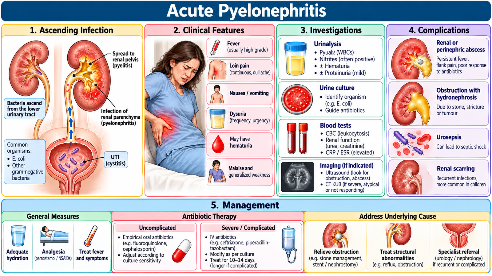
Acute pyelonephritis is an acute infection involving the renal pelvis and kidney parenchyma. It commonly develops when organisms ascend from the lower urinary tract. Because the kidney is involved, the patient usually has a more systemic illness than occurs with isolated lower urinary tract infection.
How the disease produces the clinical picture
Inflammation within and around the kidney explains the characteristic flank or loin discomfort. Cytokine-mediated systemic inflammation produces fever, malaise and rigors. Dysuria, frequency or urgency may coexist because the infection frequently begins in the lower urinary tract.
Clinical presentation
- Acute fever and systemic illness.
- Loin or flank pain.
- Renal-angle tenderness.
- Dysuria, frequency or urgency may be present.
- Nausea or vomiting may occur in more symptomatic disease.
Diagnostic workup and interpretation
Urinalysis typically supports urinary inflammation, while urine culture identifies the organism and provides antimicrobial susceptibility information. A full blood count and renal function profile help assess systemic involvement and renal function. Blood cultures may be appropriate in a patient who is significantly unwell or septic.
Imaging is not required simply to prove every uncomplicated infection. It becomes more important when the clinical course suggests obstruction, structural disease, recurrent infection, poor response to treatment or another complication.
Management
The central management principle is prompt treatment of the bacterial infection while assessing severity and correcting associated problems such as dehydration, pain or vomiting.
- Antimicrobial therapy: begin appropriate treatment and refine it according to microbiological results and local antimicrobial guidance.For uncomplicated acute pyelonephritis in clinically stable adults, oral cefalexin is an appropriate option, guided by local resistance patterns and urine culture. Severe infection, vomiting or sepsis requires hospital admission and intravenous antibiotics such as ceftriaxone. An infected obstructed urinary tract requires urgent decompression by ureteric stenting or percutaneous nephrostomy in addition to antibiotics.
- Supportive care: provide appropriate fluids, analgesia and antiemetic treatment when required.
- Assess for obstruction: infection behind an obstructed urinary tract is a more dangerous situation because antimicrobial treatment alone may be insufficient.
- Monitor clinical response: improving fever, pain and systemic status suggest response; persistent deterioration requires reassessment.
Sepsis, hypotension, deteriorating renal function, persistent vomiting, urinary obstruction or failure to improve should prompt urgent reassessment and escalation.
Complications
Important complications include sepsis, renal or perinephric abscess, renal dysfunction and infection associated with obstruction. Recognition matters because these patients may require imaging, inpatient management and specialist involvement rather than routine outpatient treatment.
E. Acute and Chronic Prostatitis
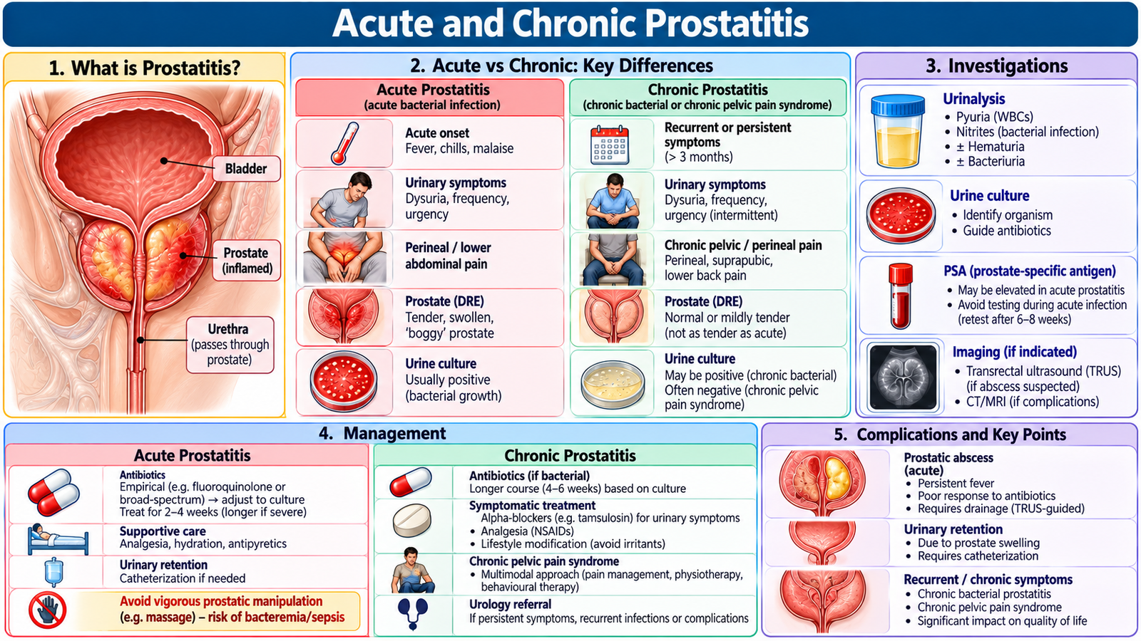
Prostatitis is inflammation of the prostate and may present as an acute bacterial illness or as a more persistent chronic syndrome. The distinction is clinically important because acute bacterial prostatitis may produce significant systemic illness, whereas chronic prostatitis tends to produce recurrent or persistent pelvic and urinary symptoms.
Acute prostatitis
Acute bacterial prostatitis typically presents with relatively sudden urinary and systemic symptoms. Prostatic inflammation may obstruct or irritate the bladder outlet, explaining dysuria, frequency, urgency and difficulty passing urine.
- Fever, chills or systemic illness.
- Dysuria, frequency and urgency.
- Pelvic, suprapubic, perineal or lower back discomfort.
- Difficulty passing urine or acute retention in severe swelling.
- A tender prostate may be found on careful rectal examination.
When acute bacterial prostatitis is suspected, examination should be gentle. Vigorous prostatic manipulation is inappropriate in a severely inflamed gland.
Diagnostic workup
Urinalysis and urine culture are central because they demonstrate urinary inflammation and help identify the infecting organism. Blood tests assess systemic illness and renal function where indicated. Blood cultures may be useful when the patient is febrile or systemically unwell. Imaging is reserved for suspected complications, obstruction or failure to respond as expected.
Management of acute prostatitis
Appropriate antimicrobial therapy is the main specific treatment. The patient also requires analgesia, hydration where needed and monitoring for systemic deterioration. Urinary retention requires urgent assessment because instrumentation through an acutely inflamed prostate may be difficult and the drainage strategy may require urological input. For acute bacterial prostatitis, oral ciprofloxacin may be used when appropriate according to susceptibility and fluoroquinolone safety considerations; trimethoprim is an alternative when suitable. Review antibiotic treatment after 14 days and continue for up to 28 days if clinically indicated. Severe infection or sepsis requires hospitalization and intravenous antibiotics such as ceftriaxone.
Chronic prostatitis
Chronic prostatitis is considered when pelvic, perineal or urinary symptoms persist or recur over a longer period. Recurrent urinary infection may be present in chronic bacterial prostatitis, while some patients have chronic pelvic symptoms without a consistently demonstrated bacterial cause.
The diagnostic approach is therefore based on the symptom pattern, examination and appropriate urine testing, with further evaluation where the presentation suggests another urological cause. Management is directed by the identified subtype and includes appropriate antimicrobial treatment when bacterial infection is demonstrated, symptom control and urological assessment when symptoms persist or complications are suspected.
Confirmed chronic bacterial prostatitis generally requires 4–6 weeks of culture-guided antibiotics, commonly ciprofloxacin when appropriate. Chronic pelvic pain syndrome without confirmed bacterial infection is managed primarily through symptom control, including analgesics, alpha-blockers such as tamsulosin for urinary symptoms, and supportive care rather than repeated empirical antibiotics.
Important complications
- Urinary retention.
- Prostatic abscess.
- Recurrent urinary infection.
- Sepsis in severe acute infection.
- Persistent pelvic and urinary symptoms in chronic disease.
Important Comparison
Urinary symptoms overlap, so the most useful distinction is the anatomical level and systemic severity of the presentation.
| Feature | Lower UTI Pattern | Acute Pyelonephritis | Acute Prostatitis |
|---|---|---|---|
| Main symptoms | Dysuria, frequency, urgency | Fever, loin pain ± urinary symptoms | Urinary symptoms + pelvic/perineal pain |
| Systemic illness | Usually limited | Common | Common in acute bacterial disease |
| Key examination clue | Suprapubic tenderness may occur | Renal-angle tenderness | Tender prostate on gentle examination |
| Important complication | Ascending infection | Sepsis / obstructed infection | Retention / abscess / sepsis |
Integrated Mechanism Flow
AIM High-Yield Review
- ⭐ Hematuria is a clinical sign; management depends on identifying its source and underlying cause.
- Hematuria + significant proteinuria or renal findings favors renal parenchymal disease.
- Hematuria + clots usually suggests a nonglomerular urinary source.
- ⭐ Painless visible hematuria requires appropriate evaluation for important structural urinary tract disease.
- Dysuria + frequency + urgency suggests lower urinary tract irritation or infection.
- Dysuria + fever + loin pain + renal-angle tenderness strongly supports acute pyelonephritis.
- Urine culture identifies the infecting organism and helps direct antimicrobial treatment.
- ⭐ In pyelonephritis, sepsis or urinary obstruction indicates potentially complicated disease requiring urgent escalation.
- Colicky loin-to-groin pain suggests ureteric obstruction rather than uncomplicated urinary infection.
- Acute prostatitis commonly causes urinary symptoms, fever and pelvic/perineal discomfort.
- A tender prostate supports acute prostatitis, but vigorous manipulation should be avoided in acute inflammation.
- ⭐ Persistent or recurrent pelvic and urinary symptoms suggest a chronic prostatic process and require cause-directed evaluation.
- Across this topic, the clinical sequence is: presentation → localization → urine testing → targeted imaging when indicated → cause-specific management.
Topic 4: Hematuria, Dysuria, Loin Pain & Urinary Tract/Prostatic Infection
Watch this supporting urology video to reinforce the clinical approach to urinary symptoms, hematuria, urinary tract infection and related urological presentations.