
This chapter follows the supplied KMU outcomes for electrolyte and acid–base disorders. First understand how each disturbance changes body function; then use the AIM High-Yield Review for rapid revision.
Topic 2. Electrolyte & Acid–Base Disorders
Renal Module — recognition, interpretation and management of clinically important electrolyte disturbances and acid–base abnormalities.
Topic Introduction
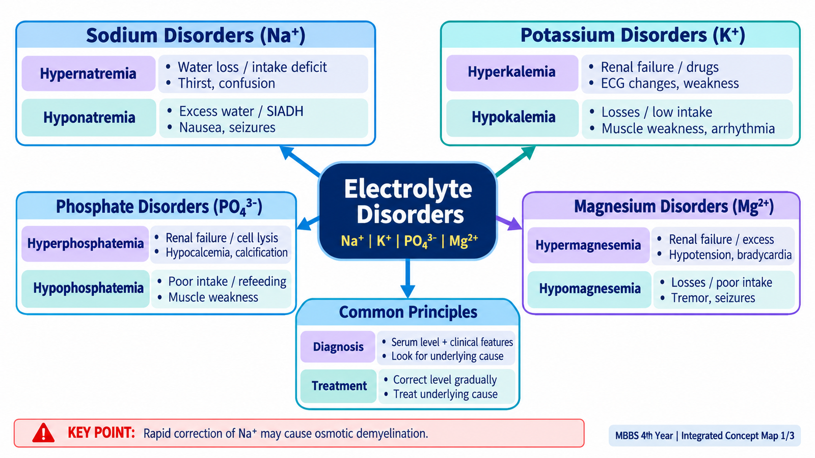
Electrolytes and hydrogen ions are tightly controlled because small disturbances can alter brain function, muscle activity, cardiac conduction and cellular metabolism. In clinical practice, the important task is not simply to recognize an abnormal laboratory value. The student must understand why it occurred, whether the patient is clinically stable, what associated abnormalities are present, and which problem requires urgent correction. This chapter covers sodium, potassium, phosphate and magnesium disorders together with metabolic and respiratory acid–base disturbances. The aim is to build a simple clinical sequence: recognize the pattern → identify the likely mechanism → interpret investigations → judge severity → apply the appropriate treatment principle. The KMU curriculum specifically requires these electrolyte disorders and acid–base compensatory responses.
A. Clinical Framework for Electrolyte & Acid–Base Interpretation
Before learning individual disorders, it helps to understand one central principle: an abnormal electrolyte concentration is usually the visible result of a disturbance in water balance, intake, loss, redistribution or renal handling. Acid–base disorders similarly reflect changes in bicarbonate, carbon dioxide, or both. Interpretation therefore begins with the patient and then moves to the laboratory result.
Start with clinical stability
A laboratory abnormality becomes urgent when it is producing neurological, cardiac, respiratory or neuromuscular dysfunction. The first question is therefore not “What is the exact cause?” but “Is this abnormality threatening the patient now?”
- Neurological danger: confusion, seizures, marked reduction in consciousness.
- Cardiac danger: arrhythmia or significant ECG abnormality, especially with potassium disturbance.
- Respiratory or neuromuscular danger: severe weakness or respiratory muscle impairment.
- Hemodynamic danger: severe dehydration, shock or major volume disturbance.
Interpret the abnormality in context
For each electrolyte abnormality, consider four questions:
- Is the concentration high or low?
- Is the patient volume depleted, euvolemic or fluid overloaded?
- Is the abnormality caused by altered intake, loss, renal handling or redistribution?
- Does it require immediate stabilization before the cause is fully defined?
Acid–base reasoning
For an arterial blood gas, use a consistent sequence:
The lungs regulate carbon dioxide relatively rapidly. The kidneys regulate bicarbonate and hydrogen-ion handling more slowly. This explains why respiratory disorders are compensated metabolically and metabolic disorders are compensated through ventilation.

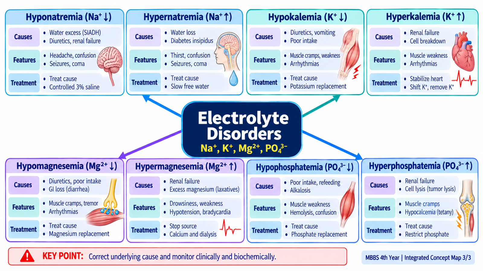
B. Sodium Disorders — Hyponatremia & Hypernatremia
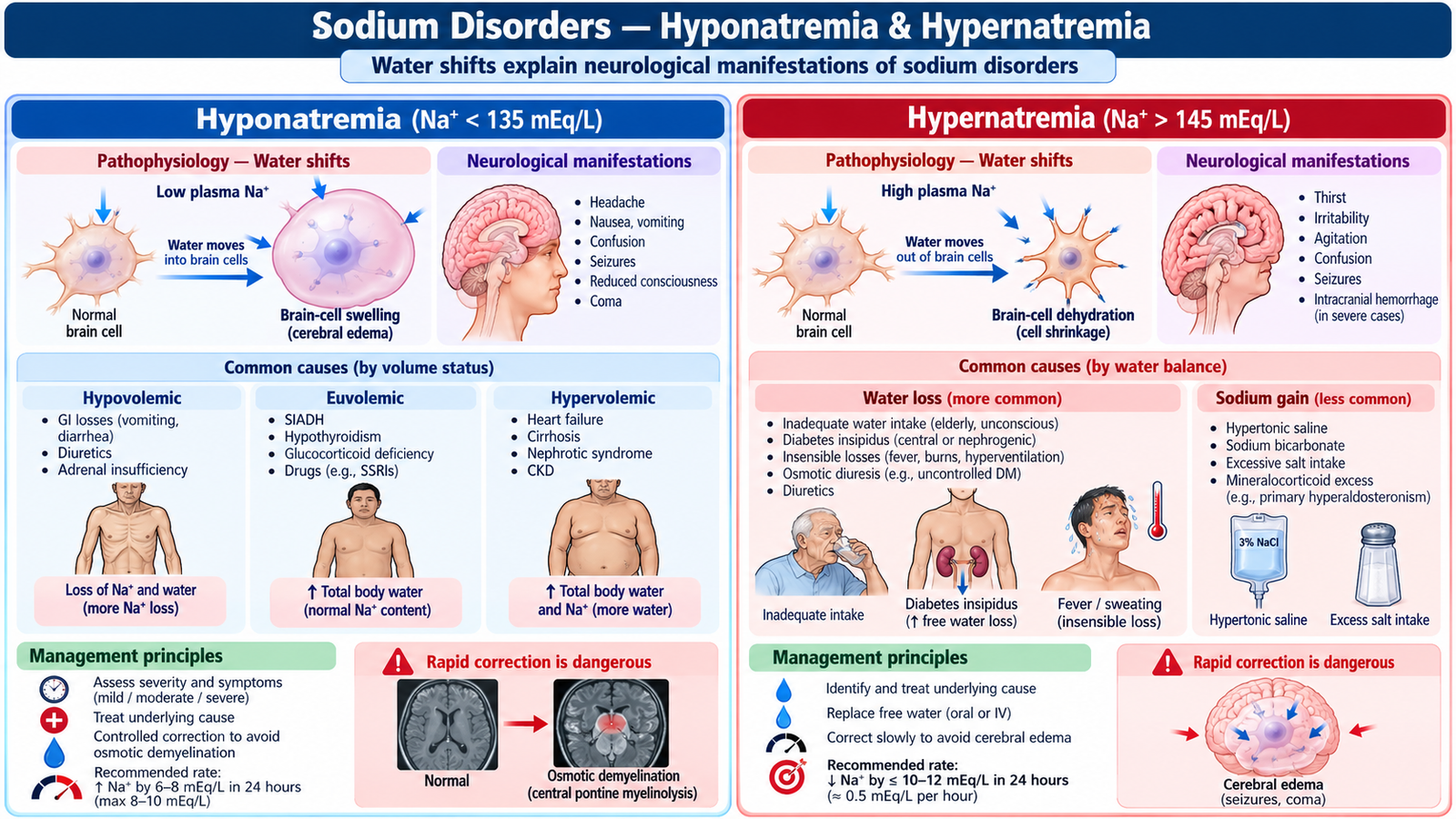
Serum sodium is best understood as a marker of the relationship between body sodium and body water, rather than simply the amount of sodium in the body. Many sodium disorders are therefore fundamentally disorders of water balance. The brain is especially vulnerable because abnormal extracellular tonicity causes water to move into or out of brain cells.
Hyponatremia
Hyponatremia means that serum sodium concentration is lower than normal. In the common hypotonic form, extracellular fluid becomes relatively dilute, so water tends to move into cells. Cerebral cellular swelling explains the most important acute neurological manifestations.
Important etiological patterns
- Sodium and water loss with proportionally greater sodium loss: vomiting, diarrhea, diuretic use or renal salt loss.
- Excess water retention: inappropriate antidiuretic hormone activity is an important mechanism.
- Edematous states: total body sodium may be increased, yet effective circulating volume is reduced and water retention produces hyponatremia.
Clinical features
Mild or slowly developing hyponatremia may produce few symptoms. Faster or more severe falls are more likely to cause nausea, headache, lethargy, confusion, seizures and coma. Neurological symptoms indicate clinically important cerebral involvement.
Diagnostic approach
Confirm the sodium abnormality, then interpret it with the patient’s fluid status and relevant laboratory data. History should look for gastrointestinal losses, diuretics, excessive fluid intake, renal disease and disorders associated with excess antidiuretic hormone activity.
Urine sodium and urine concentration can help determine whether the kidneys are appropriately conserving sodium and water, although interpretation must always be linked to the clinical setting.
Treatment principles
- Treat the underlying cause.
- Correct volume depletion where present.
- Restrict free water when inappropriate water retention is the major mechanism.
- Severe symptomatic hyponatremia requires urgent controlled correction, commonly using hypertonic saline under close monitoring.
- Avoid unnecessarily rapid correction of chronic hyponatremia because rapid osmotic shifts can cause serious neurological injury.
Hypernatremia
Hypernatremia usually means that the body has too little water relative to sodium. Extracellular fluid becomes hypertonic and draws water out of cells. Brain-cell dehydration is therefore responsible for many neurological features.
Important causes
- Inadequate water intake, especially in patients unable to access or request water.
- Excessive gastrointestinal or insensible water loss.
- Renal water loss, including diabetes insipidus or osmotic diuresis.
- Less commonly, excessive sodium gain.
Diagnosis and treatment
Assess hydration, urine output, recent fluid loss, access to water and possible renal water loss. Management is based on restoring water while also correcting circulatory volume if the patient is significantly volume depleted. Chronic hypernatremia should be corrected gradually because the brain adapts to persistent hypertonicity and overly rapid correction can produce cerebral edema.
| Feature | Hyponatremia | Hypernatremia |
|---|---|---|
| Main water shift | Water tends to enter cells | Water tends to leave cells |
| Brain effect | Cellular swelling | Cellular dehydration |
| Core treatment concept | Treat mechanism and control water balance | Replace water and treat ongoing loss |
| Major correction hazard | Too-rapid rise in chronic disease | Too-rapid fall in chronic disease |
C. Potassium Disorders — Hyperkalemia & Hypokalemia
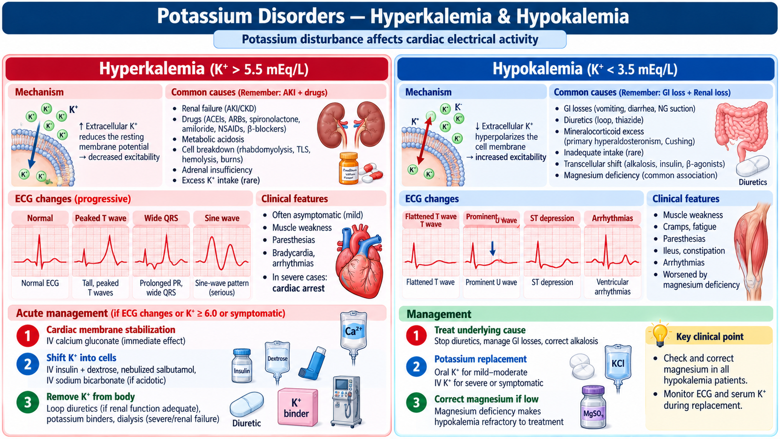
Potassium is the major intracellular cation and is essential for maintaining the resting membrane potential of nerve and muscle cells. This is why potassium disorders are particularly important in the heart. A serum potassium result must therefore be interpreted together with symptoms, renal function, drug history and the ECG.
Hyperkalemia
Hyperkalemia occurs when potassium is increased in the extracellular fluid. Important mechanisms include reduced renal excretion, release of potassium from cells and redistribution from the intracellular to the extracellular space.
Important causes
- Reduced renal potassium excretion, particularly in significant renal dysfunction.
- Drugs that reduce renal potassium excretion or interfere with the renin–angiotensin–aldosterone system.
- Cellular breakdown or tissue injury releasing intracellular potassium.
- Potassium shift out of cells, including in metabolic acidosis in appropriate clinical settings.
- Apparent elevation caused by specimen-related hemolysis should be considered when the result does not fit the patient.
Clinical features and diagnosis
Patients may have weakness or paresthesia, but the most dangerous effect is disturbed cardiac conduction. Obtain an ECG when hyperkalemia is clinically significant or severe. ECG abnormalities can progress from peaked T waves and conduction slowing to severe arrhythmias.
Treatment logic
- Stabilize the myocardium: intravenous calcium is used when significant cardiac toxicity is present. It does not remove potassium; it reduces the immediate electrical danger.
- Shift potassium into cells: insulin with glucose is an important temporary measure; other intracellular-shift therapies may be used where clinically appropriate. Nebulized salbutamol may be added to insulin–glucose therapy in severe hyperkalemia but should not be used alone. Monitor blood glucose following insulin administration because delayed hypoglycemia may occur.
- Remove potassium: stop exogenous potassium and offending drugs where appropriate, promote potassium removal, and consider renal replacement therapy when severe hyperkalemia cannot be adequately controlled, especially in major renal failure.
- Monitor: repeat potassium measurement and ECG assessment because temporary intracellular shifting does not eliminate total body potassium.
Hypokalemia
Hypokalemia occurs when serum potassium is reduced because of loss from the body or movement of potassium into cells. Gastrointestinal loss, renal loss and certain medications are common clinical settings.
Important causes
- Vomiting and diarrhea.
- Renal potassium loss, including diuretic-associated loss.
- Mineralocorticoid excess states.
- Intracellular shift caused by insulin or beta-adrenergic stimulation.
- Magnesium deficiency, which can make potassium replacement difficult.
Clinical features and treatment
Hypokalemia can cause fatigue, muscle weakness, constipation or ileus, and cardiac electrical instability. ECG changes may include T-wave flattening, ST depression and prominent U waves. Treatment is potassium replacement together with correction of the underlying cause. Oral replacement is generally suitable when the patient is stable and the deficit is not severe; intravenous replacement is reserved for more severe or clinically significant deficiency and requires careful monitoring.
D. Phosphate & Magnesium Disorders
Phosphate and magnesium abnormalities may be less immediately obvious than sodium or potassium disorders, but they are clinically important because they influence cellular energy, bone mineral balance, neuromuscular function and cardiac electrical stability. Renal function is particularly important in both.
Hyperphosphatemia
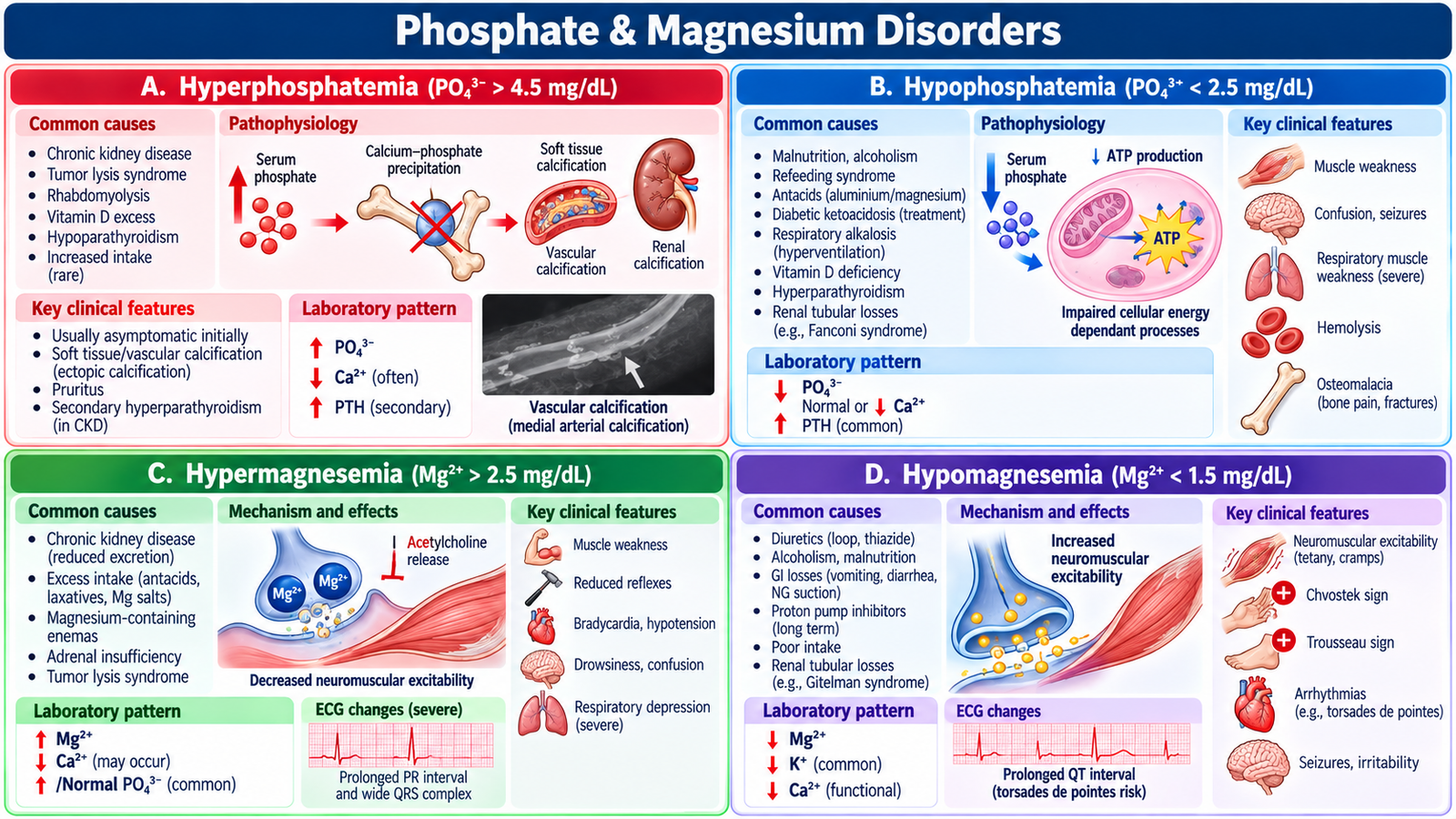
Hyperphosphatemia most commonly develops when renal phosphate excretion is reduced or when large amounts of phosphate are released from damaged cells. Increased phosphate can bind circulating calcium, so clinically important hyperphosphatemia may contribute to hypocalcemic manifestations and abnormal tissue mineral deposition.
Important contexts: significant renal impairment, major cell breakdown and excessive phosphate load. Management is directed at the cause, limiting excessive phosphate exposure and reducing phosphate burden when clinically required.
In chronic kidney disease, dietary phosphate restriction and oral phosphate binders such as sevelamer or calcium carbonate may be used when indicated. Severe refractory hyperphosphatemia in renal failure may require dialysis.
Hypophosphatemia
Phosphate is required for ATP production and normal cellular function. Marked depletion can therefore impair energy-dependent tissues.
- Important causes include poor intake, gastrointestinal loss, renal phosphate loss and movement of phosphate into cells.
- Severe deficiency can produce muscle weakness and may impair respiratory muscle function.
- Neurological dysfunction and impaired cellular function can occur when deficiency is substantial.
- Treatment requires phosphate replacement while identifying and correcting the underlying mechanism. Mild to moderate hypophosphatemia is generally treated with oral phosphate replacement. Severe or symptomatic deficiency, particularly with respiratory muscle weakness, requires monitored intravenous phosphate replacement, with assessment of calcium, potassium and renal function.
Hypermagnesemia
Magnesium is largely excreted by the kidneys. Hypermagnesemia therefore becomes particularly important when magnesium intake is increased in a patient whose renal excretion is impaired.
As magnesium rises, neuromuscular and cardiac activity can become depressed. Clinical findings may include reduced reflexes, weakness, hypotension, bradycardia and, in severe cases, respiratory or cardiac compromise.
Management includes stopping the magnesium source, supporting the patient, using intravenous calcium when significant magnesium toxicity is affecting neuromuscular or cardiac function, and enhancing magnesium removal when required.
Hypomagnesemia
Low magnesium commonly results from gastrointestinal loss, poor intake or renal magnesium loss. It increases neuromuscular excitability and can cause tremor, cramps, tetany, seizures and cardiac arrhythmias.
Hypomagnesemia is clinically linked with both hypokalemia and hypocalcemia. This means that an apparently resistant potassium or calcium abnormality may improve only after the magnesium deficit is corrected.
Mild hypomagnesemia is treated with oral magnesium salts such as magnesium citrate or magnesium glycerophosphate. Severe or symptomatic hypomagnesemia, including seizures or cardiac arrhythmias, requires intravenous magnesium sulfate with ECG and electrolyte monitoring. Adjust replacement in renal impairment.
🩺 AIM Clinical Bridge
A patient has several days of persistent vomiting and now reports weakness and palpitations. Examination suggests volume depletion. Blood tests show reduced potassium and increased bicarbonate, while the arterial blood gas shows alkalemia with an elevated bicarbonate level.
Think clinically:
- The vomiting has caused both volume and electrolyte loss.
- The raised bicarbonate with alkalemia points toward metabolic alkalosis.
- Potassium replacement and correction of the underlying fluid/chloride loss are connected parts of management.
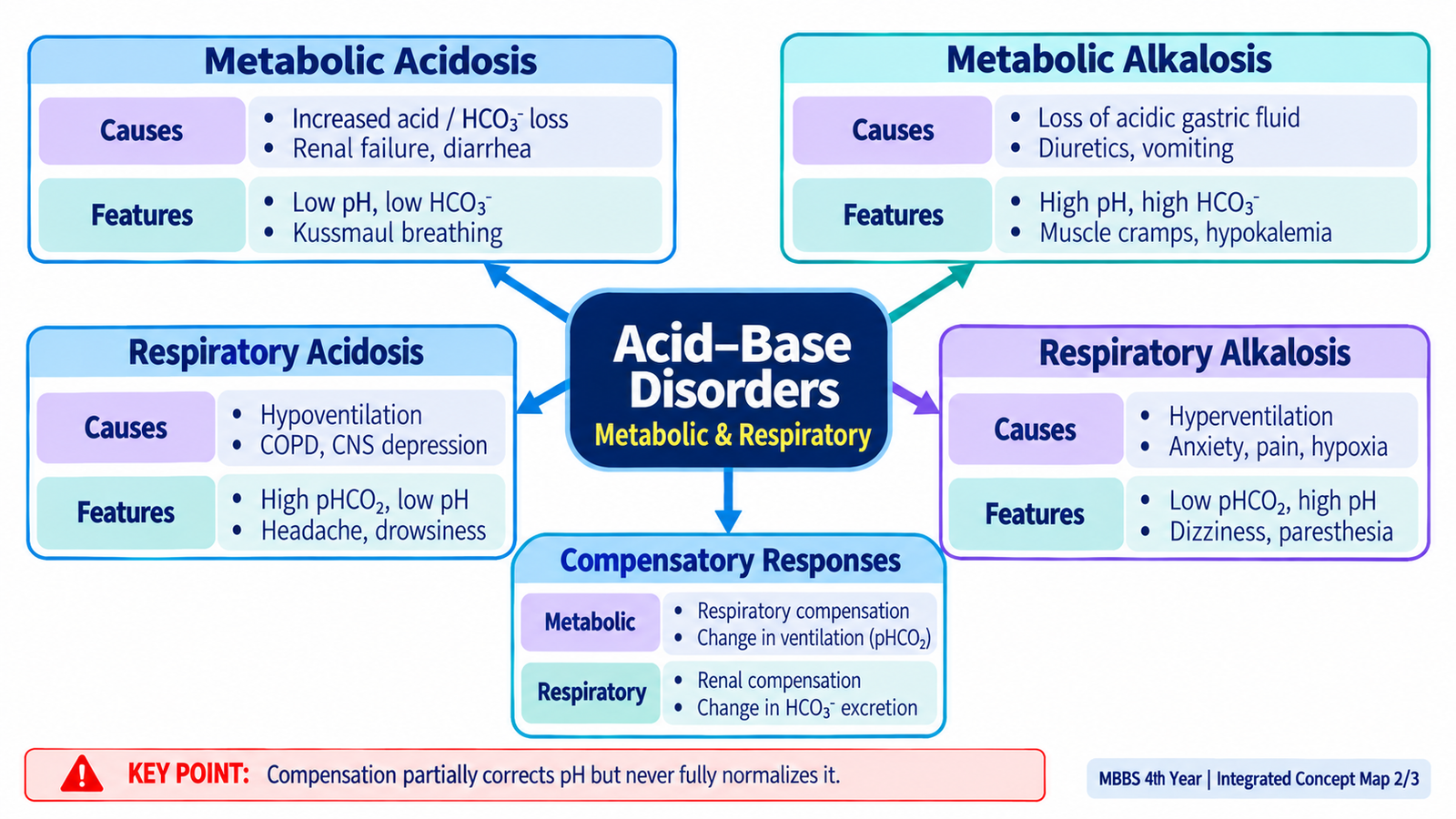
E. Metabolic Acidosis & Metabolic Alkalosis
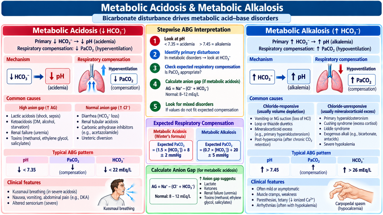
A metabolic acid–base disorder begins with a primary change in bicarbonate. The lungs respond by changing ventilation and therefore PaCO₂. Compensation reduces the change in pH but does not normally return the pH completely to normal. If the measured respiratory response is substantially different from the expected response, suspect a mixed acid–base disorder.
Metabolic acidosis
Metabolic acidosis is a primary reduction in bicarbonate that lowers blood pH. It develops because acid accumulates, bicarbonate is lost, or renal acid excretion is impaired.
Important mechanisms
- Increased acid generation: for example lactate or ketone accumulation.
- Reduced renal acid excretion: significant renal dysfunction.
- Bicarbonate loss: gastrointestinal or renal bicarbonate loss.
Anion gap
The anion gap helps organize metabolic acidosis by estimating the presence of unmeasured anions:
An increased gap suggests accumulation of unmeasured acids. A normal-gap metabolic acidosis more often reflects bicarbonate loss accompanied by a compensatory increase in chloride.
Expected respiratory compensation
In uncomplicated metabolic acidosis, PaCO₂ should fall because ventilation increases. A useful estimate is:
If measured PaCO₂ is higher than expected, an additional respiratory acidosis is likely. If it is lower than expected, an additional respiratory alkalosis is likely.
Metabolic alkalosis
Metabolic alkalosis is a primary increase in bicarbonate that raises blood pH. It commonly results from loss of hydrogen ions, contraction of extracellular fluid volume, potassium deficiency or increased mineralocorticoid activity.
Important clinical settings
- Persistent vomiting or gastric fluid loss.
- Diuretic-associated volume and electrolyte loss.
- Mineralocorticoid excess.
- Associated potassium depletion.
Respiratory compensation occurs through hypoventilation, causing PaCO₂ to rise. Compensation is limited because excessive hypoventilation would compromise oxygenation.
Management principle
Treatment is directed at the mechanism rather than at the pH alone. For example, metabolic alkalosis associated with vomiting and volume depletion improves when the extracellular-volume, chloride and potassium deficits are corrected and the ongoing cause is treated.
F. Respiratory Acidosis & Respiratory Alkalosis
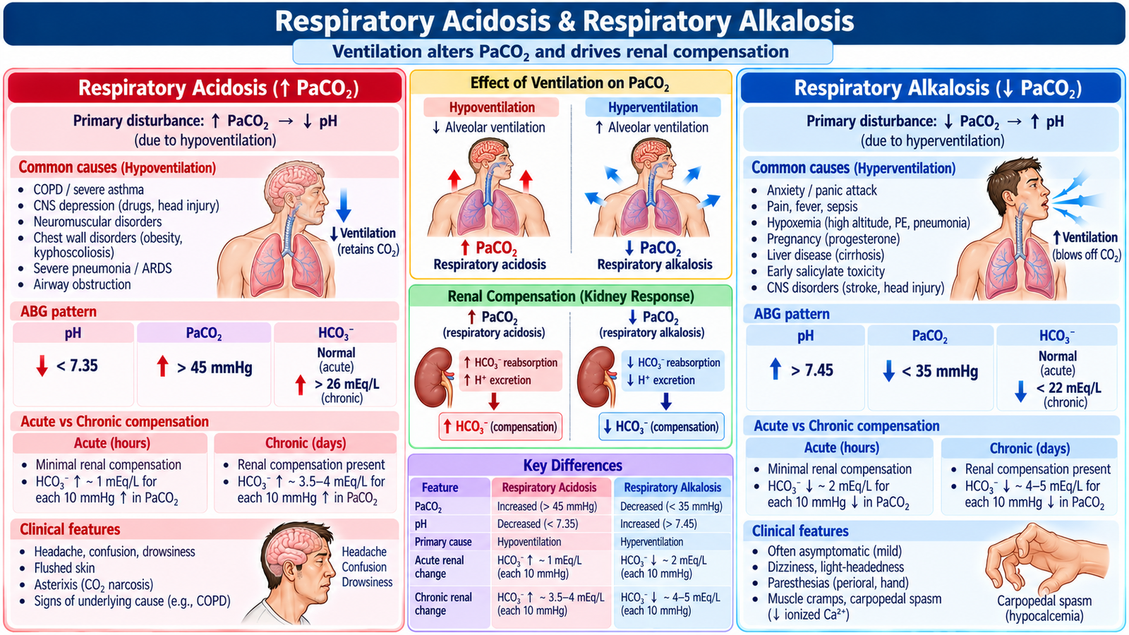
Respiratory acid–base disorders begin with a primary change in PaCO₂ caused by altered alveolar ventilation. Carbon dioxide behaves as an acid-producing component in the bicarbonate buffer system. The kidneys provide the compensatory response by altering bicarbonate retention and hydrogen-ion excretion. Because renal adaptation takes time, compensation differs in acute and chronic respiratory disorders.
Respiratory acidosis
Respiratory acidosis develops when alveolar ventilation is inadequate and carbon dioxide accumulates.
Clinical settings
- Severe airway or lung disease causing inadequate ventilation.
- Depression of respiratory drive.
- Neuromuscular weakness affecting ventilation.
- Other causes of alveolar hypoventilation.
Acute CO₂ retention produces relatively little rise in bicarbonate because the kidneys have not had time to adapt. In chronic respiratory acidosis, renal bicarbonate retention is greater and therefore pH is closer to normal.
- Acute respiratory acidosis: HCO₃⁻ rises by about 1 mmol/L for each 10 mmHg rise in PaCO₂.
- Chronic respiratory acidosis: HCO₃⁻ rises by about 3.5–4 mmol/L for each 10 mmHg rise in PaCO₂.
Respiratory alkalosis
Respiratory alkalosis develops when ventilation is excessive relative to carbon dioxide production, causing excessive CO₂ removal.
Clinical settings
- Hypoxemia stimulating ventilation.
- Pain or anxiety with sustained hyperventilation.
- Central stimulation of ventilation.
- Other disorders producing excessive alveolar ventilation.
Symptoms may include light-headedness, paresthesia and neuromuscular irritability. These are related partly to alkalemia and associated changes in ionized calcium.
- Acute respiratory alkalosis: HCO₃⁻ falls by about 2 mmol/L for each 10 mmHg fall in PaCO₂.
- Chronic respiratory alkalosis: HCO₃⁻ falls by about 4–5 mmol/L for each 10 mmHg fall in PaCO₂.
How to recognize a mixed disorder
Compensation follows predictable physiological limits. If the measured bicarbonate or PaCO₂ differs substantially from the expected compensatory response, the patient probably has more than one primary acid–base disturbance.
Integrated Mechanism Flow
Important Comparison — Primary Acid–Base Disorders
| Disorder | Primary change | pH tendency | Compensation |
|---|---|---|---|
| Metabolic acidosis | ↓ HCO₃⁻ | ↓ pH | Hyperventilation → ↓ PaCO₂ |
| Metabolic alkalosis | ↑ HCO₃⁻ | ↑ pH | Hypoventilation → ↑ PaCO₂ |
| Respiratory acidosis | ↑ PaCO₂ | ↓ pH | Renal HCO₃⁻ retention |
| Respiratory alkalosis | ↓ PaCO₂ | ↑ pH | Renal HCO₃⁻ loss / reduced retention |
AIM High-Yield Review
- ⭐ Serum sodium mainly reflects the relationship between body water and sodium; neurological manifestations arise from abnormal water movement across brain-cell membranes.
- Acute severe hyponatremia can cause cerebral edema and seizures; treatment requires controlled correction rather than indiscriminate normalization.
- Hypernatremia usually represents water deficit and causes cellular dehydration; chronic cases should also be corrected gradually.
- ⭐ Hyperkalemia is dangerous because of cardiac electrical toxicity. Significant ECG changes require immediate myocardial stabilization before potassium is definitively removed.
- Insulin with glucose shifts potassium into cells but does not remove potassium from the body.
- Hypokalemia can cause weakness, ileus and arrhythmias; persistent hypokalemia should prompt assessment for ongoing losses and magnesium deficiency.
- Severe hypophosphatemia can impair ATP-dependent tissues and cause significant muscular, including respiratory-muscle, weakness.
- Hypermagnesemia tends to depress reflexes and neuromuscular activity, while hypomagnesemia increases neuromuscular excitability.
- ⭐ Metabolic acidosis is primarily a fall in bicarbonate; appropriate respiratory compensation lowers PaCO₂.
- The anion gap helps distinguish accumulation of unmeasured acids from bicarbonate-loss patterns in metabolic acidosis.
- Metabolic alkalosis is characterized by increased bicarbonate with compensatory elevation of PaCO₂; vomiting and diuretic-associated losses are important clinical settings.
- ⭐ Respiratory acidosis is caused by CO₂ retention from inadequate ventilation; chronic disease produces greater renal bicarbonate retention than an acute episode.
- Respiratory alkalosis results from excessive ventilation and low PaCO₂; renal bicarbonate reduction provides compensation.
- Compensation reduces the pH disturbance but does not normally completely normalize pH.
- If the measured compensatory response is substantially different from the expected response, think of a mixed acid–base disorder.
Electrolyte & Acid–Base Disorders
Review sodium, potassium, phosphate, magnesium and major metabolic and respiratory acid–base disturbances.