
Station 13 — Nose and Paranasal Sinus Imaging and Investigations
🎯 Candidate Task
Interpret the provided X-ray and CT images of the nose/nasopharynx, identifying the important radiological abnormalities. State the appropriate further investigation where required and complete the relevant investigation request.
⚡ Model Station Answer
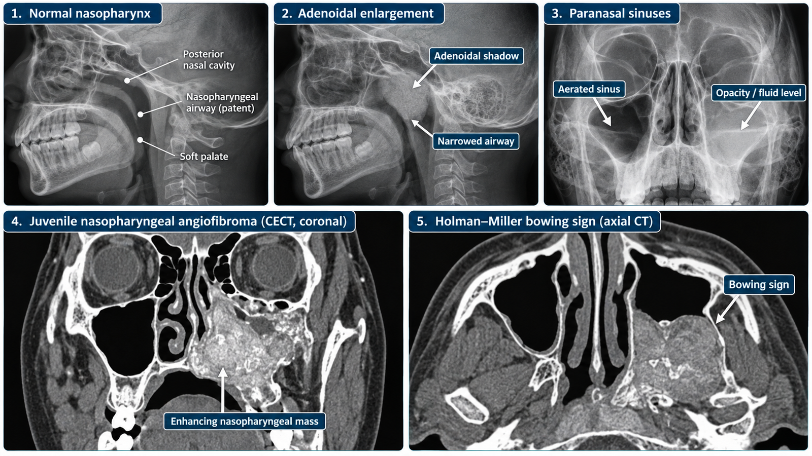
X-ray: I would assess the nasopharyngeal airway and paranasal sinuses systematically. An enlarged posterior nasopharyngeal soft-tissue shadow with narrowing of the airway is consistent with adenoidal enlargement.
CT: I would review the nasal cavity, nasopharynx, paranasal sinuses, surrounding bone and adjacent spaces systematically on appropriate planes.
Key finding: A strongly enhancing nasopharyngeal mass in the typical region with anterior bowing of the posterior wall of the maxillary sinus suggests juvenile nasopharyngeal angiofibroma.
Bowing sign: Anterior bowing of the posterior wall of the maxillary antrum is the characteristic Holman–Miller sign.
Investigation request: I would select imaging according to the suspected nasal or sinus disease and clearly document patient identification, clinical indication, anatomical region, side where relevant, requested investigation, date and authentication.
🔎 How to Perform / Approach
- Step 1 — Identify the investigation.
State whether the image is an X-ray or CT and identify the anatomical region and available view or plane. - Step 2 — Review the X-ray systematically.
Assess the nasopharyngeal airway, soft-tissue shadow and visible paranasal sinuses for opacity, air-fluid level or abnormal soft-tissue enlargement. - Step 3 — Recognize adenoidal enlargement.
Look for an enlarged soft-tissue shadow arising from the posterior nasopharyngeal region with narrowing of the nasopharyngeal air column. - Step 4 — Approach the CT systematically.
Review the nasal cavity and septum, nasopharynx, individual paranasal sinuses, surrounding bony walls and adjacent deep spaces. Identify any mass and assess its extent and bony effects. - Step 5 — Identify the characteristic JNA pattern.
Look for a strongly enhancing nasopharyngeal mass and assess for smooth anterior bowing of the posterior wall of the maxillary sinus—the Holman–Miller or bowing sign. - Step 6 — Select the appropriate investigation.
Use imaging according to the suspected pathology—for example, appropriate radiography for selected basic assessment and CT for detailed evaluation of sinonasal disease, bony anatomy or a suspected nasopharyngeal mass. - Step 7 — Complete the requisition.
Enter correct patient details, relevant clinical information, anatomical region, requested study and indication, then check the form and add the date and required authentication.
🖼️ Visual Learning
Adenoidal Enlargement, CT Nasopharynx and the Bowing Sign
📝 Important Viva Questions
1. What is the important X-ray finding in enlarged adenoids?
Answer: There is enlargement of the posterior nasopharyngeal soft-tissue shadow with narrowing of the nasopharyngeal airway.
2. How should you approach a CT of the nose and nasopharynx?
Answer: Review the nasal cavity, septum, nasopharynx, paranasal sinuses, surrounding bones and adjacent spaces systematically, then identify any lesion and assess its extent.
3. What CT features suggest juvenile nasopharyngeal angiofibroma?
Answer: A strongly enhancing nasopharyngeal mass in the characteristic location with extension into adjacent spaces and smooth pressure-related bony remodeling is suggestive.
4. What is the Holman–Miller or bowing sign?
Answer: It is anterior bowing of the posterior wall of the maxillary antrum caused by a nasopharyngeal mass, classically juvenile nasopharyngeal angiofibroma.
5. What essential information should be included on an imaging requisition?
Answer: Include correct patient identification, relevant clinical findings and indication, anatomical region and side where applicable, the requested investigation, date and authentication.