
Station 9 — Ear Examination: History, Otoscopy and Examination Under Microscope
Rapid Revision • Practical Skills • Viva
🎯 Candidate Task
Take a focused history from a patient presenting with ear discharge, hearing difficulty or dizziness, then demonstrate systematic local examination of the ear and safe otoscopy. State the important structures/findings you would assess and explain the basic role of examination under microscope (EUM) and how you may assist.
⚡ Model Station Answer
- I would take a focused history of the presenting ear symptom, associated otological symptoms and relevant red flags.
- I would inspect the pinna, surrounding skin, pre- and post-auricular areas and external auditory opening before performing otoscopy.
- During otoscopy I would examine the external auditory canal and tympanic membrane systematically for normal landmarks and common abnormalities.
- EUM provides a magnified, well-illuminated binocular view of the ear canal and tympanic membrane; I may assist with patient positioning, equipment preparation and instruments under supervision.
🔎 How to Perform / Approach
1. Introduce and prepare: Wash hands, introduce yourself, confirm the patient, explain the examination, obtain consent and ask about current ear pain.
- 2. Take a focused history: For discharge ask onset, duration, amount, colour, smell, blood, pain and fever; for hearing difficulty ask onset, side, progression and tinnitus; for dizziness clarify true vertigo, duration, triggers, nausea and associated hearing symptoms.
- 3. Check relevant background: Ask briefly about previous ear infections, trauma or surgery, water exposure, significant medication history and important symptoms such as facial weakness or neurological complaints.
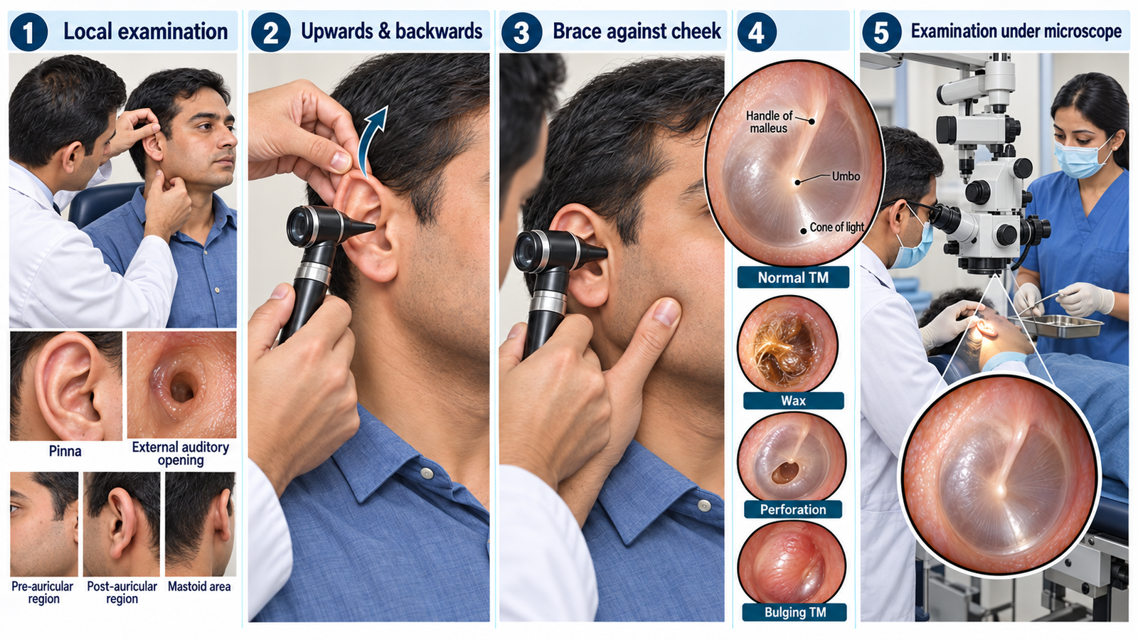
- 4. Inspect the ear: Compare both pinnae and inspect the skin, deformity, scars, swelling, discharge, external auditory opening and pre- and post-auricular regions. Examine the less painful or apparently normal ear first where appropriate.
- 5. Prepare the otoscope: Check the light and fit a clean appropriately sized speculum. In an adult, gently pull the pinna upwards and backwards to straighten the external auditory canal.
- 6. Perform safe otoscopy: Hold the otoscope like a pen in the hand corresponding to the ear being examined, brace the hand against the patient’s cheek, and advance gently under direct vision. Inspect the canal for wax, discharge, swelling or lesions, then assess the tympanic membrane for integrity, colour, position and visible landmarks.
- 7. Explain EUM: Examination under microscope gives magnified binocular illumination for detailed assessment of the canal and tympanic membrane and may facilitate precise cleaning or instrumentation. As a student, assist with positioning, preparation and passing equipment while the procedure is performed under appropriate supervision.
🖼️ Visual Learning
Systematic Ear Examination, Otoscopy and Examination Under Microscope
🎥 Practical Demonstration
Otoscopy (Ear Examination) — Oxford Medical Education
A practical demonstration produced by Oxford University medical students with ENT faculty, showing correct otoscope handling, examination technique and tympanic-membrane anatomy.
📝 Important Viva Questions
1. How do you straighten the external auditory canal during otoscopy in an adult?
Answer: Gently pull the pinna upwards and backwards before inserting the otoscope.
2. Why should the hand holding the otoscope be braced against the patient’s head?
Answer: Bracing stabilizes the instrument and reduces the risk of trauma if the patient suddenly moves.
3. Which two main structures are assessed during otoscopy?
Answer: The external auditory canal and the tympanic membrane.
4. Name important normal landmarks visible on the tympanic membrane.
Answer: Important landmarks include the handle of the malleus, umbo and cone of light on an intact translucent tympanic membrane.
5. What is the main advantage of examination under microscope compared with routine otoscopy?
Answer: EUM provides a magnified, well-illuminated binocular view of the external auditory canal and tympanic membrane, allowing more detailed examination and precise instrumentation when required.