
AIM OSCE / OSPE • MBBS
Station 2 — Oral Cavity, Oropharynx, Nasopharynx and Hypopharynx Examination
🎯 Candidate Task
Perform a systematic examination of the oral cavity and oropharynx, and demonstrate how you would examine the nasopharynx and hypopharynx using appropriate mirror techniques and palpate an oral/oropharyngeal mass. Describe the basic steps involved in assisting with biopsy of an oral or tongue ulcer and report important abnormal findings.
⚡ Model Station Answer
- I would systematically inspect the lips, buccal mucosa, teeth and gums, tongue, floor of mouth, palate, tonsillar region and posterior oropharynx.
- I would perform posterior mirror examination of the nasopharynx and indirect mirror examination of the hypopharyngeal region using a warmed mirror and correct positioning.
- Any oral or oropharyngeal mass would be palpated gently with gloves, assessing its site, size, consistency, tenderness, mobility or fixation and surrounding tissue.
- I would identify and report ulcers, mucosal lesions, asymmetry, masses, bleeding, fixation, pooling of secretions or other abnormal upper aerodigestive tract findings.
- For biopsy assistance, I would prepare the required equipment, aid exposure and suction, handle the specimen correctly, assist haemostasis and ensure accurate specimen labelling.
🔎 How to Perform / Approach
1
Prepare the patient. Introduce yourself, confirm identity, explain the examination, obtain consent, perform hand hygiene and seat the patient upright in good illumination.
2
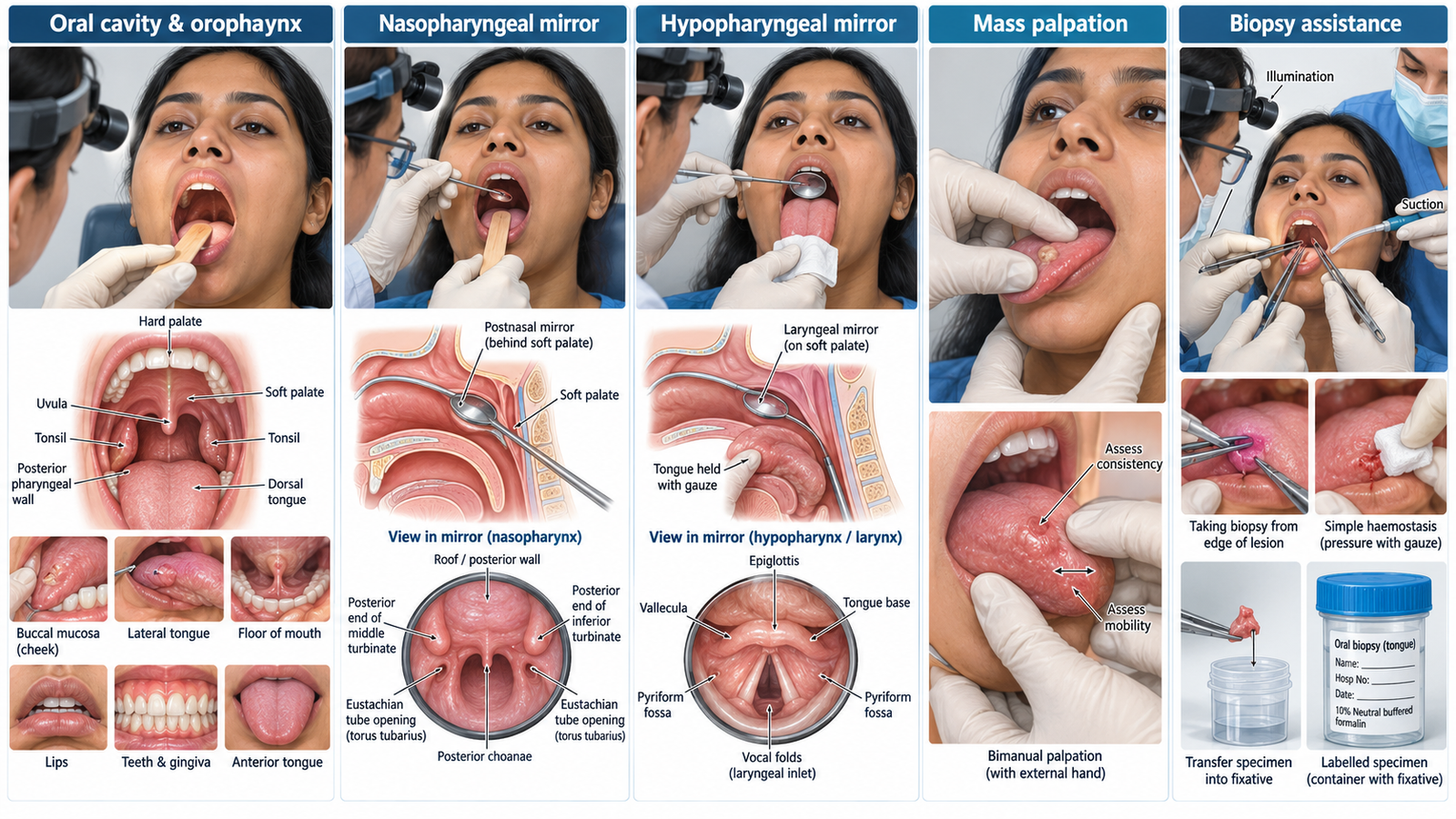
Inspect the oral cavity and oropharynx. Using a light and tongue depressor, systematically inspect the lips, buccal mucosa, teeth and gums, tongue including its undersurface, floor of mouth, palate, uvula, tonsillar regions and posterior pharyngeal wall.
3
Palpate any mass. Wear gloves and gently palpate the lesion directly; use bimanual palpation where appropriate and assess site, extent, consistency, tenderness, mobility/fixity and contact bleeding.
4
Examine the nasopharynx. Warm the postnasal mirror and check that it is not hot. Depress the tongue and introduce the mirror gently behind the soft palate without touching the posterior pharyngeal wall; inspect the choanae, posterior nasal structures, roof/posterior wall and Eustachian tube regions.
5
Examine the hypopharyngeal region. Ask the patient to protrude the tongue, hold it gently with gauze and position the warmed mirror against the soft palate. Obtain a reflected view while the patient breathes or phonates, inspecting the tongue base, valleculae, epiglottic region, pyriform fossae and laryngeal inlet for masses or pooling.
6
Recognize and report abnormalities. Comment on suspicious ulceration, mucosal irregularity, asymmetry, swelling or mass, bleeding, fixation and abnormal pooling of secretions.
7
Describe biopsy assistance and finish. Prepare PPE, illumination, suction, local-anaesthetic and biopsy equipment; assist exposure and specimen collection, support haemostasis, place the specimen promptly in the correctly labelled container, thank the patient and perform hand hygiene.
🖼️ Visual Learning
VISUAL 1
Upper Aerodigestive Tract Examination and Oral Lesion Assessment
🎥 Practical Demonstration
ENT Teaching: Examination I — Mirror Examination
University Hospital Münster teaching resource. Particularly useful from approximately 05:38 onward for nasopharyngeal, oral cavity and indirect laryngeal mirror examination.
📝 Important Viva Questions
1. Why is the examination mirror warmed before use?
Warming reduces fogging from the patient’s breath. The temperature must be checked before insertion to avoid thermal injury.
2. What important structures are assessed during posterior mirror examination of the nasopharynx?
The posterior choanae, posterior ends of the turbinates, nasopharyngeal roof and posterior wall, and regions around the Eustachian tube openings are inspected.
3. How can you minimize gagging during mirror examination?
Use gentle controlled movements and avoid touching the tongue base or posterior pharyngeal wall with the mirror.
4. What features should you assess when palpating an oral or oropharyngeal mass?
Assess its site, size or extent, consistency, tenderness, mobility or fixation and any tendency to bleed on contact.
5. How should an oral-biopsy specimen be handled after removal?
Transfer it promptly to the appropriate correctly labelled specimen container, usually containing formalin for routine histopathology, and clearly document the biopsy site.