
📖 AIM Learning Material
Topic 5. Diagnostic Imaging & Radiation Protection
Foundation Module · How images are made, when to request them and how to use radiation safely.
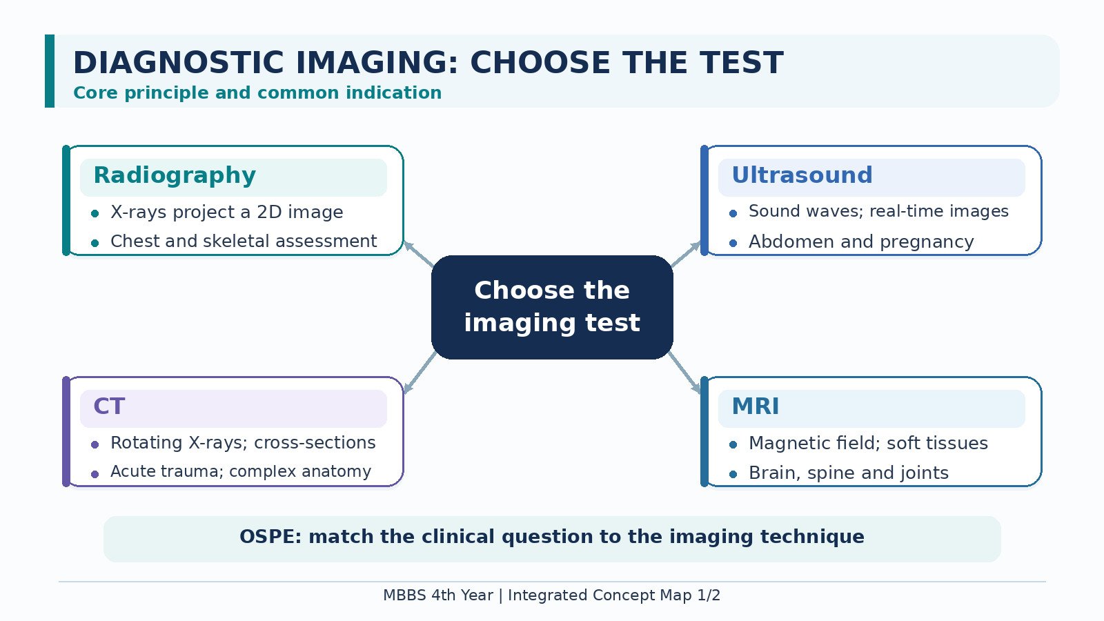
Diagnostic imaging helps answer a clinical question that history and examination cannot settle on their own. Different techniques reveal different information: an X-ray can show a fracture, ultrasound can examine the gallbladder, CT can rapidly display internal structures in cross-section, and MRI can show detailed soft tissues. The best test is therefore the one that answers the patient’s question in time to guide care. Some methods use ionizing radiation; others do not. You will learn how each method works, its common indications and limitations, and how to balance diagnostic benefit with radiation safety. Choosing an image is a clinical decision, not simply requesting the most detailed scan.
A. Begin with the Clinical Question
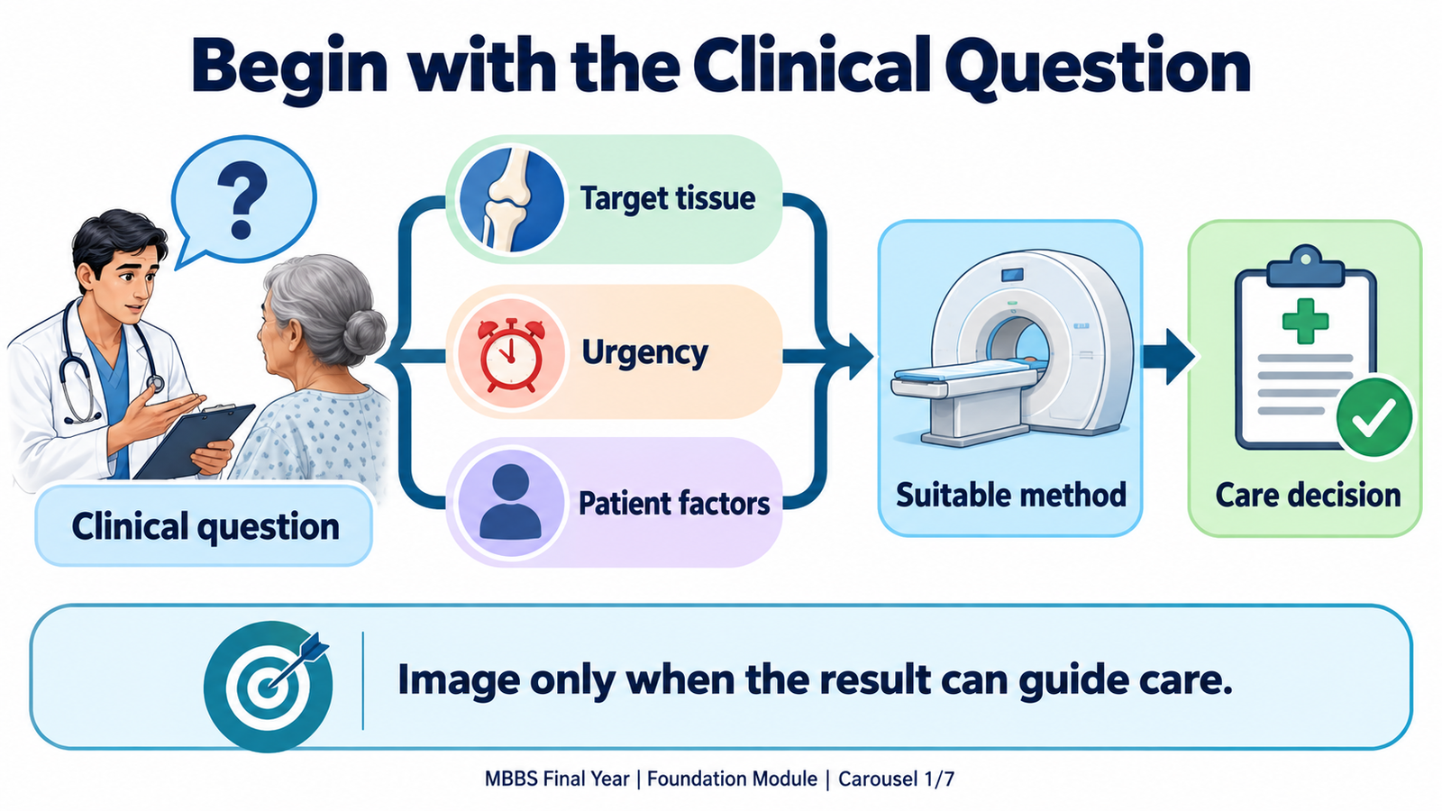
An imaging request should say what the clinician suspects and what decision depends on the result. “Abdominal pain” alone is less helpful than asking whether a patient’s symptoms and examination suggest gallstones or an acute abdominal complication. The radiologist can then select or refine a test that examines the right region with the right technique.
Choose an investigation by considering four connected questions:
- What must be seen? Bone, lung, blood vessels, soft tissue, a moving structure or organ function may require different methods.
- How quickly is the answer needed? An unstable patient may need immediate bedside assessment; a stable patient can undergo more detailed imaging when appropriate.
- Will the result change care? A scan with no useful effect on the next decision may expose the patient to avoidable risk or delay.
- What makes this patient different? Pregnancy, age, kidney function, previous contrast reaction and implanted devices may affect the choice or preparation.
Imaging complements clinical assessment. A technically good image may still miss an early, small or poorly visualized abnormality. If the result conflicts with important clinical findings, reconsider the diagnosis and discuss whether another view or a different modality is needed.
B. Plain Radiography and Fluoroscopy: Images Made with X-Rays
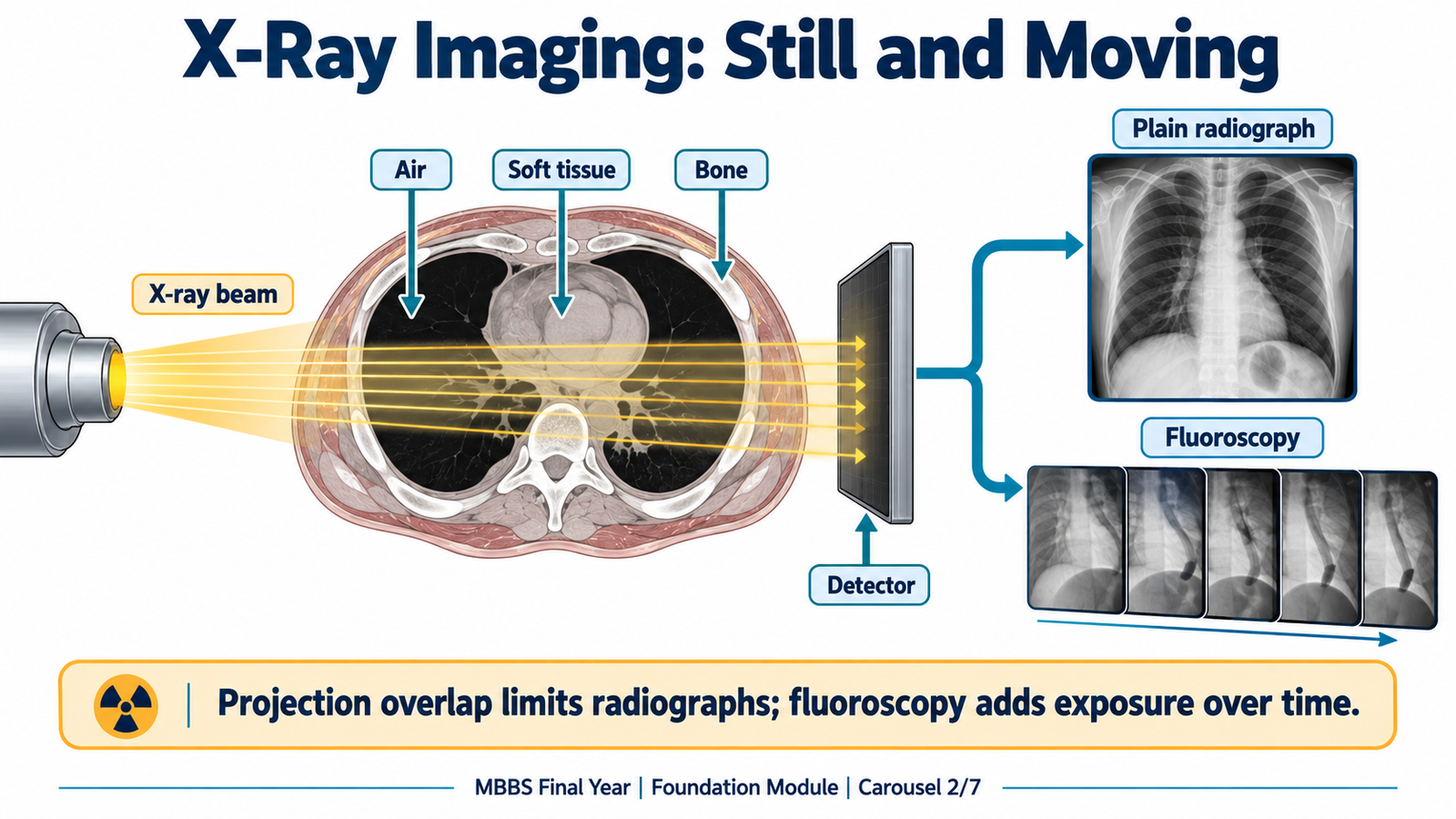
An X-ray beam passes through the body to a detector. Dense structures such as bone absorb more of the beam and usually appear lighter; air absorbs less and appears darker. A plain radiograph is a two-dimensional projection, so structures in front of and behind one another overlap. This makes radiography quick and useful, but limits its ability to separate soft tissues.
When a plain radiograph helps: a suspected fracture, a chest problem such as pneumothorax, or a question about the position of a medical device. The requested views should match the question. A normal radiograph does not automatically exclude an early or subtle abnormality; interpretation must remain linked to the patient’s findings.
Fluoroscopy uses repeated X-ray images to show movement in real time. It can follow swallowed contrast through the upper digestive tract or guide a procedure while the operator watches the image. Because exposure continues while imaging is active, procedure time and technique matter to radiation protection.
Some X-ray examinations use contrast material to outline a structure that would otherwise blend into surrounding tissue. Contrast answers a specific question; it is not required for every image. Tell the imaging team about a significant previous contrast reaction and other relevant safety concerns before an enhanced examination.
C. Ultrasound: Real-Time Imaging with Sound Waves
An ultrasound probe sends high-frequency sound waves into the body and receives their returning echoes. Differences in tissue properties create the image. Ultrasound uses no ionizing radiation and can show movement in real time. Doppler ultrasound adds information about the direction and pattern of blood flow.
It is especially useful when the clinical question concerns:
- pregnancy and pelvic structures;
- the gallbladder, kidneys or fluid within the abdomen;
- superficial masses or selected blood vessels using Doppler;
- a patient who needs a bedside examination.
The limits follow from the way sound travels. Bowel gas and bone can block the view, body habitus can make some structures harder to assess, and image quality depends partly on the examiner. A poorly seen structure is not the same as a normal structure. If ultrasound cannot answer an important question, another modality may be needed.

D. Computed Tomography: Cross-Sections Made with X-Rays
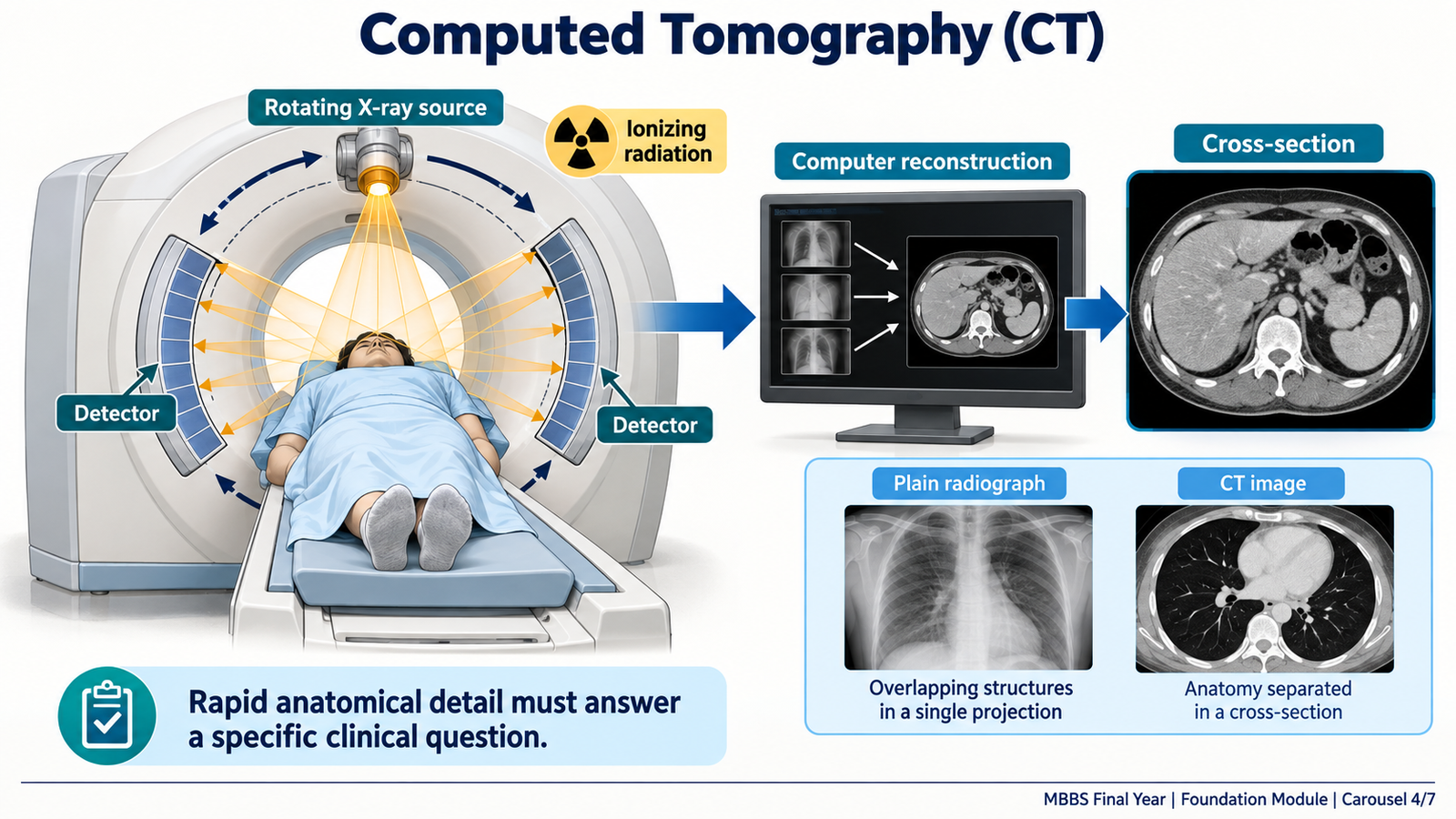
Computed tomography (CT) collects X-ray measurements from many directions as the scanner moves around the patient. A computer reconstructs these measurements into cross-sectional images and, when useful, images in other planes. Unlike a plain radiograph, CT greatly reduces the problem of overlapping structures.
CT is particularly helpful when rapid cross-sectional detail will change management. Examples include assessing suspected intracranial bleeding after head injury, defining injuries in a stable trauma patient, or examining a serious abdominal problem when clinical assessment indicates the need. The patient’s stability matters: imaging should not postpone immediate resuscitation or urgent care.
CT uses ionizing radiation. Its exposure is generally greater than that of a single plain radiograph, although actual dose varies with the examination. Request only the area and study needed to answer the clinical question. CT may be performed without contrast or with intravenous iodine-based contrast. Contrast can make vessels and tissue differences clearer, but is chosen according to the question. A previous significant contrast reaction and relevant kidney concerns should be communicated before its use.
A CT report must be interpreted in context. For example, an image that shows bleeding can explain a patient’s neurological deterioration and change the urgency of treatment. A negative scan answers only the question that the chosen scan could reliably evaluate.
E. Magnetic Resonance Imaging: Detailed Soft-Tissue Contrast
Magnetic resonance imaging (MRI) uses a strong magnetic field and radiofrequency signals to create images. It does not use ionizing radiation. Its main strength is the ability to distinguish many soft tissues clearly, such as the brain, spinal cord, joints and surrounding structures. This can answer a question that is difficult to resolve on plain radiography or CT.
MRI takes longer than many CT examinations and requires the patient to remain sufficiently still. It is therefore not automatically the best first test in an emergency. Safety screening is essential: some implanted devices or metal objects may be unsafe or may affect image quality. An implant does not automatically exclude MRI; its exact type and MRI conditions must be checked by the imaging team.
Some MRI examinations use gadolinium-based contrast to answer a particular question. This is different from iodine-based CT contrast. Relevant kidney disease and any previous contrast reaction should be disclosed so the team can decide whether contrast is appropriate. The reason to choose MRI remains the clinical information needed, rather than the absence of ionizing radiation alone.
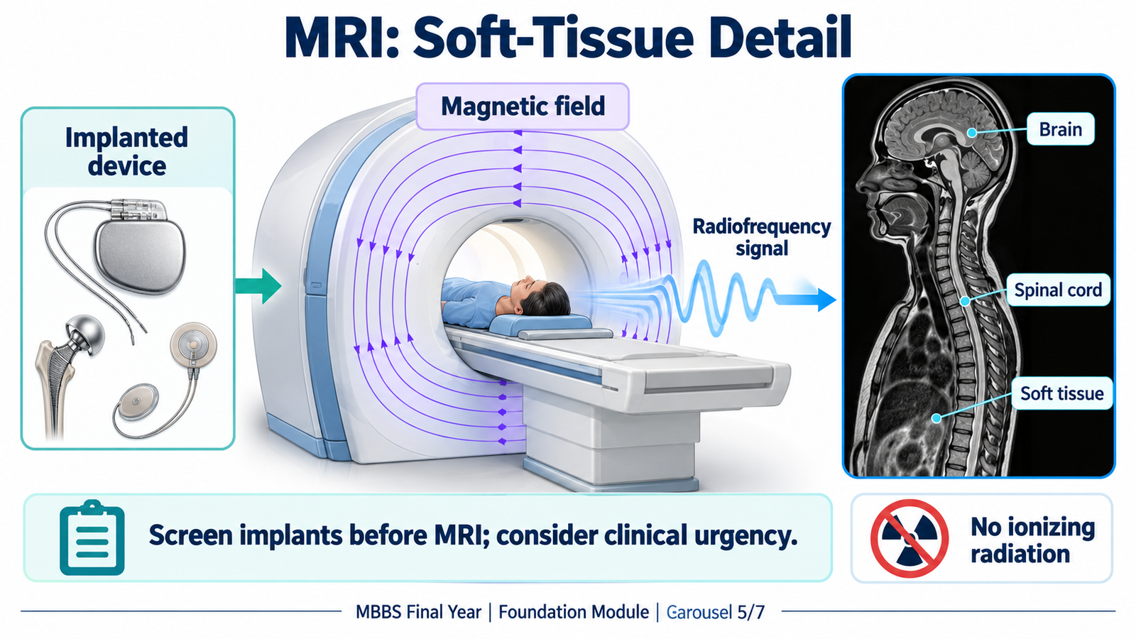
F. Nuclear Medicine: Imaging Organ Function
Nuclear medicine differs from X-ray and CT because a small amount of radioactive tracer is introduced into the patient. A camera detects radiation emitted from its distribution in the body. The pattern can show function, such as tissue activity or uptake, rather than anatomy alone.
Examples include selected bone scans or thyroid studies. Positron emission tomography (PET) is another tracer-based method used for selected clinical questions, including some cancer assessments. A CT image may be combined with a nuclear medicine study to locate the functional finding more precisely. The test should still answer a specific clinical question; a tracer study is not a substitute for every structural image.
Nuclear medicine uses ionizing radiation because the administered tracer is radioactive. Pregnancy or breastfeeding information can affect planning and must be communicated to the imaging team. As with other imaging, the expected diagnostic benefit should justify the examination.
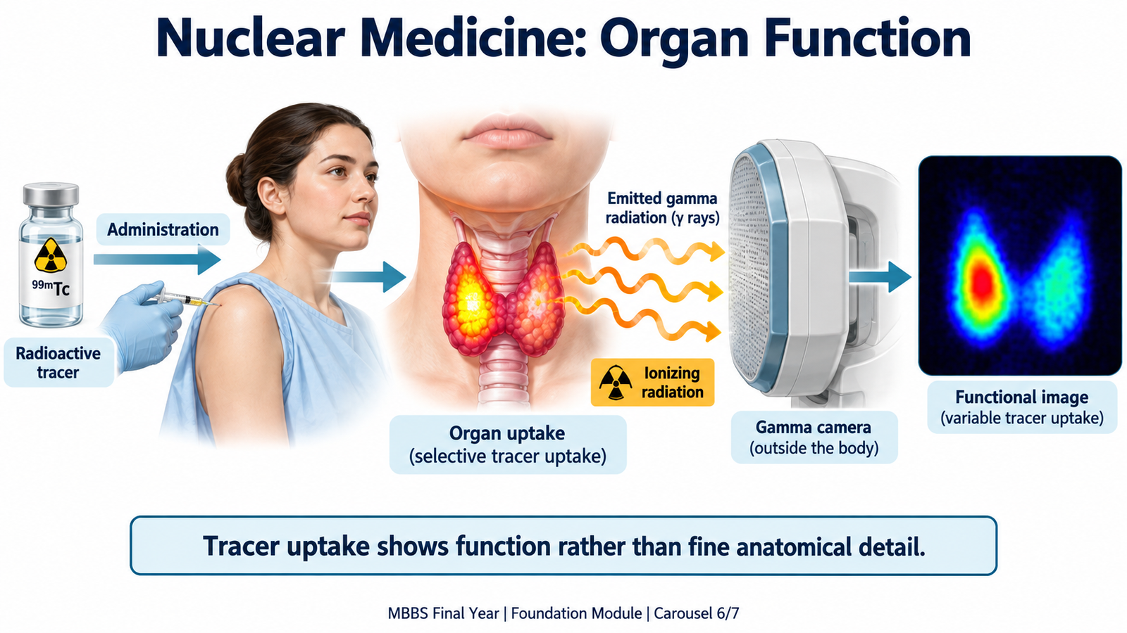
G. Radiation Protection: Benefit, Dose and Patient Safety
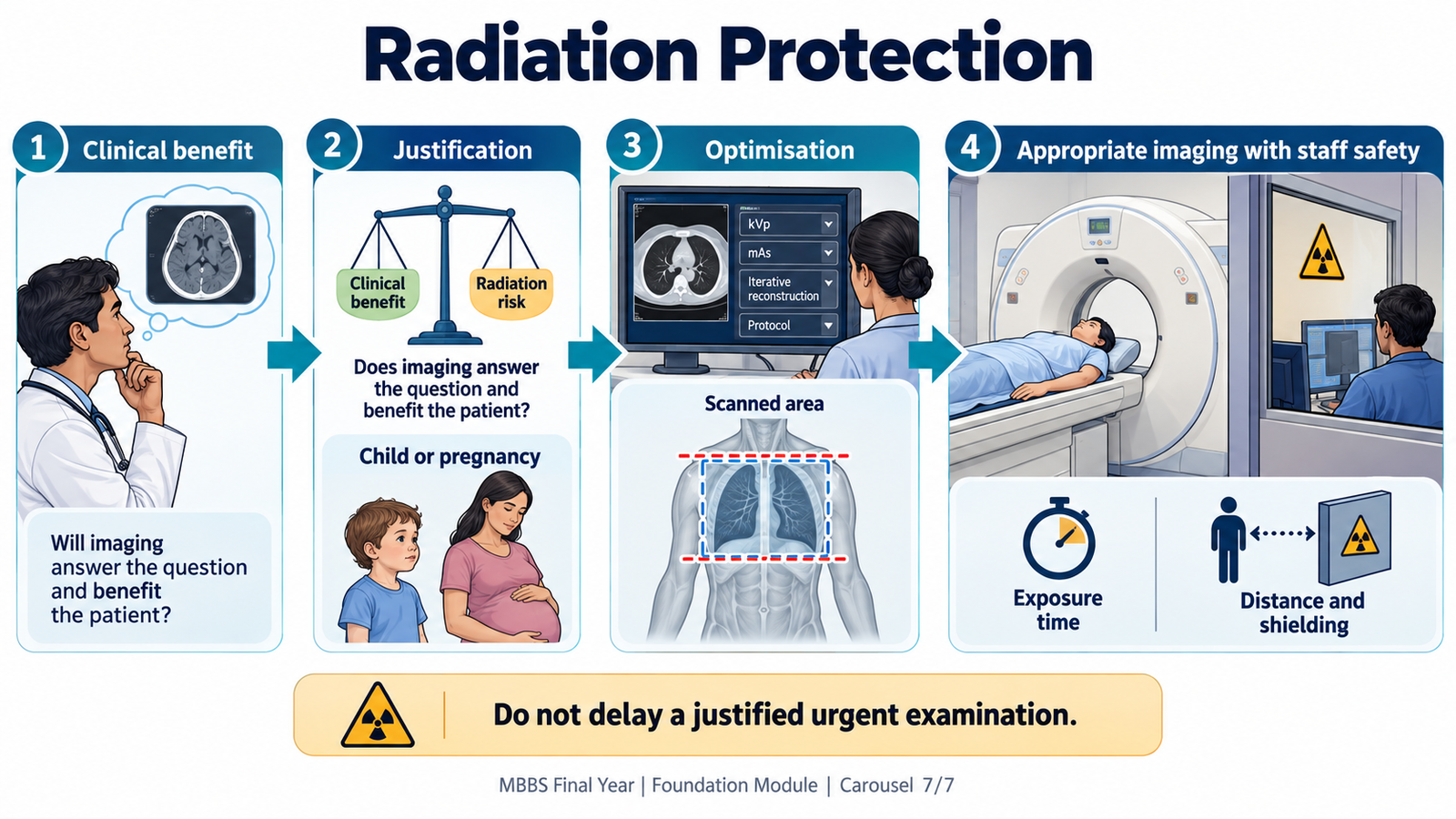
Ionizing radiation can alter molecules in tissue. Diagnostic imaging is valuable because the information gained can improve care, but unnecessary exposure brings no benefit. Radiation protection begins before the scan: decide whether imaging is needed and whether a method without ionizing radiation can answer the same question well enough.
Two principles guide medical imaging:
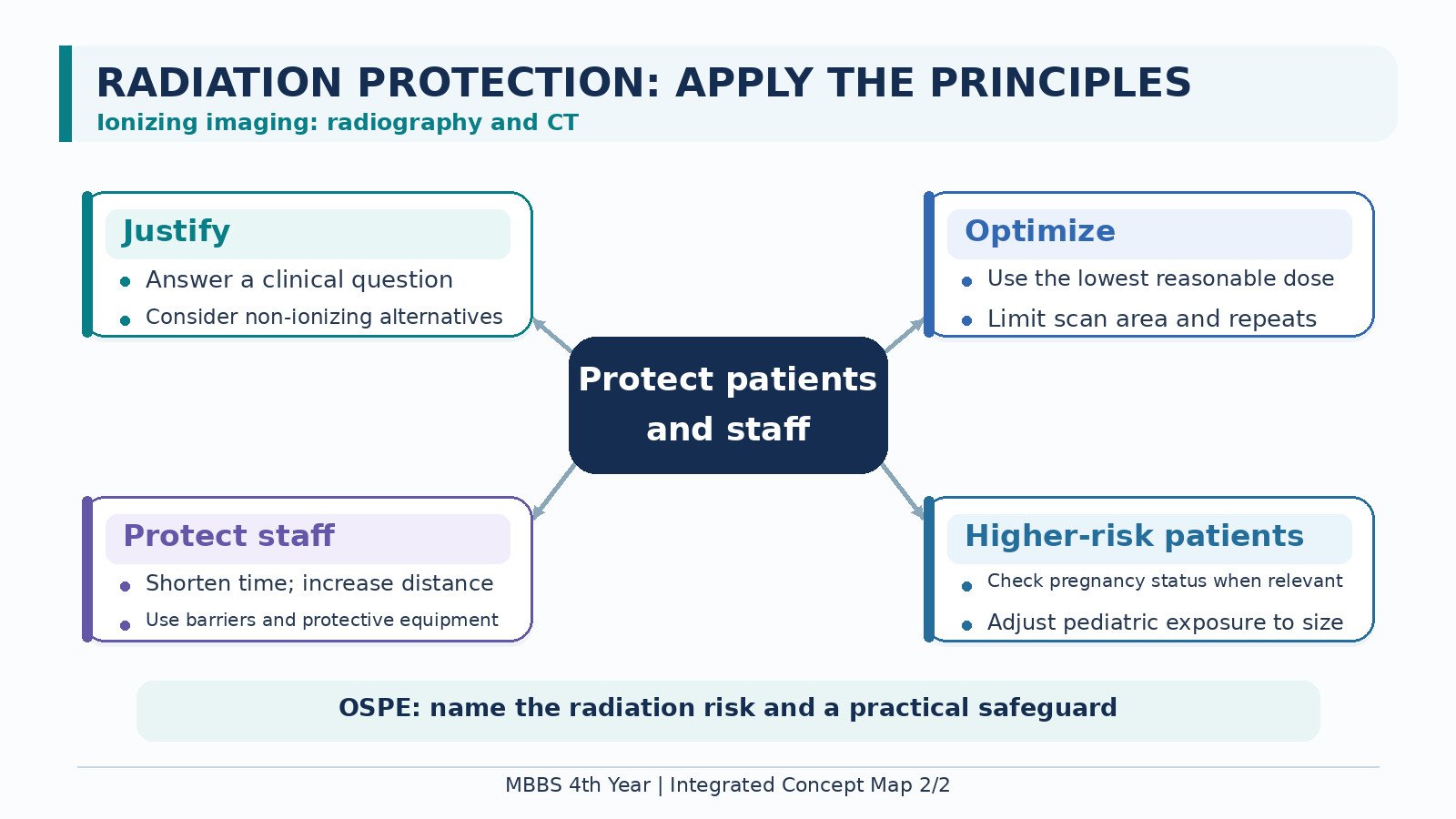
- Justification: the expected benefit of the examination outweighs its potential harm for this patient and clinical question.
- Optimisation: use a technique that obtains an image fit for diagnosis without unnecessary exposure. This includes selecting the correct study, limiting the scanned area and avoiding needless repeat images.
Children warrant particular care because they can be more sensitive to radiation and have a longer remaining lifespan in which an effect could appear. In a patient who is or may be pregnant, inform the imaging team and consider ultrasound or MRI when they can answer the question. Pregnancy does not mean that a necessary X-ray or CT must be withheld: an urgent, clinically justified examination may provide greater benefit than delaying diagnosis.
For staff working around ionizing radiation, shorter exposure time, greater distance from the source and appropriate shielding reduce occupational exposure. These measures do not replace correct equipment settings and safe working practices. MRI and ultrasound avoid ionizing radiation but still require their own safety checks, such as MRI implant screening and appropriate ultrasound use.
🩺 AIM Clinical Bridge
A pregnant patient has persistent abdominal pain. Examination suggests a condition that needs imaging, but the exact cause is unclear. Ultrasound can answer many initial abdominal questions without ionizing radiation. If it cannot show the relevant structure and concern remains, the team considers the next suitable test based on urgency, the suspected problem and imaging safety. A necessary examination should not be delayed solely because the patient is pregnant.
Think clinically: What question must imaging answer? Did the first image adequately show the suspected structure? Which method can give the needed answer safely and promptly?
Important Comparison: Choosing a Common Imaging Method
| Method | Main strength | Ionizing radiation? | Typical question |
|---|---|---|---|
| Plain X-ray | Quick projection image | Yes | Is there a fracture? |
| Ultrasound | Real-time organs and flow | No | Are gallstones visible? |
| CT | Rapid cross-sectional detail | Yes | Is there intracranial bleeding? |
| MRI | Detailed soft-tissue contrast | No | Is the spinal cord affected? |
| Nuclear medicine | Tracer-based functional information | Yes | How is an organ functioning? |
AIM High-Yield Review
- ⭐ Start with the clinical question: request the image that can change the next care decision.
- Plain X-ray is quick but projects overlapping structures into a two-dimensional image.
- Fluoroscopy shows movement with repeated X-ray images; exposure time matters.
- Ultrasound uses sound waves and no ionizing radiation; bowel gas and bone can limit the view.
- ⭐ CT uses X-rays to create cross-sections and is useful when rapid anatomical detail is needed.
- MRI uses magnetic fields and radiofrequency signals, provides strong soft-tissue contrast and requires implant screening.
- Nuclear medicine detects an administered radioactive tracer to examine selected aspects of organ function.
- ⭐ X-ray, fluoroscopy, CT and nuclear medicine use ionizing radiation; ultrasound and MRI do not.
- Contrast is chosen for a specific diagnostic purpose; communicate important previous reactions and relevant kidney disease.
- ⭐ Radiation protection begins with justification, followed by optimisation of an examination that remains diagnostically useful.
- Pregnancy and childhood affect imaging choice, but a necessary urgent examination should not be delayed without considering the clinical risk.
- A negative or limited image must be interpreted alongside symptoms, examination and the question that the test could answer.
Topic 5 | Diagnostic Imaging & Radiation Protection
Watch these videos alongside the Topic 5 learning material.
1. X-ray, CT, MRI and ultrasound
An MBBS teaching session on how the main imaging methods work and where they are used.
2. Choosing the right imaging test
The IAEA explains why imaging that uses radiation should answer a justified clinical question.
3. Radiation safety for children
A radiologist explains benefit, risk and age-appropriate imaging choices.