
This chapter connects the supplied KMU learning outcomes through clinical assessment, diagnosis and treatment. Understand the explanations first; use the AIM High-Yield Review later for revision.
📖 AIM Learning Material | GIT & Hepatobiliary Module
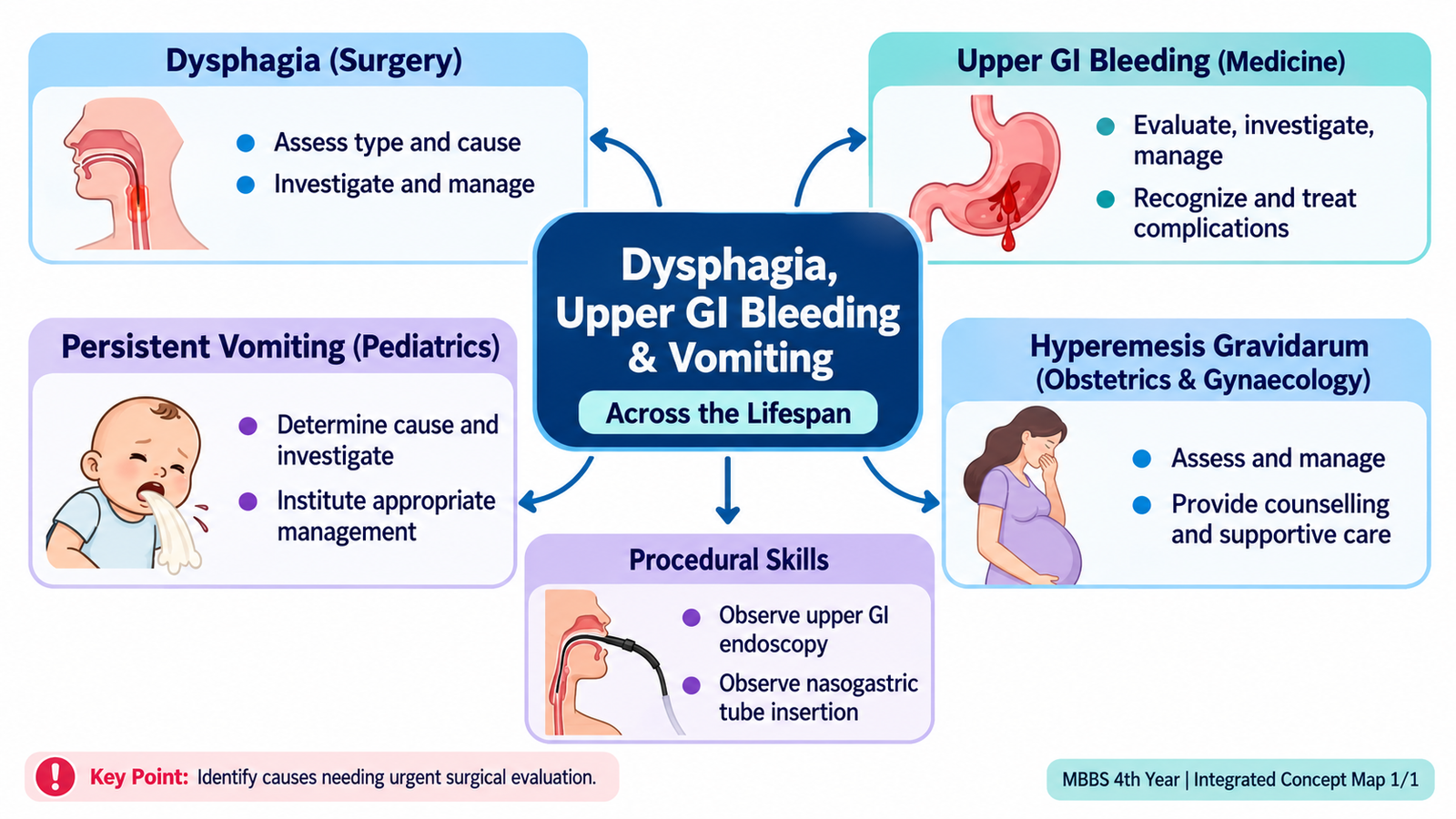
TOPIC 1. Dysphagia, Upper GI Bleeding & Vomiting Across the Lifespan
A patient-centred approach to swallowing difficulty, digestive bleeding, vomiting in infancy and severe vomiting in pregnancy.
Topic Introduction
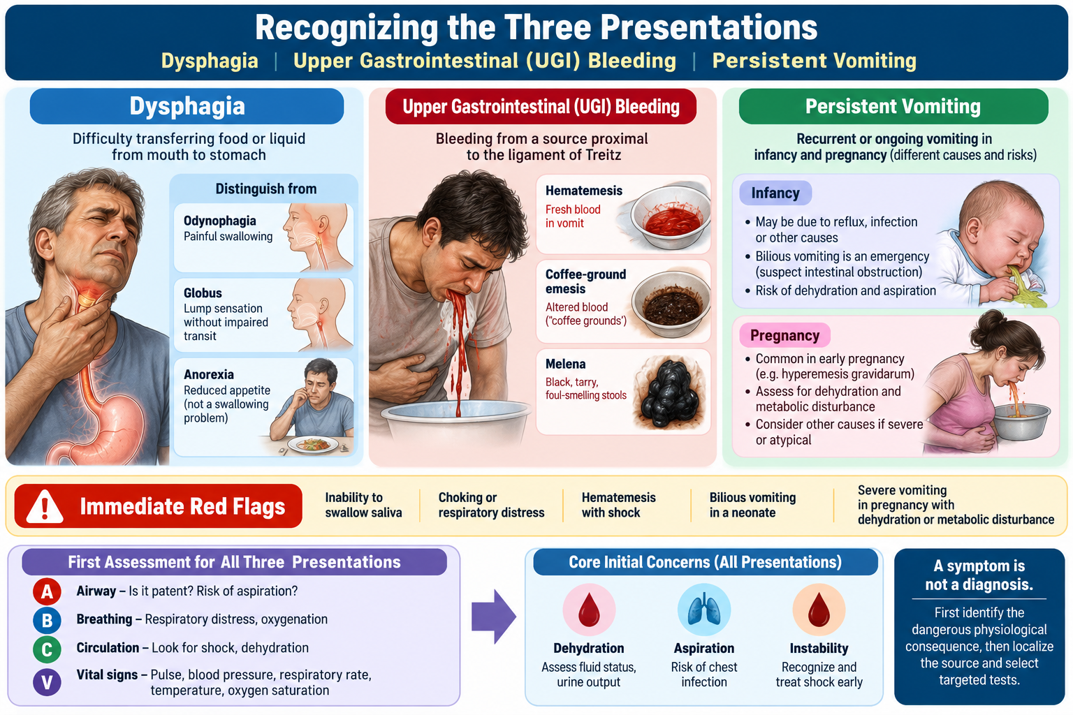
Dysphagia, upper gastrointestinal bleeding and persistent vomiting are common clinical presentations, but their significance changes with age and circumstance. Difficulty swallowing may arise from a structural narrowing or disturbed movement of food through the esophagus. Vomiting blood or passing black stools can indicate dangerous blood loss that requires immediate assessment. Repeated vomiting in a young infant may reflect a surgically correctable obstruction, whereas severe vomiting during pregnancy may cause dehydration and nutritional deficiency. This chapter teaches how symptoms arise, which details in the history and examination differentiate important causes, how investigations guide decisions, and when urgent treatment or referral is required. The same guiding principle applies throughout: recognize danger first, then investigate and treat the cause.
A. Understanding Dysphagia — Mechanisms and Clinical Patterns
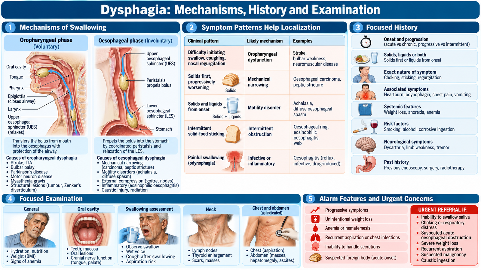
Dysphagia means difficulty transferring food or liquid from the mouth to the stomach. It is different from odynophagia (pain on swallowing) and from a sensation of a lump in the throat without true swallowing difficulty (globus). Swallowing has an oral/pharyngeal phase, requiring coordinated muscles and airway protection, followed by an esophageal phase in which peristalsis moves the bolus through the esophagus. A problem at either stage produces a different history.
Localizing the problem from symptoms
In oropharyngeal dysphagia, initiation of swallowing is difficult. Food or fluid may cause coughing, choking, nasal regurgitation or a wet voice. These findings indicate impaired coordination and possible aspiration into the airway, often in neurological or neuromuscular disease. Esophageal dysphagia produces a sensation that food sticks after swallowing, usually behind the sternum; patients may point to the neck even when disease is lower in the esophagus.
Esophageal dysphagia is further separated into mechanical obstruction and motility disturbance. A narrowing initially obstructs bulky solid food; as it worsens even soft food and liquids may become difficult. A movement disorder, such as achalasia, often interferes with both solids and liquids from the beginning because coordinated propulsion fails.
- Progressively worsening solids-first dysphagia: stricture or esophageal cancer; ask about weight loss and speed of progression.
- Intermittent dysphagia for solids: ring or other intermittent structural narrowing.
- Dysphagia to solids and liquids from onset: consider achalasia or another motility disorder.
- Difficulty initiating a swallow with choking: oropharyngeal dysfunction and aspiration risk.
Why the history matters
Ask when the problem began, whether it is progressive or intermittent, what consistency of food provokes it, and whether the patient has regurgitation, heartburn, chest discomfort, pain on swallowing, respiratory symptoms or unintentional weight loss. Chronic reflux may injure the esophageal lining and lead to a peptic stricture. Dysphagia with weight loss in an older adult is concerning for malignancy, whereas regurgitation of undigested food and night-time coughing suggest retained material in a poorly emptying esophagus.
B. Dysphagia — Focused Assessment, Investigations and Treatment
The first clinical decision is whether swallowing is unsafe or an obstructing lesion is possible. Examine hydration and nutritional status; note weight loss, pallor, cervical or supraclavicular lymph nodes, voice changes and signs of neurological weakness. Observe speech and the ability to handle saliva. Do not challenge a patient with oral fluids when aspiration or complete obstruction is suspected.
Choosing investigations for a clinical reason
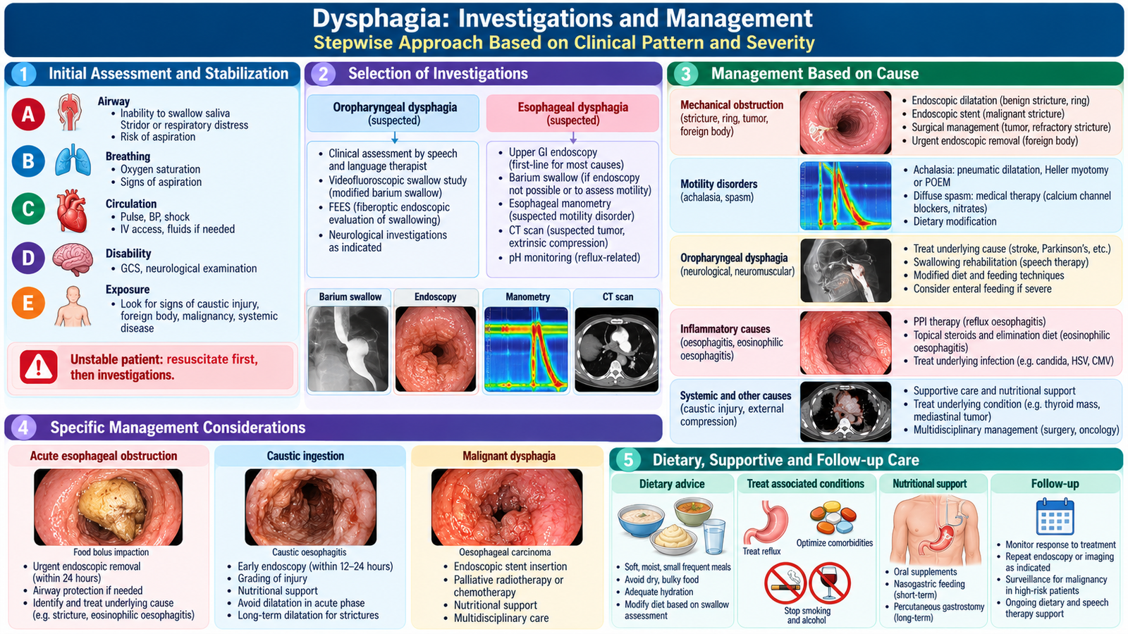
- Upper GI endoscopy (esophagogastroduodenoscopy): directly identifies esophagitis, stricture and tumor, permits biopsy of suspicious lesions, and may allow treatment of a benign narrowing. It is a key initial investigation for persistent or alarm-feature esophageal dysphagia.
- Barium swallow: displays the outline and transit of the esophagus. A smooth tapering “bird-beak” appearance supports achalasia; a focal narrowing suggests a structural lesion requiring further assessment.
- Esophageal manometry: measures pressure and contraction patterns; it confirms motility disorders such as achalasia when mechanical obstruction has been excluded.
- Swallowing assessment: patients with coughing/choking and suspected oropharyngeal dysfunction may need specialist swallowing evaluation and instrumental assessment to establish aspiration risk.
A normal-looking endoscopy does not exclude a motility disorder: a patient with persistent dysphagia to liquids and solids may still require manometry. Conversely, a suspicious stricture must not simply be dilated without adequate evaluation and biopsy where indicated.
Management follows the cause
Treat the underlying condition while protecting airway, hydration and nutrition. Reflux-related inflammation is treated with acid suppression, usually a proton-pump inhibitor (PPI), and appropriate reflux measures; an established benign stricture may need endoscopic dilation and continued treatment of reflux. Achalasia is a disorder of impaired lower-esophageal sphincter relaxation and absent normal peristalsis; specialist treatments reduce sphincter resistance, for example pneumatic dilation or myotomy. Suspected cancer requires endoscopic biopsy, staging and multidisciplinary treatment planning rather than empirical treatment alone. Oropharyngeal dysfunction needs aspiration precautions, feeding modification and management of the neurological or structural cause.
Urgent surgical/endoscopic evaluation is required when a patient cannot swallow saliva, has food bolus impaction, suspected perforation, significant aspiration or rapidly progressive dysphagia with alarming systemic features. Severe chest or neck pain with fever after instrumentation or forceful vomiting raises concern for esophageal perforation; keep the patient nil by mouth and obtain urgent specialist care.
C. Upper Gastrointestinal Bleeding — Causes, Recognition and Severity
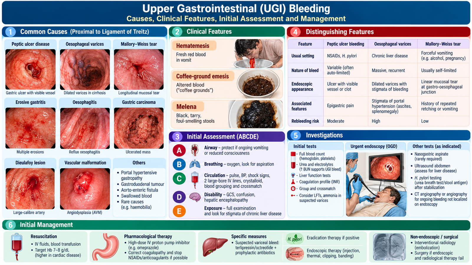
Upper gastrointestinal (UGI) bleeding originates proximal to the ligament of Treitz, principally from the esophagus, stomach or duodenum. Patients may vomit fresh red blood (hematemesis), dark “coffee-ground” material altered by gastric acid, or pass black tarry stools (melena) after blood is digested during intestinal transit. Very brisk UGI bleeding can occasionally produce red rectal bleeding. Visible blood loss does not precisely measure severity; circulation and organ perfusion are more important.
Major causes and the mechanisms behind them
Peptic ulcers may erode submucosal vessels, especially with Helicobacter pylori infection or NSAID exposure; NSAIDs diminish protective gastric prostaglandins, increasing mucosal injury. Erosive gastritis/esophagitis may cause superficial bleeding. Forceful retching can create a Mallory–Weiss mucosal tear at the gastroesophageal junction. Esophageal or gastric varices develop when portal hypertension diverts blood through fragile collateral veins; rupture can produce rapidly life-threatening hemorrhage. Malignancy is another important bleeding source, especially with weight loss or chronic anemia.
Clinical history and examination
Ask about the nature and frequency of blood loss, dizziness, syncope, black stools, abdominal pain, prior ulcer or bleeding, liver disease, alcohol history and drugs such as NSAIDs, aspirin, anticoagulants or antiplatelets. Coffee-ground vomiting suggests blood has remained in the stomach, but does not guarantee low risk. Examine pulse, blood pressure, mental state, peripheral perfusion, capillary refill and urine output. Tachycardia, cool extremities, postural symptoms, confusion and hypotension suggest significant circulatory compromise; hypotension may be a late finding.
Look for epigastric tenderness and signs of chronic liver disease or portal hypertension, including ascites, jaundice and splenomegaly. A patient with cirrhosis and hematemesis must be regarded as having possible variceal bleeding until assessed, but peptic ulcers also occur in such patients.
D. Upper GI Bleeding — Investigations, Resuscitation and Definitive Care
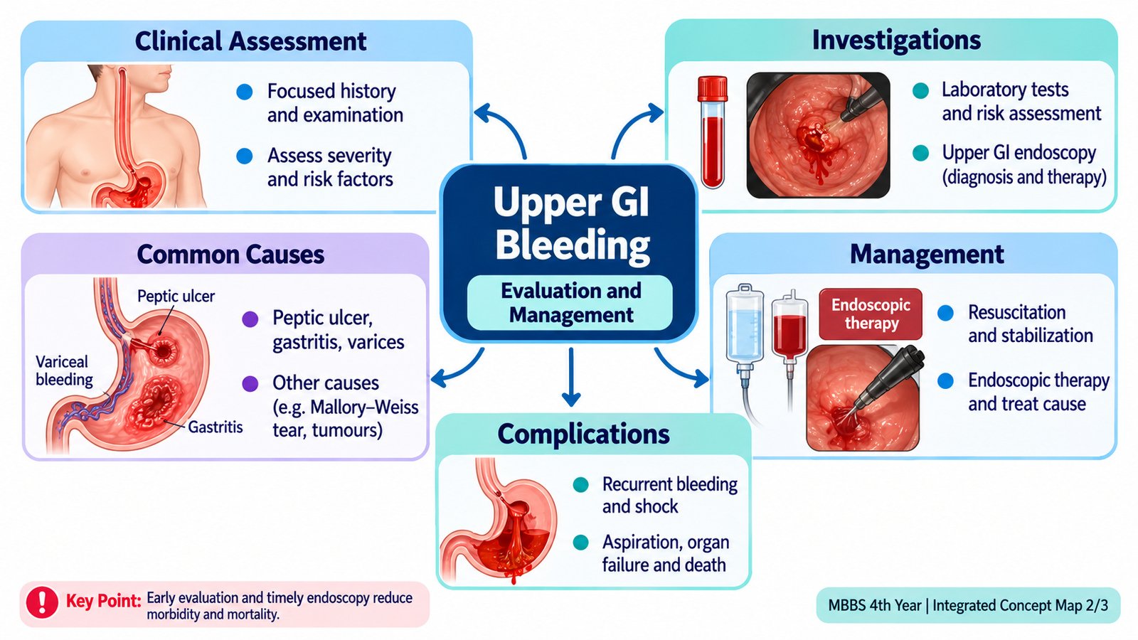
Upper GI bleeding is managed in order of urgency. In an unstable patient, begin resuscitation while gathering the focused history. Alert the senior clinical team and assess airway, breathing and circulation. Patients with major ongoing hematemesis or reduced consciousness may be unable to protect the airway. Keep the patient nil by mouth, obtain reliable intravenous access, send blood tests and arrange blood products when indicated. Balanced clinical assessment matters more than waiting for a laboratory number.
Investigations and what their findings mean
- Complete blood count: identifies anemia and platelet abnormalities. Hemoglobin can be deceptively normal early in acute hemorrhage before fluid shifts occur; serial measurements and clinical condition are essential.
- Blood group and crossmatch: prepares compatible blood for a patient who may need transfusion.
- Urea, creatinine and electrolytes: assess renal perfusion and dehydration. Blood digestion may elevate urea disproportionately in UGI bleeding; kidney function also informs resuscitation.
- Coagulation profile and liver tests: identify coagulopathy or evidence of liver dysfunction, relevant to ongoing bleeding and possible portal hypertension.
- Upper GI endoscopy: identifies the source and permits hemostasis, such as clipping, thermal treatment or injection in nonvariceal bleeding, and band ligation for esophageal varices.
Ongoing bleeding, altered mental status, repeated hematemesis or shock warrants urgent endoscopic and specialist assessment after initial stabilization. A restrictive red-cell transfusion strategy is generally used in many stable UGI-bleeding patients, but active major hemorrhage is managed according to perfusion, blood loss and overall clinical status rather than a single hemoglobin threshold.
Treat nonvariceal and variceal bleeding differently
In nonvariceal upper GI bleeding, perform endoscopy within 24 hours after resuscitation. Bleeding peptic ulcers with high-risk endoscopic findings require hemostasis followed by high-dose PPI therapy, such as pantoprazole or omeprazole, for 72 hours. Test for H. pylori and provide eradication therapy when positive. In suspected variceal hemorrhage, arrange endoscopy within 12 hours after resuscitation and initiate a vasoactive agent such as terlipressin or octreotide, with ceftriaxone prophylaxis in patients with cirrhosis.
When a peptic ulcer is suspected, a PPI reduces acid secretion by blocking the gastric proton pump, supporting clot stability and ulcer healing. High-risk ulcer bleeding commonly requires endoscopic hemostasis followed by PPI therapy. Address H. pylori where present and review ulcerogenic drugs. Rebleeding or failure of endoscopic control requires urgent specialist reassessment; interventional radiology or surgery may be needed.
Suspected variceal bleeding requires urgent resuscitation, early vasoactive treatment such as terlipressin or octreotide to reduce portal pressure, prophylactic antibiotics in patients with cirrhosis, and urgent endoscopic therapy—usually band ligation of esophageal varices. These measures work alongside, not instead of, management of shock. Patients with uncontrolled bleeding need higher-level intervention.
Recognizing complications and monitoring recovery
Monitor vital signs, mental status, urine output, repeated hemoglobin where indicated and any fresh hematemesis or ongoing melena. Important complications are hypovolemic shock, acute kidney injury from inadequate perfusion, aspiration of vomitus, recurrent hemorrhage and death in uncontrolled bleeding. After stabilization, discuss medication safety, avoidance of unnecessary NSAIDs, follow-up for ulcer healing or liver disease, and prompt return if bleeding recurs.
Endoscopy and nasogastric tube — understanding procedural roles
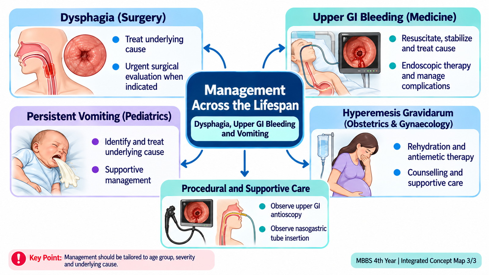
During upper GI endoscopy, a flexible camera is passed through the mouth to inspect the esophagus, stomach and duodenum; channels permit biopsy and therapeutic instruments. Students observing should understand the indication, fasting and consent requirements, sedation/airway monitoring and possible complications such as aspiration, bleeding or perforation. A nasogastric tube passes through the nostril into the stomach for selected decompression or feeding indications. Its placement must be confirmed according to local safety practice before use. Routine insertion or lavage is not required for every patient with UGI bleeding and should not delay resuscitation or endoscopy; potential injury and aspiration must be considered.
E. Persistent Vomiting in Neonates and Infants — Diagnostic Approach
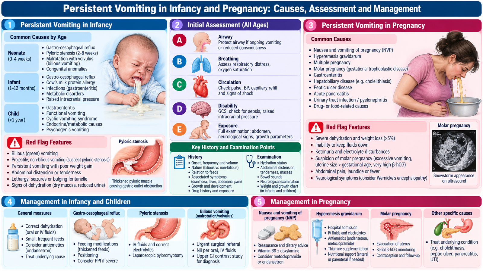
Vomiting is forceful expulsion of gastric contents, whereas regurgitation is effortless passage of milk into the mouth. Small-volume regurgitation in a thriving infant is commonly physiological because the lower esophageal sphincter is immature. Persistent or forceful vomiting needs closer evaluation because infants have limited fluid reserves and may deteriorate quickly. The most helpful early distinctions are age at onset, bilious versus non-bilious vomitus, forcefulness, feeding tolerance, stool passage and weight gain.
Clinical patterns that direct the differential diagnosis
Hypertrophic pyloric stenosis classically develops at approximately 2–8 weeks of age. Thickening of the pyloric muscle obstructs gastric emptying, causing progressive projectile non-bilious vomiting shortly after feeds. The infant may remain hungry, lose weight or become dehydrated. Persistent loss of gastric hydrochloric acid produces a hypochloremic, hypokalemic metabolic alkalosis; these biochemical changes become more marked with delay.
Bilious (green) vomiting suggests obstruction distal to the entry of bile into the duodenum and is a potential surgical emergency. Malrotation with midgut volvulus can threaten bowel blood supply rapidly; lack of distension or an initially well-looking infant does not exclude it. Other intestinal obstruction may cause distension, failure to pass meconium or stool, and feeding intolerance. In contrast, gastroesophageal reflux commonly causes effortless post-feed regurgitation in a growing child, without bilious vomiting or systemic illness.
Other important causes include gastroenteritis or systemic infection; consider infection particularly with fever, lethargy, poor feeding or abnormal examination. A congenital obstruction often presents early, and the timing of first meconium passage helps assess intestinal patency. Ask caregivers about the colour of vomit in plain language: true green bile is more concerning than yellow milk-stained material.
Focused examination
Assess alertness, hydration, capillary refill, weight and growth trajectory, abdominal distension or tenderness, bowel sounds and any palpable mass. An infant with pyloric stenosis may show visible gastric peristalsis or a small olive-like epigastric mass, but absence of that finding does not exclude the diagnosis. Poor perfusion, lethargy, a tender/distended abdomen or bilious vomiting indicates urgent hospital assessment.
F. Pediatric Vomiting — Investigation Interpretation and Management
Select investigations from the clinical pattern rather than ordering every test in every infant. An otherwise thriving infant with simple regurgitation usually needs careful feeding assessment rather than routine imaging. The child with projectile vomiting or bilious emesis needs prompt assessment for a mechanical cause and the degree of dehydration.
- Abdominal ultrasonography: the preferred imaging test for suspected pyloric stenosis. It demonstrates a thickened and elongated pyloric muscle/channel; interpret measurements with age and specialist assessment.
- Electrolytes and acid–base status: in prolonged pyloric vomiting, low chloride with metabolic alkalosis indicates gastric acid loss and guides fluid/electrolyte correction.
- Abdominal radiography: may identify a pattern of obstruction or perforation, but a normal film does not exclude malrotation/volvulus.
- Urgent upper GI contrast study or surgical imaging pathway: may be used to assess suspected malrotation when the infant is stable and the surgical team directs investigation. Imaging must not delay operative assessment in a deteriorating child.
- Infection assessment: urine and other appropriate tests are guided by fever, age, examination and signs of sepsis.
Treatment based on the clinical diagnosis
For physiological reflux, explain its usual natural improvement, avoid overfeeding and support appropriate feeding technique; feeding advice must preserve safe infant sleep practices. Routine acid-suppressing medication is not indicated for uncomplicated regurgitation. Persistent poor growth, feeding refusal or suspected complications needs pediatric review.
In pyloric stenosis, the infant first needs intravenous fluid resuscitation and correction of chloride, potassium and alkalosis; definitive treatment is pyloromyotomy after stabilization. It is unsafe to proceed directly to anesthesia while significant metabolic abnormalities remain. In bilious vomiting/suspected bowel obstruction, keep the infant nil by mouth, institute intravenous fluid support and gastric decompression when indicated, and obtain urgent pediatric surgical assessment. A possible volvulus requires immediate escalation because delay risks bowel ischemia and necrosis.
Monitor hydration, urine output, weight, feeding tolerance and electrolyte correction. Parents need simple explanations of the suspected cause, why the infant cannot feed temporarily when obstruction is suspected, and warning symptoms requiring immediate reassessment—especially green vomiting, lethargy, abdominal distension or fewer wet nappies.
G. Hyperemesis Gravidarum — Clinical Assessment and Diagnosis
Nausea and vomiting are common in early pregnancy, but hyperemesis gravidarum (HG) refers to severe pregnancy-associated nausea and vomiting that prevents normal eating and drinking and substantially impairs daily function. It may cause dehydration, weight loss and electrolyte abnormalities. HG is a clinical diagnosis after assessing severity and excluding other important causes. Symptoms usually develop in early pregnancy, when rising pregnancy-related hormonal signals contribute to nausea and vomiting.
History and examination: differentiate HG from other illness
Ask about gestational age, duration and frequency of vomiting, ability to retain fluids and medicines, urine output, weight change, previous episodes, abdominal pain, fever, urinary symptoms and any vaginal bleeding. Examine pulse, blood pressure, mucous membranes, postural symptoms, hydration, weight and abdominal findings. Persistent marked pain, fever, focal abdominal tenderness or other atypical symptoms should prompt consideration of gastrointestinal, urinary, metabolic or obstetric causes rather than attributing all vomiting to pregnancy.
Prolonged vomiting and poor intake produce volume depletion, electrolyte loss and sometimes nutritional deficiency. Severe thiamine deficiency can cause Wernicke encephalopathy, presenting with confusion, eye movement abnormalities or ataxia; the full classical triad may be absent. In women with significant prolonged vomiting and nutritional depletion, provide thiamine before dextrose-containing fluids.
Investigations and interpretation
- Urinalysis: assess for urinary infection when indicated. Ketonuria can occur with starvation but should not be used alone to grade HG severity or decide admission.
- Urea, electrolytes and creatinine: reveal dehydration, low potassium or sodium disturbances and impaired renal function; these results guide replacement and monitoring.
- Blood count: may show hemoconcentration or suggest another illness; interpret alongside the examination.
- Liver and thyroid tests: selected when symptoms are significant or atypical; modest abnormalities can accompany HG but pronounced or persistent derangement requires further evaluation.
- Obstetric ultrasonography: confirms pregnancy status and assesses multiple gestation or gestational trophoblastic disease where clinically appropriate.
H. Hyperemesis Gravidarum — Treatment, Monitoring and Counselling
Management aims to restore hydration, enable adequate nutrition, relieve symptoms and prevent maternal complications while maintaining safe care in pregnancy. The treatment setting depends on ability to drink, clinical dehydration, electrolyte changes, weight loss, comorbidity and response to initial measures. Mild or moderate cases may be supported in the community or ambulatory care; severe cases often need hospital treatment.
Stepwise supportive and drug treatment
Offer small, frequent meals and tolerated fluids without blaming the woman for inability to eat. Pregnancy-compatible antiemetics are the basis of symptom control; commonly used choices include doxylamine–pyridoxine combinations where available and agents such as cyclizine, promethazine, prochlorperazine, metoclopramide or ondansetron according to clinical circumstances and local guidance. Choice depends on prior response, adverse effects and pregnancy counselling. Metoclopramide may cause extrapyramidal symptoms and is generally used with attention to duration; ondansetron counselling should consider available pregnancy-safety information and potential QT effects in susceptible patients.
A patient unable to retain oral treatment may require parenteral antiemetics and intravenous isotonic fluids with potassium replacement when indicated. Reassess electrolytes rather than replacing potassium blindly. In prolonged poor intake, give thiamine before dextrose to reduce the risk of Wernicke encephalopathy. Consider nutritional support and multidisciplinary input when oral intake remains inadequate despite treatment. Venous thromboembolism risk and the need for prevention should be considered in hospitalized, dehydrated patients according to individual risk.
Follow-up and patient-centred counselling
Track symptoms, oral intake, weight, fluid balance and relevant electrolytes. Explain that HG is a recognized medical condition and that effective treatment is available; many pregnancies progress normally after symptoms improve. Ask sensitively about psychological distress, loss of function, family support and practical difficulties. Provide instructions for urgent reassessment if the woman cannot keep fluids down, passes very little urine, faints, develops confusion, has severe pain or develops vaginal bleeding. Arrange timely follow-up and reassess if symptoms persist beyond the expected early-pregnancy period.
Important Comparison — Vomiting Patterns in Early Life and Pregnancy
| Presentation | Most useful clue | Clinical implication |
|---|---|---|
| Infant reflux | Effortless milk regurgitation; thriving infant | Usually feeding support, not routine imaging |
| Pyloric stenosis | Progressive projectile non-bilious vomiting | Ultrasound; correct alkalosis before surgery |
| Intestinal obstruction/volvulus | Green bilious vomiting ± distension | Urgent pediatric surgery assessment |
| Hyperemesis gravidarum | Pregnancy with inability to maintain intake | Assess hydration/electrolytes; antiemetics and thiamine when indicated |
AIM High-Yield Review
- ⭐ Dysphagia with solids first suggests mechanical obstruction; difficulty with both solids and liquids from onset raises concern for impaired esophageal motility.
- Oropharyngeal dysphagia with choking and wet voice signals possible aspiration.
- ⭐ Progressive dysphagia with weight loss requires urgent evaluation for malignancy.
- Endoscopy identifies structural lesions and permits biopsy; manometry evaluates motility when obstruction has been excluded.
- Hematemesis and melena indicate possible upper GI bleeding; severity is determined by perfusion and stability, not stool colour alone.
- ⭐ Early hemoglobin can underestimate acute blood loss; serial examination and circulation are essential.
- Peptic ulcer bleeding requires stabilization, appropriate endoscopic management and acid suppression; variceal bleeding additionally requires portal-pressure-directed treatment and antibiotics in cirrhosis.
- Upper GI endoscopy can diagnose and treat bleeding; routine nasogastric lavage is not required for all bleeds.
- ⭐ Bilious green vomiting in an infant is intestinal obstruction until urgently assessed; exclude malrotation with volvulus.
- Pyloric stenosis causes projectile non-bilious vomiting and may produce hypochloremic metabolic alkalosis.
- Correct fluid and electrolyte derangements before pyloromyotomy.
- Uncomplicated infant regurgitation with normal growth usually needs feeding advice rather than drugs or imaging.
- HG severity depends on oral intake, functional impact, hydration and metabolic disturbance—not ketonuria alone.
- ⭐ Give thiamine before dextrose in prolonged severe pregnancy vomiting with poor nutritional intake.
- Clinical deterioration, aspiration, shock, bilious vomiting or neurological symptoms warrant prompt escalation.
AIM Video Learning Library
Final Year MBBS | GIT Module | Topic 1
Dysphagia, Upper GI Bleeding & Vomiting Across the Lifespan
1. Dysphagia — Ninja Nerd
Clinical assessment, important causes and diagnostic approach.
2. Upper GI Bleeding — Ninja Nerd
Causes, clinical assessment, resuscitation, investigations and management.
3. Infant Vomiting — Pyloric Stenosis
Clinical presentation of persistent projectile vomiting in infancy.
4. Hyperemesis Gravidarum — Ninja Nerd Nursing
Severe vomiting in pregnancy, diagnosis, complications and supportive treatment.