
Topic 4. Acute Abdomen, Appendicitis, Intestinal Obstruction, Peritonitis & Hernias
Integrated surgery and family medicine learning material
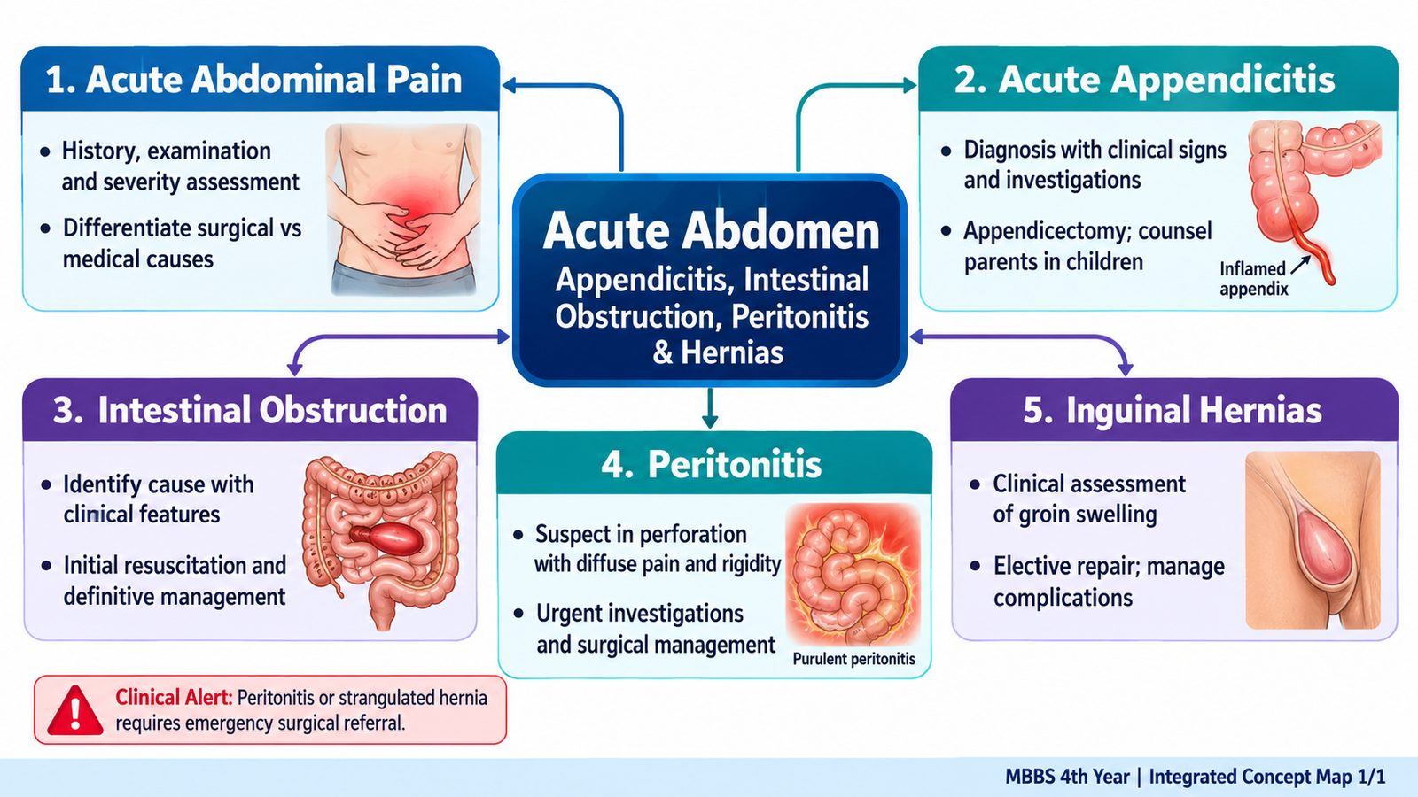
Abdominal pain becomes clinically meaningful when the history, examination and underlying disease mechanism are interpreted together. This chapter covers common surgical causes of the acute abdomen and their first-contact assessment, with particular attention to appendicitis in children, bowel obstruction, perforation, peritonitis and groin hernias. Emphasis is on safe clinical reasoning, investigation selection, emergency recognition and undergraduate-level treatment.
A. Clinical Approach to Acute and Chronic Abdominal Pain
Understanding the presentation
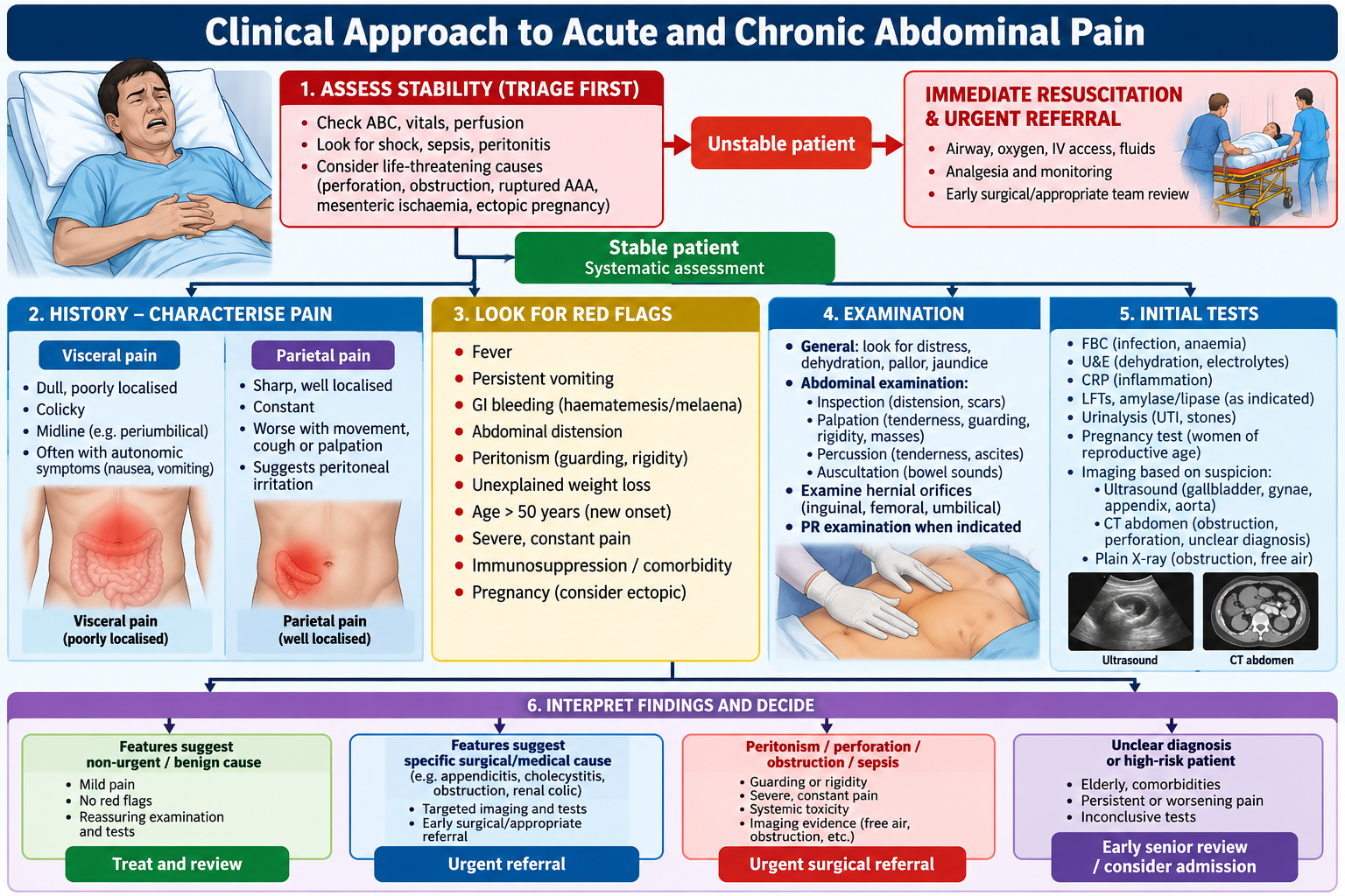
Acute abdominal pain develops over hours to days and may represent a time-critical surgical problem. Chronic or recurrent pain lasts weeks to months and requires assessment for functional, inflammatory, obstructive, urinary, metabolic and gynecological causes. The first decision is whether the patient is unstable or has peritonitis, ischemia, perforation or obstruction.
Visceral pain is dull, poorly localized and often midline because autonomic afferents convey distension or ischemia. Parietal pain is sharp, localized and aggravated by movement when the somatic innervation of parietal peritoneum is irritated. Referred pain is perceived at a site sharing spinal segments with the affected organ. Early appendicitis can therefore cause periumbilical pain before inflammation reaches the right iliac fossa.
History and examination
Clarify onset, site, migration, character, severity, progression, radiation, vomiting, bowel motions, flatus, fever, urinary symptoms, menstrual and pregnancy history, prior operations, hernias, medications and comorbidities. Sudden maximal pain raises concern for perforation, vascular catastrophe or torsion; colicky pain suggests obstruction; progressive localized pain suggests inflammation. Ask about weight loss, rectal bleeding and altered bowel habit in chronic pain.
Assess airway, breathing, circulation, mental status, pulse, blood pressure, temperature, respiratory rate, hydration and urine output. Inspect for distension, scars and visible hernias; auscultate bowel sounds without delaying urgent care; gently palpate for focal tenderness, guarding, rigidity and masses. Percussion tenderness or involuntary guarding suggests peritoneal irritation. Examine the groins and perform pelvic, rectal or testicular examination only when clinically indicated and with consent.
Clinical reasoning and first tests
Upper abdominal pain suggests biliary, gastric or pancreatic disease; right iliac fossa pain suggests appendicitis but may reflect urinary or gynecological pathology; diffuse pain with rigidity suggests generalized peritonitis. Investigations are selected by risk: CBC, electrolytes, renal function, CRP, urinalysis, pregnancy test where relevant, lipase for suspected pancreatitis, and lactate/blood gas in severe illness. Ultrasound is useful for biliary, pelvic and selected pediatric causes; CT abdomen/pelvis is valuable in uncertain adult acute abdomen, obstruction and suspected perforation when stable. Do not delay resuscitation or surgical review for imaging in a critically ill patient.
Primary care disposition: stable, mild, nonprogressive symptoms may be managed with targeted testing, hydration, analgesia, safety-netting and planned review. Urgent hospital referral is needed for shock, rigid abdomen, persistent severe or worsening pain, bilious vomiting, distension with obstipation, GI bleeding, suspected ectopic pregnancy, painful irreducible hernia, sepsis or inability to maintain hydration. Older, pregnant and immunosuppressed patients may have subtle signs.
B. Acute Appendicitis: Mechanism, Presentation and Diagnosis
Why appendicitis develops
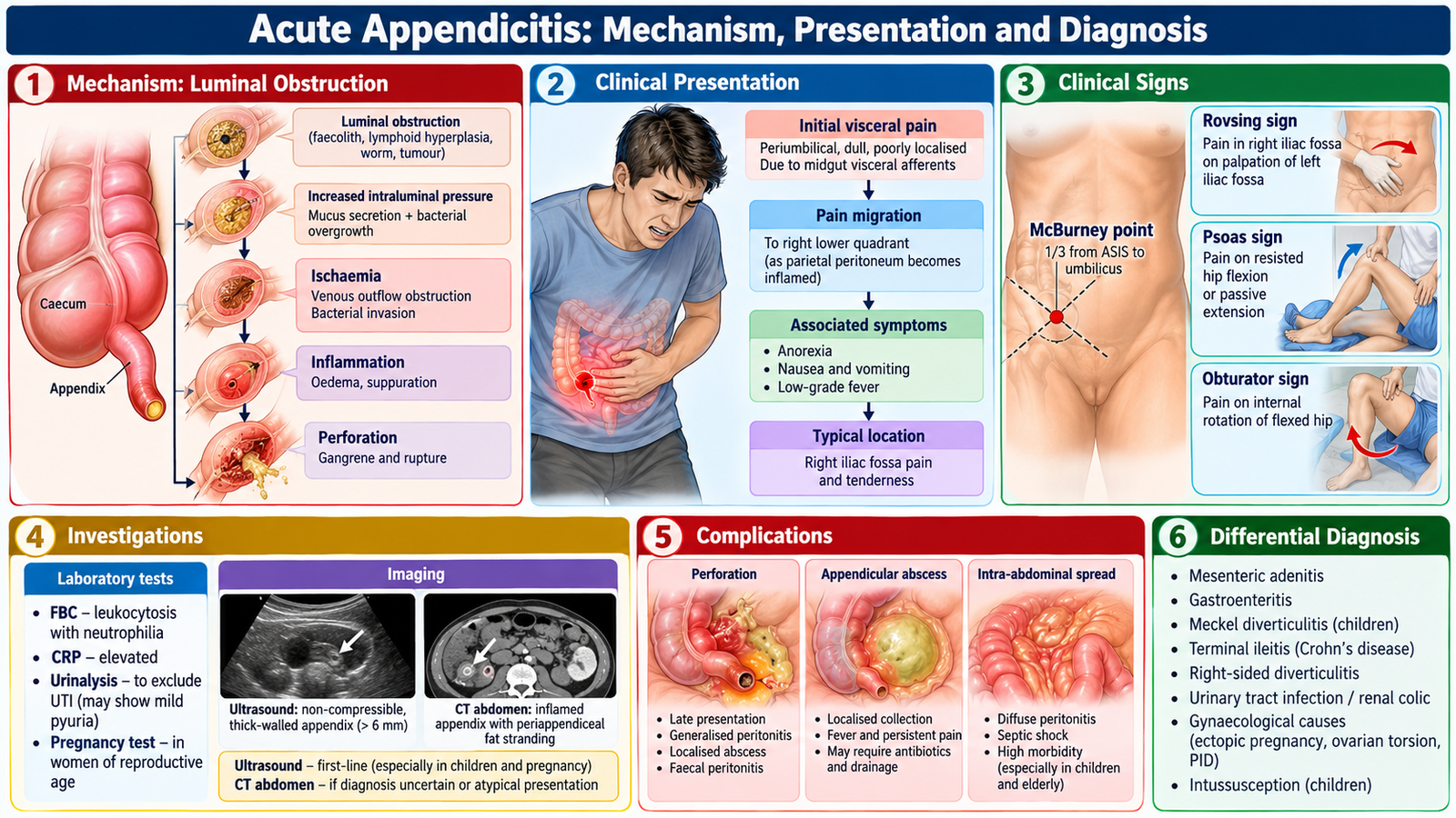
Obstruction of the appendiceal lumen by a fecalith or lymphoid hyperplasia increases intraluminal pressure, impairs venous drainage and promotes bacterial proliferation. Progressive ischemia can lead to gangrene, perforation, abscess or diffuse peritonitis. In children, lymphoid hyperplasia is an important contributor and perforation can occur earlier because diagnosis is more difficult.
Typical and atypical findings
Classically, vague periumbilical pain migrates to the right lower quadrant, followed by anorexia, nausea and low-grade fever. Focal tenderness at McBurney point, cough/percussion tenderness and localized guarding support the diagnosis. Rovsing sign is right-sided pain with left lower quadrant palpation; psoas sign may occur with a retrocecal appendix; obturator sign may occur with a pelvic appendix. None is sufficiently sensitive to rule out disease when absent. Diarrhea or urinary frequency can occur with a pelvic appendix; young children may present with fever, vomiting, lethargy or diffuse tenderness rather than a classic sequence.
Diagnosis and differentials
Appendicitis is a clinical diagnosis supported by investigations. Neutrophilic leukocytosis and elevated CRP are supportive but neither confirms nor excludes it. Clinical risk scores such as Alvarado can structure assessment but should not replace judgment. Ultrasound is generally preferred first in children and pregnancy; CT is often appropriate for equivocal nonpregnant adults. MRI may be used during pregnancy if ultrasound is nondiagnostic and available. Consider mesenteric adenitis, gastroenteritis, constipation, UTI, ureteric stone, ovarian torsion, ectopic pregnancy and inflammatory bowel disease according to context.
Management and parental counselling
Keep the patient nil by mouth if surgery is anticipated; provide IV fluids when needed, analgesia, antiemetics, antibiotics according to surgical protocol and prompt surgical assessment. Appendectomy, commonly laparoscopic, is standard definitive treatment for many patients. Selected uncomplicated cases may be considered for antibiotic-first management after specialist discussion, but recurrence or later surgery is possible. Perforation or abscess requires antibiotics and source control, sometimes drainage and staged surgery.
Explain to parents that the diagnosis may require repeated examination or imaging, that untreated inflammation may perforate, and that treatment balances urgency with the child’s condition. Discuss anesthesia, infection, bleeding, recovery, wound care and return precautions. A child with worsening pain, persistent fever, repeated vomiting or reduced responsiveness needs urgent reassessment.
C. Intestinal Obstruction: Causes, Mechanisms and Clinical Recognition
Classification and causes
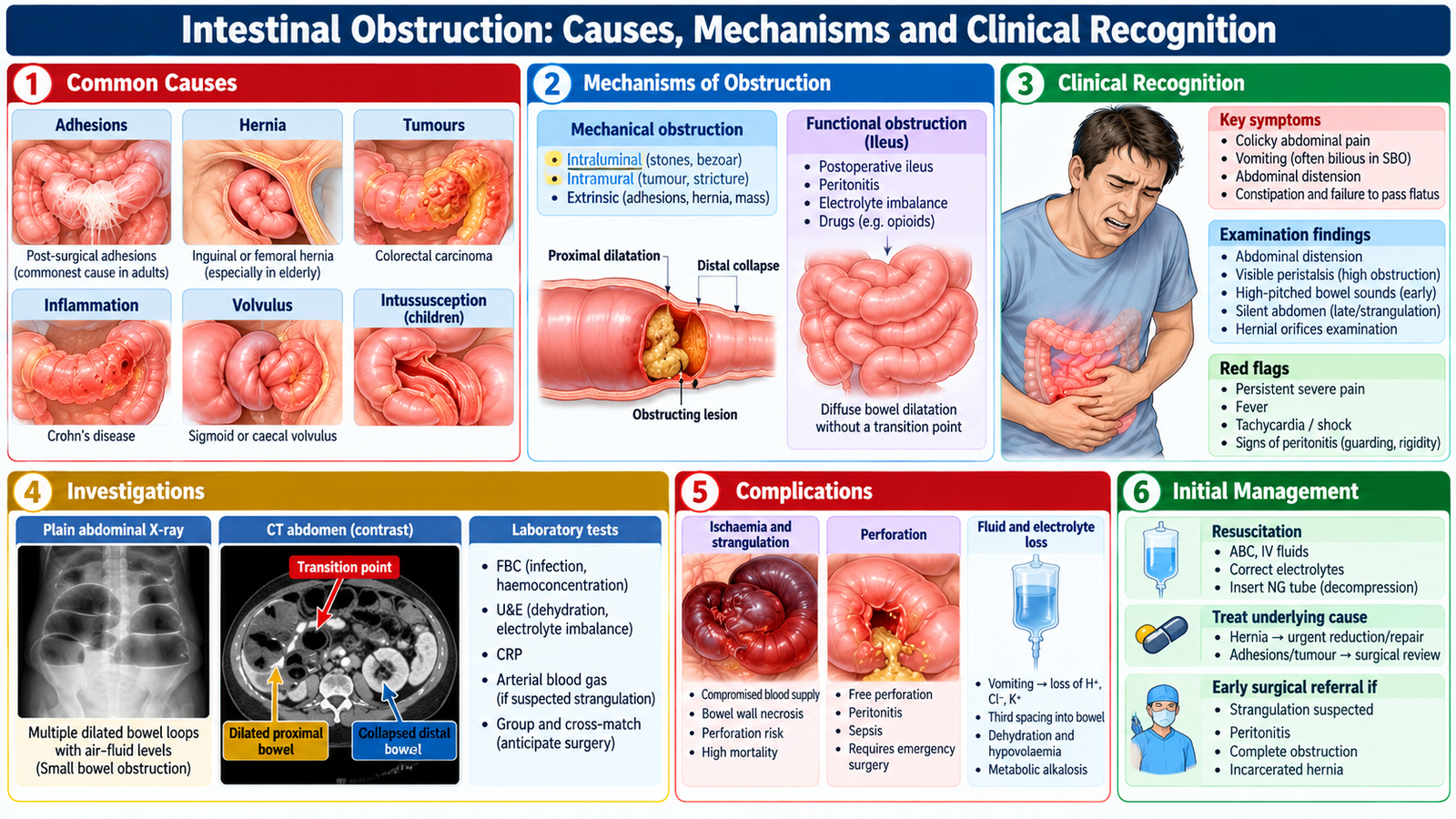
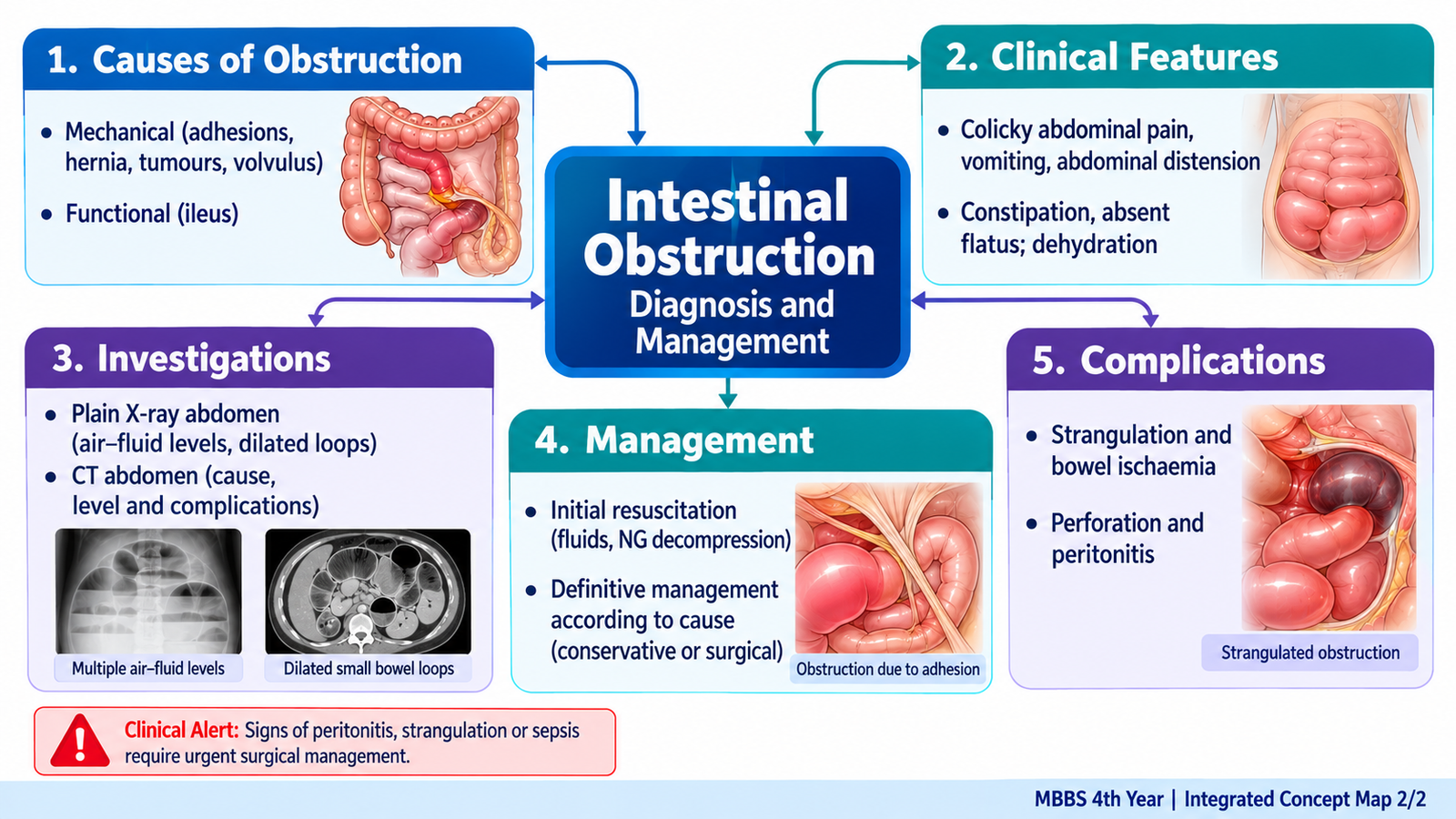
Mechanical obstruction results from a physical barrier; small-bowel causes include postoperative adhesions, incarcerated hernia, tumors and strictures. Large-bowel causes include colorectal cancer and volvulus. Functional obstruction (ileus) occurs when intestinal propulsion fails without a mechanical transition point, commonly after surgery, peritonitis, severe illness or electrolyte disturbance. Obstruction may be partial or complete, simple or strangulating.
Proximal obstruction tends to cause early, prominent vomiting and less distension; distal obstruction tends to cause marked distension, later vomiting and constipation or obstipation. Colicky pain reflects peristalsis against an obstruction. Persistent severe pain, fever, tachycardia, focal peritonism or shock raises concern for strangulation and ischemia. Passage of some stool early does not exclude obstruction.
Pathophysiology and complications
Proximal bowel dilates as gas and fluid accumulate, causing vomiting, sequestration of fluid, dehydration and electrolyte loss. Increased bowel-wall pressure can compromise venous return, then arterial perfusion, producing ischemia, necrosis, perforation and sepsis. A closed-loop obstruction can rapidly progress to strangulation.
Investigations
Check CBC, electrolytes, renal function and lactate where ischemia is suspected; laboratory findings assess severity rather than prove the cause. Erect/supine abdominal radiographs may show dilated loops and air–fluid levels but cannot reliably exclude obstruction. CT with IV contrast, when appropriate, identifies the transition point, cause and complications such as ischemia or perforation. Consider ultrasound in selected children and pregnancy.
Initial management
Resuscitate with isotonic IV fluids, correct electrolyte abnormalities, keep nil by mouth, provide analgesia and antiemetics, and monitor urine output. Nasogastric decompression is useful in significant vomiting or distension. Arrange early surgical review. Selected adhesive small-bowel obstruction without ischemia/peritonitis may resolve with supervised nonoperative care; complete obstruction, strangulation, peritonitis, perforation or failed conservative management requires urgent intervention. Antibiotics are indicated when bowel compromise, perforation or infection is suspected.
D. Intestinal Perforation and Peritonitis: Emergency Recognition and Treatment
How perforation causes peritonitis
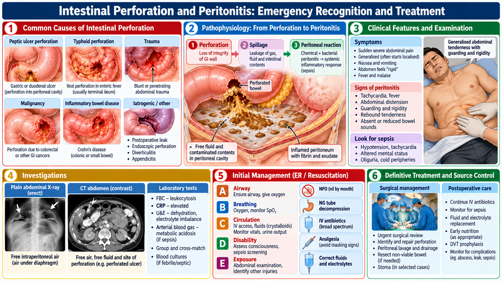
Perforation releases gastric, intestinal or infected contents into the peritoneal cavity. Chemical irritation is followed by bacterial contamination and an inflammatory response. Causes include perforated peptic ulcer, complicated appendicitis, diverticular disease, bowel ischemia, trauma and obstruction with necrosis. Peritonitis may be localized or generalized; systemic inflammation can progress to sepsis and shock.
Clinical features and differentiation
Patients may develop sudden severe pain, worsening pain with movement, abdominal tenderness, involuntary guarding, board-like rigidity, fever, tachycardia and reduced bowel sounds. Early perforation may produce abrupt pain; elderly or immunosuppressed patients may lack dramatic rigidity. A patient with free intraperitoneal gas and peritonism requires immediate surgical evaluation.
Investigation and immediate care
Obtain vital signs, IV access, CBC, renal function, electrolytes, lactate, blood cultures when septic and group/crossmatch as indicated. An erect chest radiograph can reveal subdiaphragmatic free air, but a normal film does not exclude perforation. CT is more sensitive in stable patients and may identify the source. Start rapid IV crystalloid resuscitation, nil by mouth, analgesia, broad-spectrum IV antibiotics covering enteric organisms, monitoring and urgent surgical consultation. Definitive care requires source control by repair/resection, drainage or another appropriate procedure. Delayed source control increases the risk of organ failure and death.
Post-treatment priorities
Monitor perfusion, urine output, fever, pain, bowel function, wound infection and intra-abdominal abscess. Nutritional support, mobilization and reassessment of antibiotics are part of recovery. Explain that sepsis and persistent collections may require further treatment.
E. Inguinal Hernias: Applied Anatomy, Examination and Diagnosis
Relevant anatomy
An inguinal hernia is protrusion of abdominal contents through a weakness in the groin. The deep inguinal ring lies lateral to the inferior epigastric vessels. Indirect inguinal hernias enter through the deep ring lateral to these vessels and may follow a patent processus vaginalis into the scrotum. Direct inguinal hernias protrude through the posterior wall in Hesselbach triangle, medial to the inferior epigastric vessels. Femoral hernias lie below the inguinal ligament and are particularly prone to incarceration and strangulation.
History and examination
Ask about a groin bulge, discomfort on coughing or lifting, reducibility, duration, prior repair, vomiting and bowel symptoms. Examine standing and supine, with consent and appropriate privacy. Inspect and palpate the groin and scrotum; ask the patient to cough to elicit an expansile impulse. Determine whether the swelling is reducible and whether the examiner can get above a scrotal swelling. Examine both groins and consider femoral, lymph-node, saphena varix, hydrocele and testicular causes.
Transillumination: a fluid-filled hydrocele may transmit light, while a bowel-containing hernia generally does not. This is supportive rather than definitive; do not attempt forceful reduction or painful examination of a tender irreducible mass. Bedside distinction between direct and indirect hernia is not consistently reliable, and management should not depend solely on cough impulse or deep-ring occlusion tests.
Investigations and treatment
Many inguinal hernias are diagnosed clinically. Ultrasound helps when examination is equivocal; CT may be appropriate for complex or occult pathology. Symptomatic hernias generally merit elective repair, usually mesh-based in adults when suitable. Watchful waiting can be considered in selected minimally symptomatic adult men after discussion of risks and follow-up; women with groin hernias require timely assessment because femoral hernias may be overlooked. Pediatric inguinal hernias usually need surgical repair because incarceration risk is significant.
F. Complicated Hernias: Incarceration, Obstruction and Strangulation
Recognizing the progression
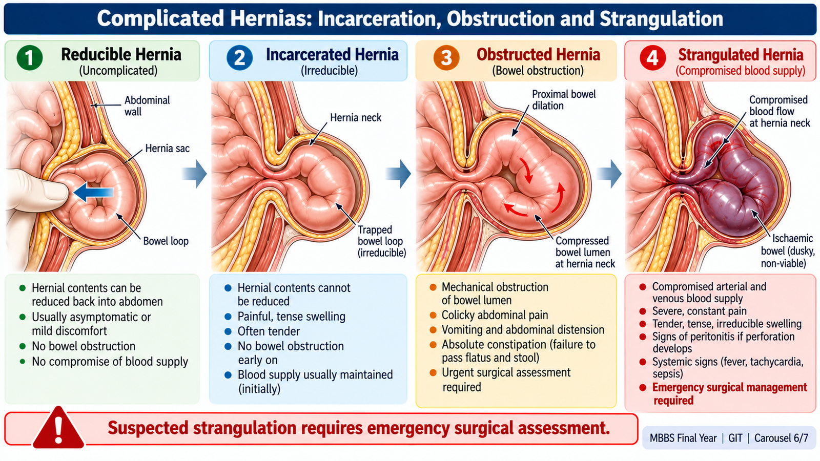
A reducible hernia returns into the abdomen spontaneously or with gentle pressure. An incarcerated hernia cannot be reduced; it may cause intestinal obstruction. A strangulated hernia has compromised blood supply and risks ischemia, necrosis and perforation. A tender irreducible swelling with persistent pain, vomiting, distension, skin changes, fever or tachycardia is an emergency until proven otherwise.
Clinical decision-making
Assess circulation, hydration and peritoneal signs; establish IV access, provide fluids and analgesia, keep nil by mouth and seek urgent surgical review. Do not force reduction when strangulation is suspected. Antibiotics are appropriate when ischemia, perforation or sepsis is suspected. Emergency surgery may require reduction, assessment of bowel viability and resection of nonviable intestine, followed by repair tailored to contamination and patient condition.
Prevention and counselling
Explain that progressive pain, a previously reducible lump becoming irreducible, vomiting, abdominal distension or discoloration over the swelling requires immediate hospital attendance. Elective repair prevents many emergency presentations in appropriately selected symptomatic patients. Address smoking, cough, constipation and heavy-straining concerns while avoiding false reassurance that lifestyle measures can close an established hernia.
G. Integrated Primary-Care Decisions, Follow-Up and Patient Safety
Putting the conditions together
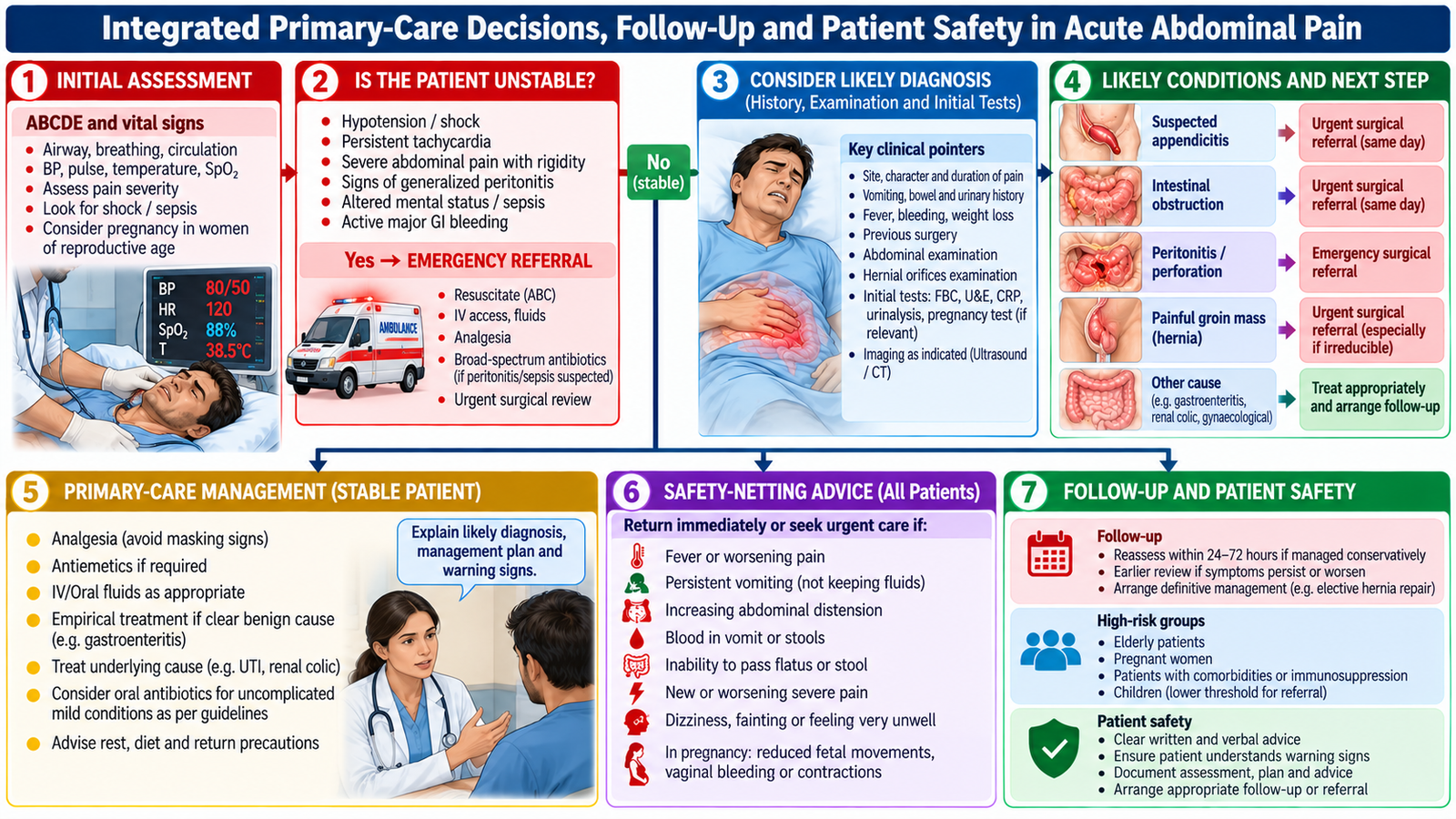
The same complaint—abdominal pain—can arise from inflammation, luminal obstruction, perforation or a groin defect. Pattern recognition directs urgency: migrating pain and focal right iliac fossa tenderness favor appendicitis; colic with vomiting and distension suggests obstruction; generalized rigidity suggests peritonitis; a painful irreducible groin mass suggests a complicated hernia. Reassess when findings evolve, rather than relying on a single early examination.
Safe first-contact management
In primary care, record vital signs and red flags, give appropriate analgesia, assess hydration, and use pregnancy testing when applicable. Stable patients without alarming features may receive targeted investigations, symptom control, explicit return precautions and a time-defined review. Persistent unexplained pain, weight loss, anemia, rectal bleeding or altered bowel habit warrants further evaluation. Severe pain with shock, sepsis, peritoneal signs, bowel obstruction or a tender irreducible hernia requires emergency transfer and early communication with the surgical team.
Communication and continuity
Explain the working diagnosis and remaining uncertainty in plain language. Advise against eating or drinking when emergency surgery is likely. During referral, document onset, progression, examination, observations, administered fluids/medications, allergies and relevant test results. For discharged patients, give specific instructions to return for worsening pain, fever, persistent vomiting, fainting, inability to pass stool or flatus, or a new painful groin lump. Arrange follow-up after appendectomy, obstruction or hernia repair to assess recovery and complications.
AIM Clinical Bridge
Case pattern 1: An 11-year-old with migrating abdominal pain, anorexia and right iliac fossa tenderness needs urgent assessment for appendicitis; a normal early blood count does not safely exclude it.
Case pattern 2: An older patient with previous abdominal surgery, colicky pain, vomiting and distension needs assessment for adhesive bowel obstruction and fluid depletion.
Case pattern 3: A patient with a painful irreducible groin lump, vomiting and tachycardia needs emergency surgical review for strangulation.
Important Comparison: Common Surgical Pain Patterns
| Condition | Key pattern | Priority |
|---|---|---|
| Appendicitis | Migrating pain → focal RIF tenderness | Surgical evaluation; ultrasound often first in children |
| Obstruction | Colic + vomiting + distension/obstipation | Fluids, decompression when indicated, surgical review |
| Perforation/peritonitis | Severe pain + guarding/rigidity ± shock | Antibiotics, resuscitation, urgent source control |
| Strangulated hernia | Painful irreducible groin swelling ± obstruction | Emergency surgery assessment |
AIM High-Yield Review
- Visceral pain is poorly localized; parietal irritation produces localized tenderness and guarding.
- Appendicitis may progress from luminal obstruction to perforation; children can present atypically.
- No single clinical sign or inflammatory marker excludes appendicitis.
- Distension, vomiting and obstipation suggest obstruction; persistent severe pain suggests ischemia.
- Resuscitation and surgical assessment take priority over delayed imaging in unstable patients.
- Perforation with peritonitis requires antibiotics and timely source control.
- Indirect inguinal hernias lie lateral and direct hernias medial to inferior epigastric vessels.
- Transillumination supports hydrocele rather than bowel-containing hernia.
- A tender irreducible hernia may be strangulated: avoid forceful reduction.
- Safe primary-care management requires red-flag recognition, referral and clear return precautions.
🎥 AIM Clinical Video Learning
Topic 4: Acute Abdomen, Appendicitis & Peritonitis
Supplementary clinical teaching video covering the approach to acute abdominal emergencies, with emphasis on appendicitis and peritonitis.
Source: Lecturio Medical | Supplementary video for AIM Learning Material.