
TOPIC 7. Lower GI Bleeding, Inflammatory Bowel Disease & Colorectal Cancer
From bleeding per rectum and chronic bowel symptoms to interpretation of investigations, severity-based treatment and colorectal malignancy planning.
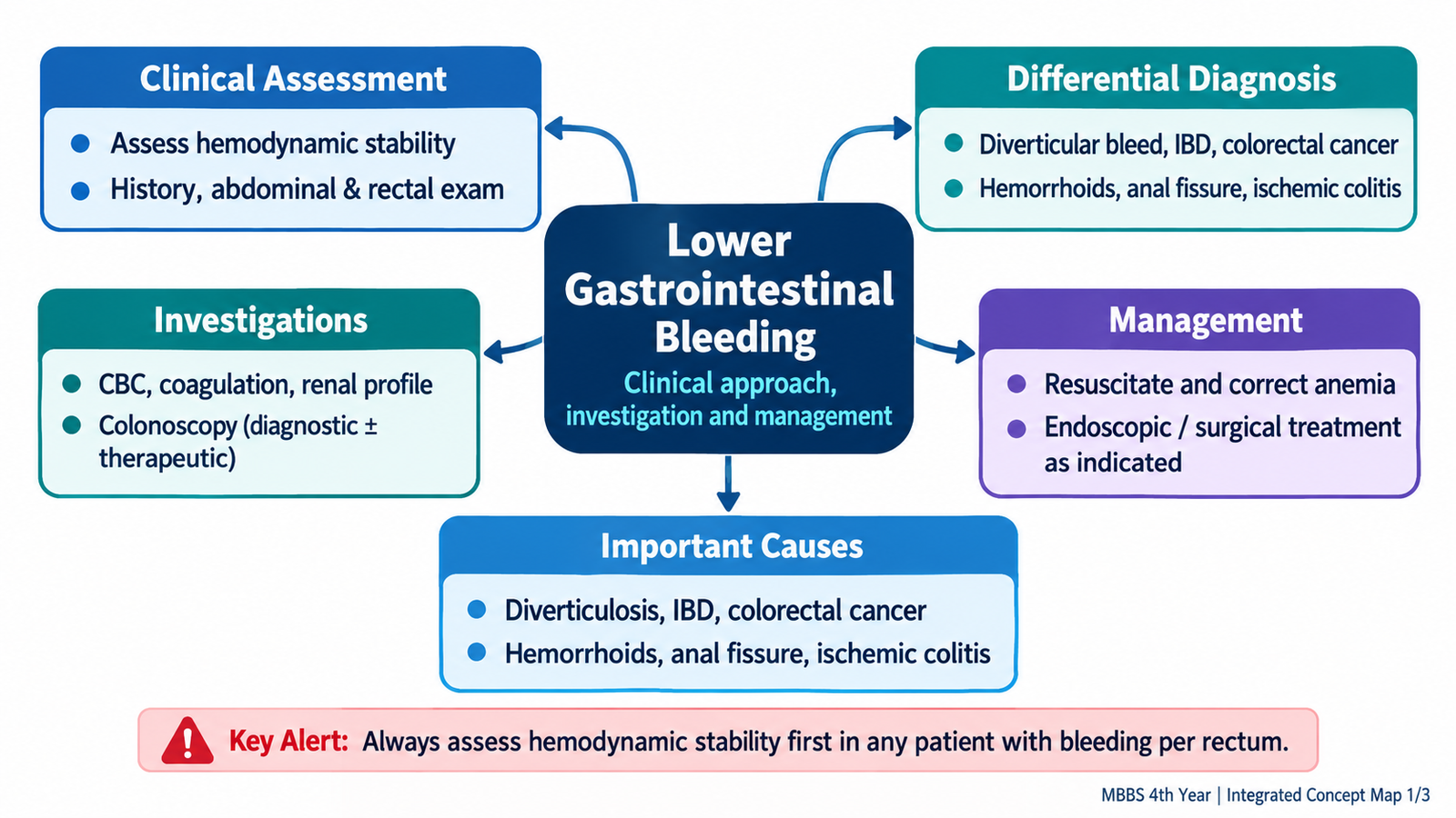
Lower gastrointestinal (GI) bleeding is blood loss arising most commonly from the colon, rectum or anus. It may represent a minor local disorder or major hemorrhage requiring emergency resuscitation. Two important causes of chronic bowel symptoms are inflammatory bowel disease (IBD)—ulcerative colitis and Crohn’s disease—and colorectal cancer. These conditions overlap in symptoms but differ in their mechanisms, investigation findings and management. The central clinical task is to assess safety first, recognize the likely source, obtain an appropriate diagnosis and choose treatment according to disease severity. This chapter explains common treatment drugs, when to escalate care and why long-term surveillance matters.
A. Lower Gastrointestinal Bleeding — Presentation and Clinical Assessment
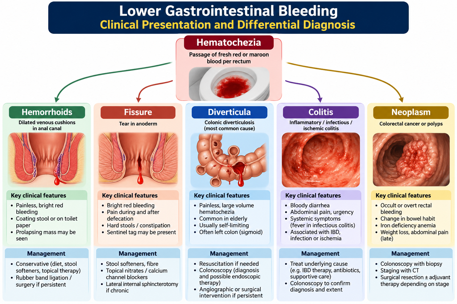
Bleeding per rectum means passage of visible blood through the anus; it is a symptom, not a diagnosis. Fresh red blood (hematochezia) commonly comes from a distal source, while darker maroon blood may indicate more proximal colonic bleeding. A brisk upper GI hemorrhage can occasionally cause red rectal bleeding, especially with hemodynamic instability. Distinguishing these patterns helps select investigations, but the appearance alone cannot safely identify the source.
The important first distinction is whether the patient is unstable. Tachycardia, hypotension, postural dizziness, syncope, confusion, pallor, cold extremities or ongoing large-volume bleeding suggest major blood loss. A patient with these findings requires immediate resuscitation rather than routine outpatient investigation.
Focused history and examination
Ask whether blood is mixed with stool, coats the stool or appears on toilet paper; whether it is painful; the amount and recurrence; diarrhea, urgency or mucus; altered bowel habit; abdominal pain; fever; weight loss; anemia symptoms; previous episodes; anticoagulant or antiplatelet use; family history of colorectal cancer; and known IBD. These questions narrow the cause and reveal danger.
- Painless blood after defecation: internal hemorrhoids are possible, but this pattern never excludes another lesion.
- Severe pain during defecation with bright blood: suggests anal fissure.
- Bloody diarrhea with urgency and tenesmus: suggests inflammatory colitis, especially ulcerative colitis.
- Sudden painless substantial bleeding in an older adult: consider diverticular bleeding or angiodysplasia.
- Bleeding with a persistent bowel-habit change, iron-deficiency anemia or weight loss: requires assessment for colorectal malignancy.
Examination includes vital signs and perfusion, assessment of abdominal tenderness or mass, perianal inspection and, when appropriate and safe, digital rectal examination to identify blood, lesions or a palpable rectal mass. A normal rectal examination does not rule out proximal disease. Chronic illness, cachexia or pallor may signal anemia, IBD or cancer.
B. Lower GI Bleeding — Investigations, Interpretation and Management
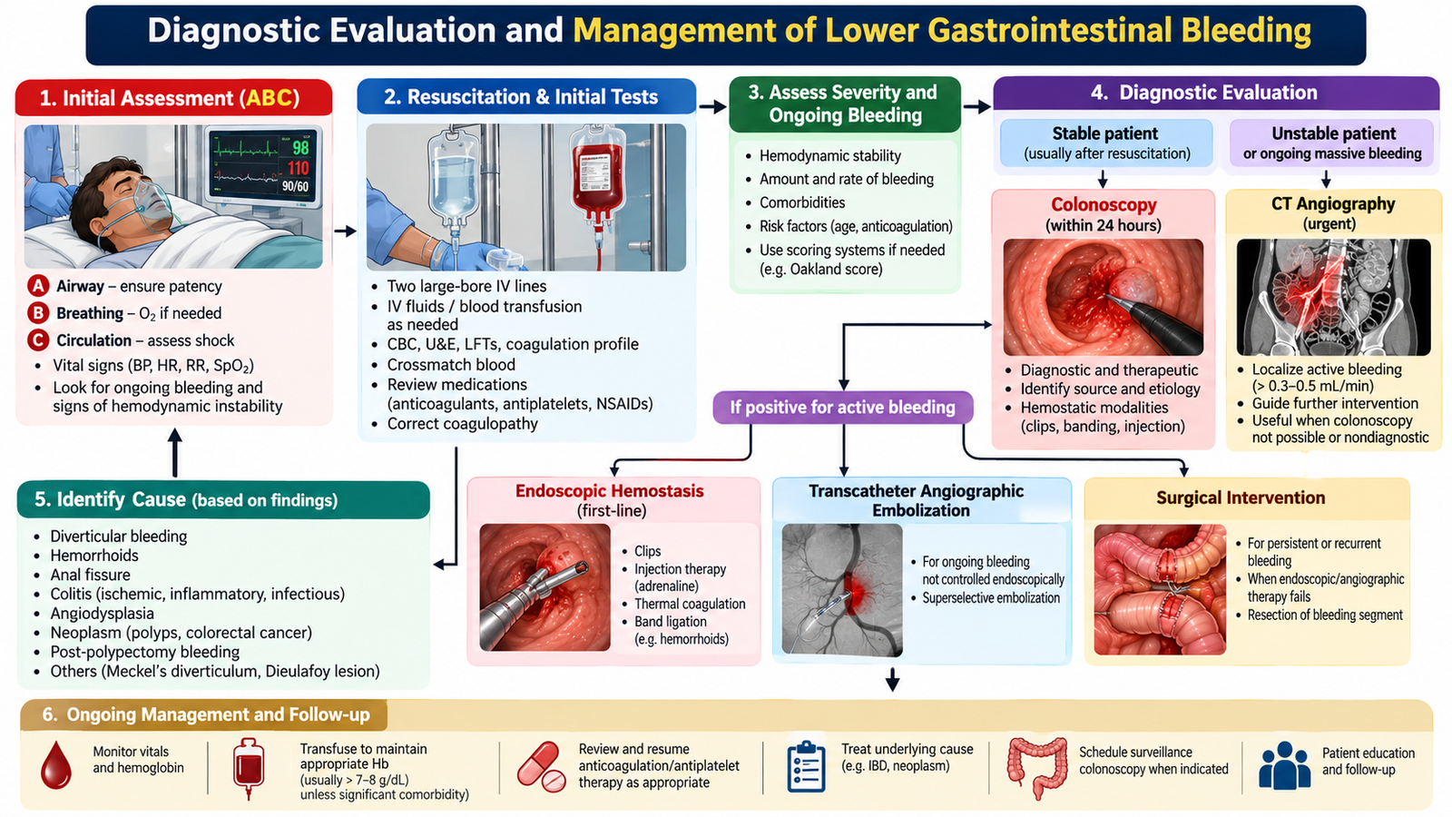
The investigation pathway depends on hemodynamic status, ongoing bleeding and the suspected cause. Stabilization and diagnostic evaluation may occur simultaneously in a severely bleeding patient. Stable patients usually undergo a focused investigation for the suspected colonic or anorectal source.
What to request and what the findings mean
- Complete blood count: estimates anemia and platelet count. Early hemoglobin may not reflect the full extent of acute blood loss; repeat tests and clinical assessment are important.
- Blood group and crossmatch: prepare compatible blood when major bleeding or transfusion is likely.
- Urea, creatinine and electrolytes: help assess dehydration, renal perfusion and fitness for investigations or treatment.
- Coagulation tests: useful in major hemorrhage and patients with liver disease or anticoagulant exposure.
- Colonoscopy: directly examines the colon, may identify inflammation, polyps, tumors or bleeding lesions, allows biopsy and can sometimes provide endoscopic hemostasis. Adequate bowel preparation is important in stable patients.
- CT angiography: especially useful for hemodynamically significant ongoing bleeding when active hemorrhage needs localization; a positive study may guide interventional radiological embolization.
- Biopsy: histology distinguishes inflammatory changes from dysplasia or cancer when suspicious mucosa or a mass is encountered.
Management according to severity
Unstable or major ongoing bleed: use an ABC approach, obtain intravenous access, monitor pulse, blood pressure, mental status and urine output, send urgent blood tests and crossmatch, and give appropriate crystalloid while arranging blood products when indicated. Transfusion is based on instability, ongoing blood loss, hemoglobin and comorbidities; a restrictive threshold around 7 g/dL is commonly used in stable hospitalized adults, but it is not a rule for active shock or significant cardiac disease. Review anticoagulants and antiplatelets with senior input: reversal or temporary interruption may be necessary in life-threatening bleeding, but cardiovascular and thrombotic risks must be considered. Seek urgent gastroenterology, surgery and interventional radiology input. CT angiography can localize ongoing major bleeding; embolization or endoscopic treatment may control the source. Surgery is reserved for persistent severe bleeding that cannot be controlled otherwise.
Stable bleed: investigate the suspected source, correct iron deficiency where present and treat the cause. Internal hemorrhoids are commonly managed with fiber, hydration and avoidance of straining; persistent symptoms may require office procedures such as rubber-band ligation. Anal fissures benefit from stool-softening measures and topical smooth-muscle relaxants in suitable patients. Diverticular or angiodysplastic bleeding may stop spontaneously but can require endoscopic hemostasis, angiographic embolization or rarely surgery. Colitis-related bleeding requires treatment of the underlying inflammation. Suspected colorectal cancer must proceed to colonoscopic evaluation and biopsy, not empirical hemorrhoid treatment alone.
Monitoring: repeated vital signs, bleeding frequency, hemoglobin and perfusion indicate whether resuscitation and hemostasis are working. Continued tachycardia, recurrent large-volume stool blood or falling hemoglobin requires reassessment and escalation.
A 28-year-old patient reports several weeks of bloody loose stools, urgency and lower abdominal cramps. He is not shocked, but the symptoms are persistent and affect daily activity. Inflammatory colitis is more likely than an isolated anal fissure because the bleeding accompanies diarrhea and urgency.
Think clinically: Assess severity and anemia; exclude stool infection; use colonoscopy with biopsies to establish the pattern of inflammation before deciding disease-specific treatment.
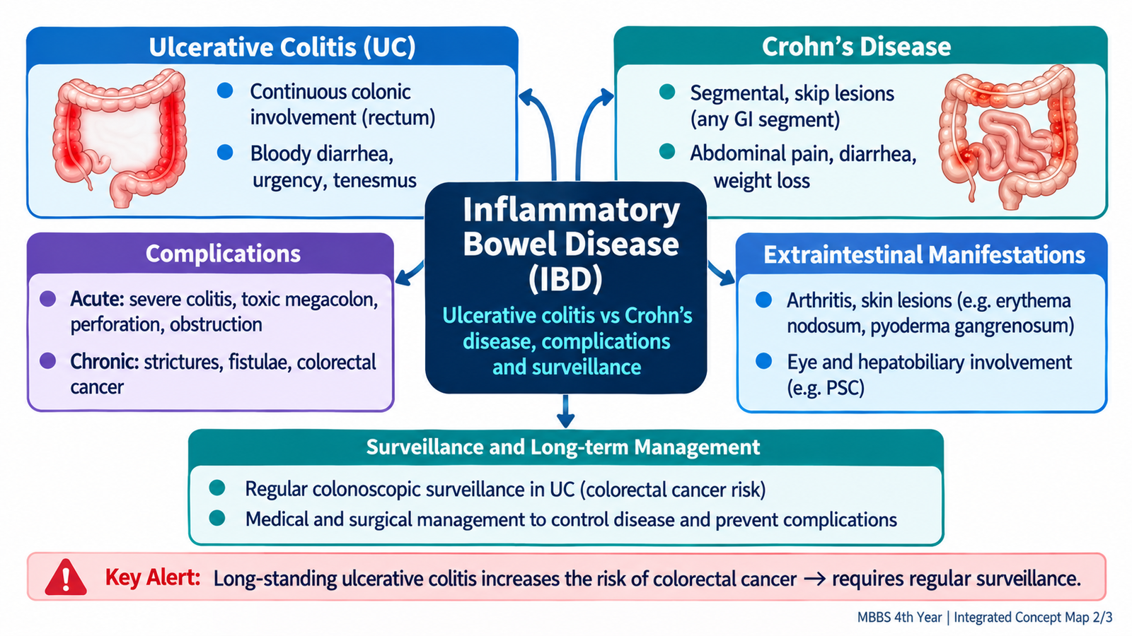
C. Inflammatory Bowel Disease — Mechanisms and Clinical Recognition
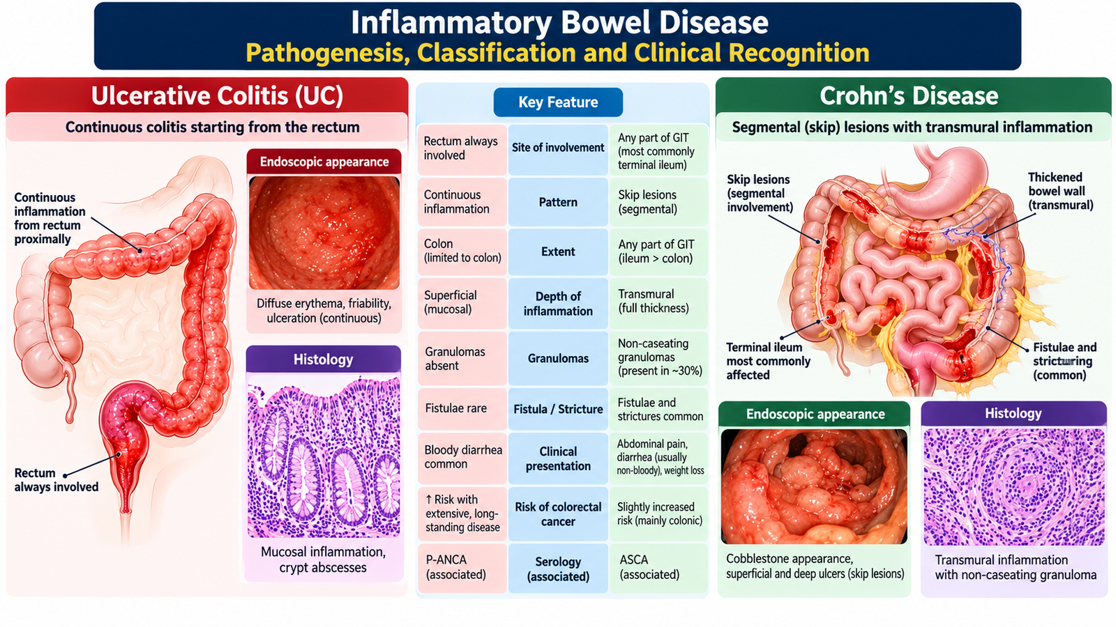
Inflammatory bowel disease is chronic, relapsing inflammation of the gastrointestinal tract resulting from an inappropriate intestinal immune response in a genetically susceptible person, influenced by gut microbes and environmental factors. Persistent inflammation injures mucosa, causing diarrhea, bleeding, pain and impaired bowel function. The two principal forms are ulcerative colitis (UC) and Crohn’s disease (CD). Distinguishing them is important because the site and depth of disease determine complications and treatment.
UC affects the colon, begins in the rectum and usually extends proximally in a continuous pattern. Inflammation is mainly mucosal, so bleeding, urgency and tenesmus are prominent. Crohn’s disease can affect any part of the gastrointestinal tract, often the terminal ileum or colon, with discontinuous ‘skip’ areas and transmural (full-wall) inflammation. Deep inflammation explains strictures, fistulas and abscesses; ileal involvement can produce malabsorption and weight loss.
| Clinical distinction | Ulcerative colitis | Crohn’s disease |
|---|---|---|
| Distribution | Colon; rectum usually involved; continuous | Mouth to anus; often ileocolonic; skip lesions |
| Depth | Predominantly mucosal | Transmural |
| Typical symptoms | Bloody diarrhea, urgency, tenesmus | Pain, diarrhea, weight loss; bleeding variable |
| Distinctive complications | Acute severe colitis, toxic megacolon | Stricture, fistula, abscess, perianal disease |
| Effect of resection | Removing colon/rectum can cure intestinal UC | Disease may recur elsewhere or after resection |
Extraintestinal inflammation can affect joints (peripheral arthritis or sacroiliitis), eyes (uveitis or episcleritis) and skin (erythema nodosum or pyoderma gangrenosum). These findings support the systemic nature of IBD and require coordinated care. A careful drug and travel/infection history helps distinguish IBD from infective colitis and medication-related symptoms.
D. Ulcerative Colitis — Assessment and Severity-Based Treatment
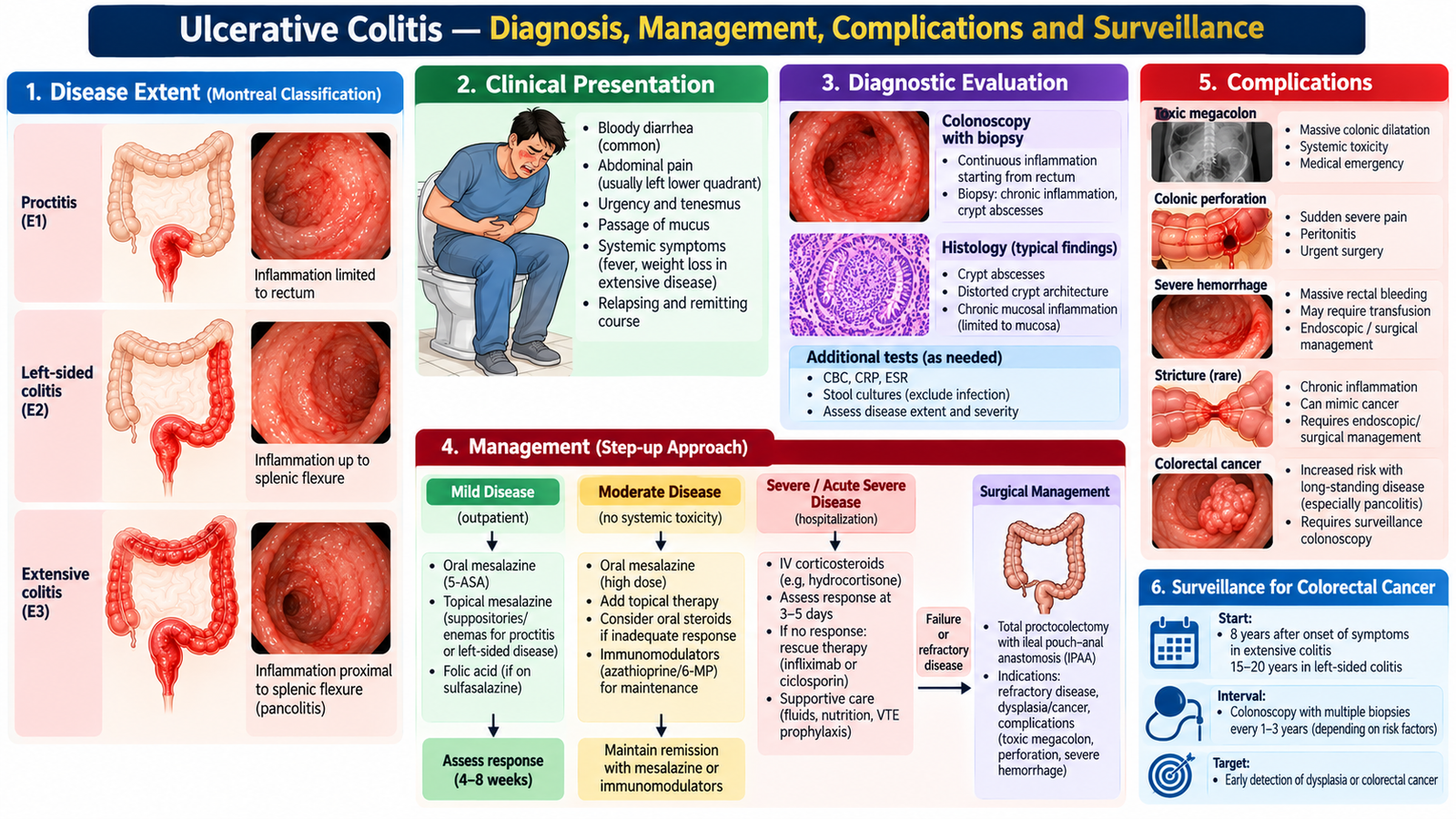
UC typically causes recurrent bloody diarrhea, urgency, mucus passage, tenesmus and crampy lower abdominal pain. The symptom burden depends on the extent of colonic involvement and the degree of inflammation. In severe attacks, fever, tachycardia, anemia, dehydration and abdominal distension are warning findings.
Confirming the diagnosis and assessing severity
Request a complete blood count (anemia or leukocytosis), CRP (inflammation), electrolytes and albumin (nutrition and systemic effects). Send stool testing for relevant pathogens, including Clostridioides difficile where appropriate; infection can mimic or worsen UC. Fecal calprotectin supports intestinal inflammation but does not by itself distinguish UC from infection or Crohn’s disease. Colonoscopy with multiple biopsies generally confirms continuous mucosal inflammation starting in the rectum; in acute severe colitis, limited flexible sigmoidoscopy is usually preferred over full colonoscopy because of perforation risk.
For management, classify disease by extent (proctitis, left-sided or extensive colitis) and activity (mild-to-moderate versus severe). Repeated frequent bloody stools, systemic toxicity, elevated inflammatory markers or marked anemia suggest severe disease. Classic acute severe UC is defined by at least six bloody stools daily together with systemic features such as fever, tachycardia, anemia or raised ESR.
How the medicines work
Mesalazine (5-aminosalicylic acid, 5-ASA) acts locally on intestinal mucosa to reduce inflammatory mediator activity. It is particularly useful for mild-to-moderate UC and can maintain remission. Corticosteroids, such as prednisolone or intravenous hydrocortisone/methylprednisolone, suppress inflammatory immune activity more rapidly and are used to induce remission during flares; prolonged maintenance is avoided because of infection risk, osteoporosis, hyperglycemia and other steroid toxicity. Thiopurines such as azathioprine may help selected steroid-dependent patients maintain remission but have a delayed onset and require blood-count and liver-test monitoring. Biological therapies such as infliximab (anti-TNF) or vedolizumab (gut-selective anti-integrin) are specialist treatments for moderate-to-severe or refractory disease. Screening for infections, particularly tuberculosis and hepatitis B, and vaccination review are important before significant immunosuppression.
Treatment selected by disease extent and activity
- Mild ulcerative proctitis: rectal mesalazine suppositories deliver treatment to the rectum and are generally preferred as first-line induction treatment. Rectal treatment also supports maintenance when needed.
- Mild-to-moderate left-sided UC: combine oral mesalazine with rectal mesalazine enemas or other suitable topical preparation. Oral therapy reaches more proximal inflammation, while rectal therapy treats distal inflammation effectively.
- Mild-to-moderate extensive UC: oral mesalazine is important; adding rectal mesalazine can improve symptom control. If optimized 5-ASA fails, an oral corticosteroid course is considered to induce remission.
- Persistent or moderate-to-severe UC: specialist assessment for biologic or other advanced treatment is needed, especially after steroid dependence or failure. Azathioprine can be used in selected maintenance strategies but does not rapidly treat an acute attack.
- After remission: maintain with appropriate mesalazine or steroid-sparing therapy, monitor adherence and symptoms, and avoid long-term systemic corticosteroids.
Acute severe ulcerative colitis is a hospital emergency
Admit the patient, assess for hypovolemia and sepsis, correct fluids/electrolytes, monitor stool frequency, vital signs, hemoglobin and inflammatory markers, and investigate for infection. Intravenous corticosteroids are first-line anti-inflammatory treatment. Give venous thromboembolism prophylaxis unless contraindicated: active UC itself increases clot risk even when stools contain blood. Provide nutritional support, avoid unnecessary antidiarrheal or opioid drugs that may worsen colonic dilatation, and obtain early surgical input.
Clinical response is reassessed early, commonly around day 3. Failure to improve requires a time-critical multidisciplinary decision regarding rescue infliximab or ciclosporin or colectomy. Perforation, uncontrolled major hemorrhage, worsening toxic megacolon or refractory severe disease makes emergency surgery necessary. Colectomy is also considered electively for medically refractory disease or colorectal neoplasia. Definitive options include proctocolectomy with ileal pouch–anal anastomosis or permanent ileostomy, depending on patient circumstances; detailed operative technique is not required at undergraduate level.
Long-term complications and surveillance
UC may cause severe bleeding, dehydration, fulminant colitis, toxic megacolon, perforation and venous thrombosis. Chronic colonic inflammation increases colorectal dysplasia/cancer risk, particularly with extensive and long-standing disease. Surveillance colonoscopy generally begins about 8 years after symptom onset for colonic IBD, with subsequent intervals tailored to risk and local guidance; isolated proctitis usually does not require the same IBD-specific surveillance program. Primary sclerosing cholangitis is a higher-risk association for which surveillance usually begins at diagnosis. Dysplasia findings guide enhanced surveillance versus surgical referral.
E. Crohn’s Disease — Clinical Assessment and Medical/Surgical Management
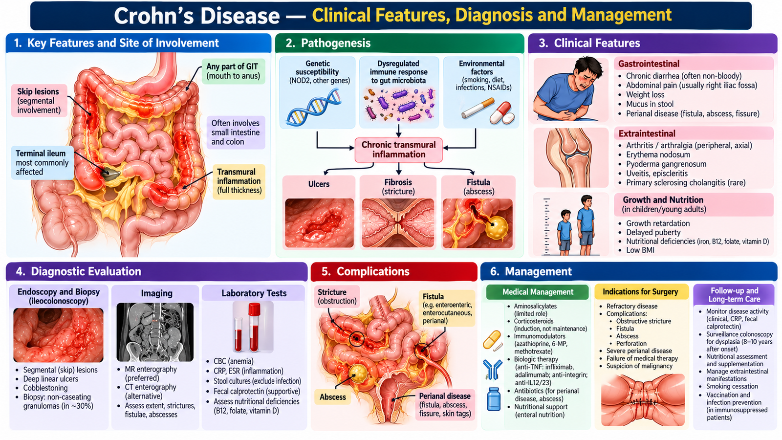
Crohn’s disease frequently causes recurrent abdominal pain, prolonged diarrhea, weight loss, fatigue and poor appetite. Terminal ileal disease may produce right iliac fossa pain and vitamin B12 deficiency; perianal fistulas or abscesses are valuable diagnostic clues. Inflammation passing through the bowel wall produces narrowing (fibrotic stricture), penetrating fistulas and abscess formation. These complications explain why not all symptoms respond to anti-inflammatory medication alone.
Look for fever, weight loss, malnutrition, palpable abdominal mass, perianal discharge or painful swelling, and extraintestinal manifestations. CBC may show anemia, CRP and fecal calprotectin may indicate inflammation, while albumin and nutritional indices assess disease effects. Ileocolonoscopy with biopsies is a major diagnostic test; segmental inflammation, aphthous/deep ulcers or histological chronic inflammation support CD. Non-caseating granulomas can support diagnosis but are often absent. MR enterography or CT enterography assesses small-bowel inflammation and complications; MRI pelvis is especially helpful for complex perianal fistulas. Exclude relevant intestinal infections, especially intestinal tuberculosis when clinically plausible, before substantial immunosuppression.
Induction, maintenance and medication selection
Induction of remission means controlling an active flare; maintenance means preventing recurrence once inflammation is controlled. These aims require different drug strategies.
- Mild active ileocecal Crohn’s: controlled-release budesonide may be suitable because it has predominantly local intestinal glucocorticoid activity and less systemic exposure than conventional oral steroids. It is not an appropriate universal treatment for every disease location.
- Moderate-to-severe active inflammation: systemic corticosteroids such as prednisolone can induce short-term remission. They are not used as long-term maintenance because toxicity increases while sustained disease control is poor.
- Maintenance and steroid-sparing treatment: azathioprine or methotrexate may be appropriate in selected patients under specialist supervision; onset is delayed, so these drugs are not relied upon for immediate induction. Azathioprine requires CBC/liver monitoring and appropriate pre-treatment assessment; methotrexate is contraindicated in pregnancy and requires liver and blood-count monitoring.
- Moderate-to-severe, recurrent, high-risk or fistulizing disease: biologics such as infliximab or adalimumab (anti-TNF), ustekinumab (IL-12/23 pathway) or vedolizumab may be selected by the specialist based on disease phenotype, previous treatment and patient risks. Anti-TNF therapy is particularly important in selected fistulizing disease. Assess TB, hepatitis B, infections and vaccination status before biologics.
- Mesalazine: unlike UC, 5-ASA is generally not an effective core treatment for Crohn’s disease; do not apply a UC treatment pathway automatically.
When a complication changes the plan
Abscess: an intra-abdominal or perianal collection needs imaging, antibiotics when indicated and drainage/source control; simply increasing immunosuppression before controlling infection may be hazardous. Fistula: treatment may combine drainage or a seton with biological therapy, depending on anatomy and disease activity. Stricture: active inflammatory narrowing may improve with medical therapy, but fixed fibrotic obstruction frequently requires endoscopic dilatation or surgery. Acute obstruction or perforation: hospital admission, resuscitation, surgical evaluation and urgent intervention when appropriate.
Resection or strictureplasty may relieve localized obstruction, control perforation, drain sepsis or address disease refractory to medication. Unlike colectomy for UC, surgery does not cure Crohn’s disease, because inflammation may recur in remaining bowel. Postoperative surveillance and maintenance strategy remain important.
Monitoring and long-term care
Follow symptoms, weight, nutritional status, anemia, CRP/fecal calprotectin where useful and objective bowel inflammation. Assess drug toxicity, infections and disease complications. Counsel patients about adherence, smoking cessation, nutrition and seeking help for fever, obstruction symptoms or perianal sepsis. Treatment goals are sustained remission, preservation of bowel function and avoidance of repeated steroid courses.
F. Inflammatory Bowel Disease — Complications, Recognition and Escalation
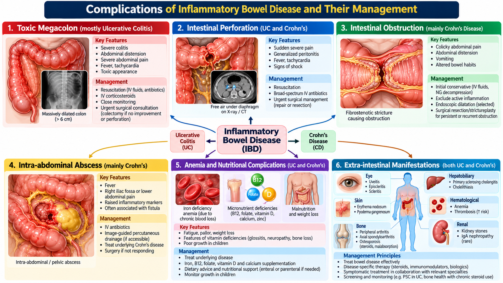
Complications of IBD arise from active inflammation, structural bowel injury, chronic blood/nutrient losses and treatment-related immunosuppression. Their management is not a separate drug list: the important decision is whether the problem represents uncontrolled inflammatory disease, infection, obstruction, perforation, thrombosis or malignancy.
Recognize the complication and respond appropriately
- Acute severe UC / toxic megacolon: bloody diarrhea, systemic toxicity, distended tender abdomen and radiological colonic dilatation indicate danger. Admit urgently, resuscitate, give IV steroids for severe UC when appropriate, provide thromboprophylaxis and arrange early surgical review; perforation or failure of treatment requires surgery.
- GI hemorrhage and iron-deficiency anemia: check hemoglobin and hemodynamic status, control inflammation or another bleeding lesion, replace iron orally or intravenously according to tolerance, severity and disease activity, and transfuse when clinically indicated.
- Crohn’s obstruction: colicky pain, distension, vomiting and inability to pass stool/flatus require admission, fluid/electrolyte correction, imaging and surgical input. Differentiate inflammatory narrowing from fixed fibrosis when choosing medicines versus intervention.
- Abscess and fistula: evaluate with imaging; drain infected collections and consider antibiotics. Surgical and gastroenterology teams coordinate ongoing fistula care and control intestinal inflammation.
- Venous thromboembolism: active IBD increases clot risk through systemic inflammation, immobility and hospitalization. Prevent with hospital thromboprophylaxis when appropriate; suspected thrombosis needs urgent diagnostic and treatment pathways.
- Extraintestinal disease: inflammatory arthritis, uveitis and important skin lesions may accompany bowel activity or occur independently. Treat intestinal inflammation and seek joint specialist care, especially for painful red eye or vision symptoms.
- Dysplasia and cancer: long-standing colonic inflammation increases risk; surveillance colonoscopy and assessment of suspicious lesions support early detection and management planning.
Drug safety is part of complication prevention. Corticosteroids increase infection and metabolic risks; azathioprine can suppress bone marrow or cause liver toxicity; methotrexate is teratogenic; and biological therapies increase susceptibility to selected serious infections. Before or during immunosuppressive therapy, confirm appropriate screening, immunization advice and monitoring arrangements.
G. Colorectal Cancer — Diagnosis, Staging and Treatment Planning
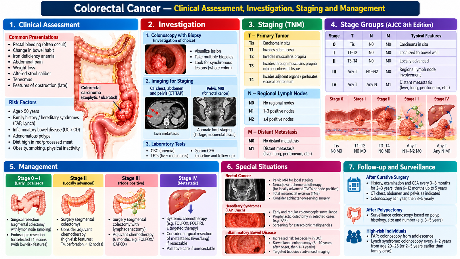
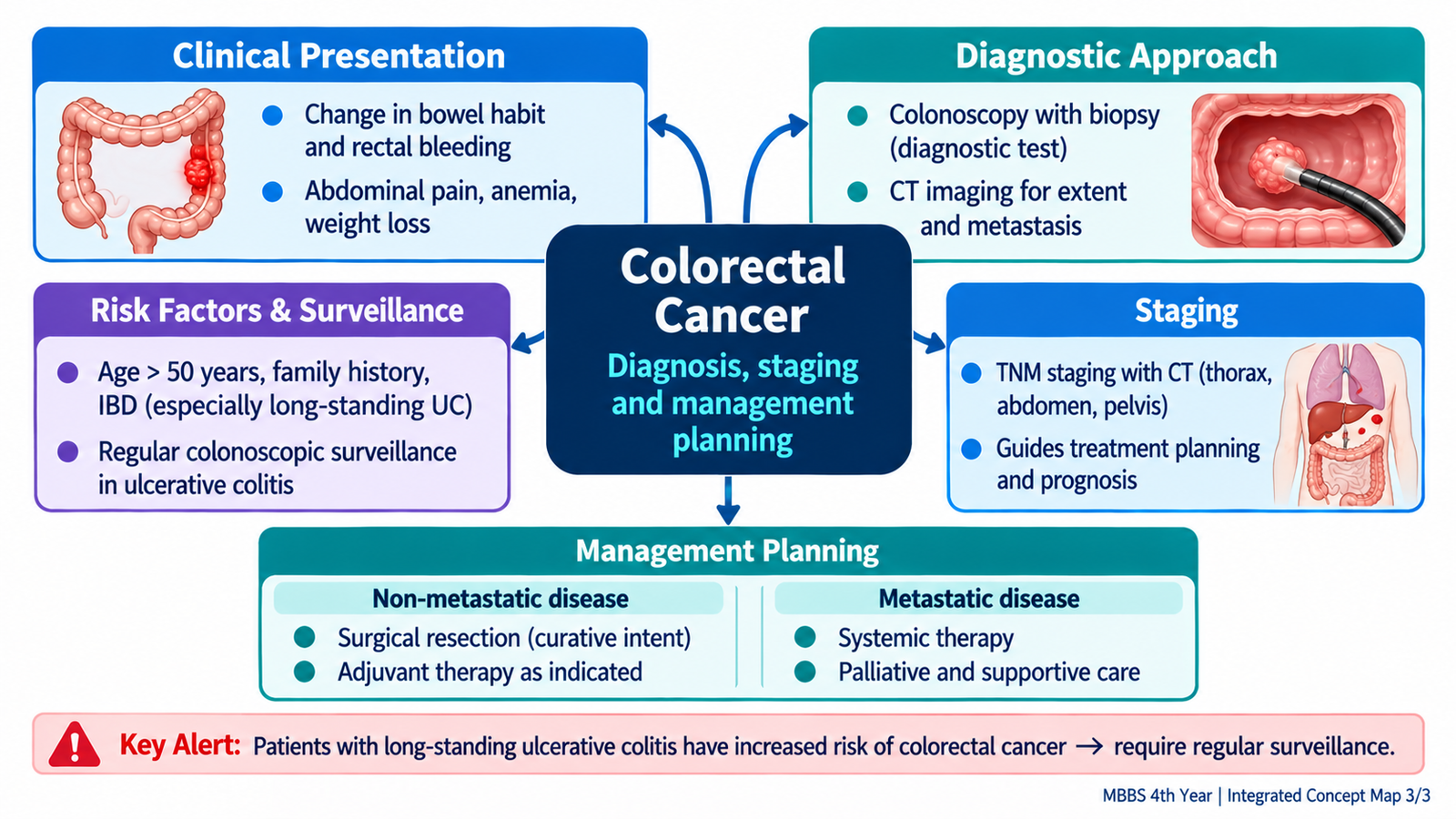
Colorectal cancer commonly develops from neoplastic glandular epithelium (adenocarcinoma). Some cancers progress from adenomatous polyps through accumulated cellular changes; chronic colonic inflammation in IBD provides a different important risk context. Risk increases with age, family history, inherited predisposition, previous advanced polyps and long-standing colitis.
How it presents and what clinical examination adds
Patients may present with rectal bleeding, a persistent change in bowel habits, abdominal discomfort, unexplained iron-deficiency anemia, weight loss or bowel obstruction. Right-sided colon cancers may cause occult blood loss and iron-deficiency anemia without visible bleeding; left-sided lesions more often cause altered bowel habit or obstructive symptoms. Rectal cancer may produce visible bleeding, tenesmus and a sensation of incomplete evacuation. Examination includes nutritional state, pallor, abdominal mass or distension, and digital rectal examination for accessible rectal tumors. A normal rectal examination cannot exclude colon cancer.
Investigations: diagnosis is different from staging
- Complete colonoscopy with biopsy: confirms a lesion histologically and checks for synchronous colonic lesions where feasible. Cancer diagnosis requires tissue confirmation.
- CBC and iron indices: identify chronic blood-loss anemia; renal and liver tests contribute to treatment fitness and metastatic assessment.
- Carcinoembryonic antigen (CEA): useful as a baseline and in follow-up after treatment, but not sufficiently accurate for diagnosis or population screening.
- Contrast-enhanced CT chest, abdomen and pelvis: evaluates spread to lymph nodes, liver, lungs and other sites and supports treatment planning.
- Pelvic MRI in rectal cancer: characterizes local tumor invasion, mesorectal fascia/circumferential margin risk and pelvic nodes; this information strongly influences neoadjuvant treatment and surgery.
Staging uses TNM: T describes local depth of invasion, N indicates regional lymph nodes and M indicates distant metastases. Broad stages I–IV summarize these patterns: stage I usually localized early disease; stage II deeper bowel-wall invasion without regional nodal involvement; stage III regional nodal spread; stage IV distant metastasis. Stage and exact tumor location shape the treatment plan.
Colon cancer — stage-directed management
Resectable stage I disease: surgery is generally definitive; selected very early lesions may be adequately treated endoscopically if favorable pathological criteria are met. Stage II: oncological surgical resection is the principal treatment; adjuvant chemotherapy is considered for selected high-risk features rather than automatically for every patient. Stage III: surgical resection followed by adjuvant chemotherapy is standard for many fit patients because lymph-node spread raises recurrence risk. Common adjuvant chemotherapy uses a fluoropyrimidine (such as capecitabine or 5-fluorouracil) with oxaliplatin in suitable patients (CAPOX or FOLFOX). Fluoropyrimidines interfere with DNA synthesis in dividing cancer cells; oxaliplatin damages DNA. Important toxicities include diarrhea, myelosuppression and hand–foot syndrome with capecitabine, and peripheral neuropathy with oxaliplatin. Exact regimens and cycles are decided by the oncology team.
Stage IV: management is multidisciplinary. Systemic chemotherapy is often central, with targeted or immunotherapy options in selected tumors based on molecular features and specialist assessment. A limited, resectable liver or lung metastasis may sometimes be treated with curative intent; more extensive disease may require symptom-directed systemic and palliative measures. Bowel obstruction, perforation or bleeding may necessitate urgent surgery, stenting or stoma formation.
Rectal cancer — why treatment differs
The rectum lies within the pelvis, close to the sphincters and other structures, so local staging and surgical margins are particularly important. Early rectal cancer may be managed by local excision in carefully selected cases or formal oncologic resection. Locally advanced rectal cancer often receives neoadjuvant radiotherapy/chemoradiotherapy or total neoadjuvant treatment before surgery, according to pelvic MRI findings and multidisciplinary planning. This aims to reduce local recurrence and improve resectability. The appropriate operation depends on tumor position, stage and sphincter involvement: anterior resection may preserve anal continuity, while abdominoperineal resection removes the anus/sphincter complex and requires a permanent colostomy. These are principles, not operative technique instructions.
Surgical planning, perioperative care and follow-up
For colon tumors, operation selection follows the segment involved: right hemicolectomy for suitable right-sided cancers, left hemicolectomy for selected left-sided cancers, and sigmoid colectomy for sigmoid lesions. Oncologic resection includes appropriate bowel margins and regional mesenteric lymph nodes for staging. Patients need preoperative fitness and anemia/nutrition assessment and an explanation of whether an anastomosis or temporary/permanent stoma may be required. Postoperatively, watch for bleeding, infection, anastomotic leak, ileus and thrombosis. Histopathology determines final stage and need for adjuvant therapy.
Follow-up after curative treatment includes clinical review, CEA when appropriate, colonoscopy and stage-specific imaging. Patients should understand warning symptoms of recurrence, bowel-function changes, possible stoma care, treatment adverse effects and why surveillance matters. In advanced cancer, symptom control, nutritional support and patient priorities are essential alongside anticancer treatment.
AIM High-Yield Review
- Shock with hematochezia demands resuscitation immediately; a brisk upper GI source also remains possible.
- Bloody diarrhea with urgency and tenesmus suggests distal colonic inflammation, especially UC, but infection must be excluded.
- Colonoscopy with biopsy establishes IBD pattern or colorectal cancer; fecal calprotectin alone is not diagnostic.
- ⭐ UC is continuous and predominantly mucosal; Crohn’s disease is often discontinuous and transmural, explaining fistulas and strictures.
- ⭐ Mild distal UC: rectal mesalazine; more extensive UC often needs oral plus rectal mesalazine.
- Corticosteroids induce remission during IBD flares but should not be continued for long-term maintenance.
- ⭐ Acute severe UC: admit, give IV corticosteroids, monitor closely and consider rescue therapy or colectomy if response fails.
- Azathioprine is a delayed-onset steroid-sparing maintenance option in selected IBD patients; monitor blood counts and liver tests.
- ⭐ Crohn’s abscess requires source control; fibrotic obstruction may need intervention. Surgery does not cure Crohn’s disease.
- Long-standing colonic IBD increases colorectal cancer risk and requires risk-adapted surveillance.
- ⭐ Colorectal cancer: colonoscopy/biopsy confirms diagnosis; CT stages spread; pelvic MRI guides rectal cancer planning.
- Stage III colon cancer commonly requires surgery plus adjuvant fluoropyrimidine-based chemotherapy, often with oxaliplatin.
- Locally advanced rectal cancer often needs neoadjuvant treatment before carefully planned oncologic surgery.
- CEA is a follow-up marker, not a diagnostic substitute; treatment and surveillance should match stage, fitness and patient goals.
Video Learning Resources — Topic 7
Lower GI Bleeding, Inflammatory Bowel Disease & Colorectal Cancer
Video 1 — Lower Gastrointestinal Bleeding
Clinical causes of bleeding per rectum, diagnostic evaluation and management principles. The lecture covers both upper and lower GI bleeding; the lower GI sections are especially relevant.
Source: Ninja Nerd | GI Bleed — Clinical Medicine
Video 2 — Inflammatory Bowel Disease
Ulcerative colitis and Crohn’s disease, including pathophysiology, clinical presentation, investigations, complications and management.
Source: Ninja Nerd | Inflammatory Bowel Disease — Clinical Medicine
Video 3 — Colorectal Cancer
Risk factors, clinical features, diagnostic investigations, staging and treatment principles in colorectal malignancy.
Source: Ninja Nerd | Colorectal Cancer — Podcast