
📖 AIM Learning Material
TOPIC 2. Hepatocellular Disease, Liver Failure, Cirrhosis & Hepatic Encephalopathy
GIT Module • Integrated Medicine • From liver injury to cirrhosis, failure and encephalopathy
Topic Introduction
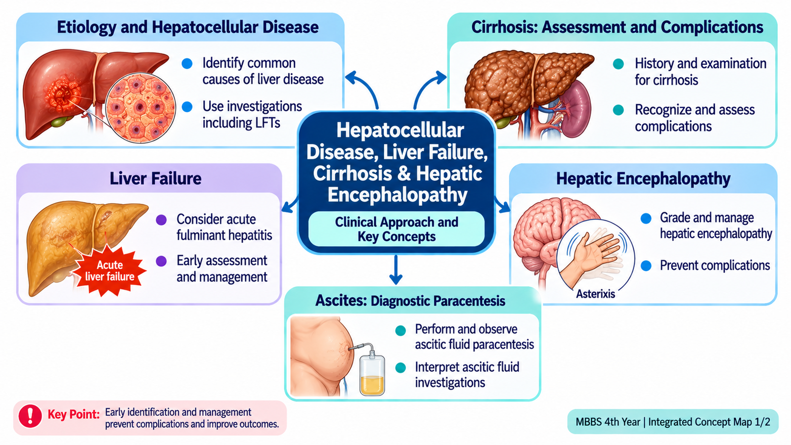
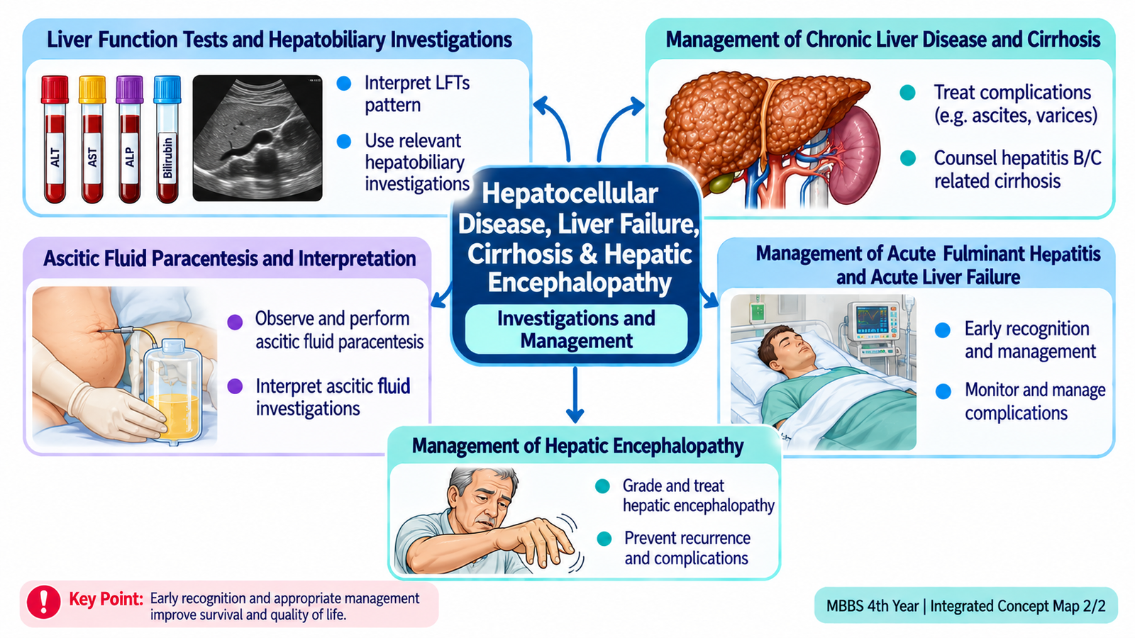
The liver can be injured without immediately losing its function, but sustained injury may cause fibrosis, cirrhosis and portal hypertension. Severe acute injury can instead produce acute liver failure within days or weeks. These conditions are connected by changes in hepatocyte function, blood flow, detoxification and protein synthesis. This chapter explains how to recognize clinical patterns, take a focused history, examine patients with chronic liver disease, interpret liver and ascitic fluid tests, manage dangerous complications and counsel patients with hepatitis-related cirrhosis. Hepatic encephalopathy receives particular attention because early recognition and correction of its triggers can prevent deterioration.
A. Clinical Framework of Hepatocellular Disease
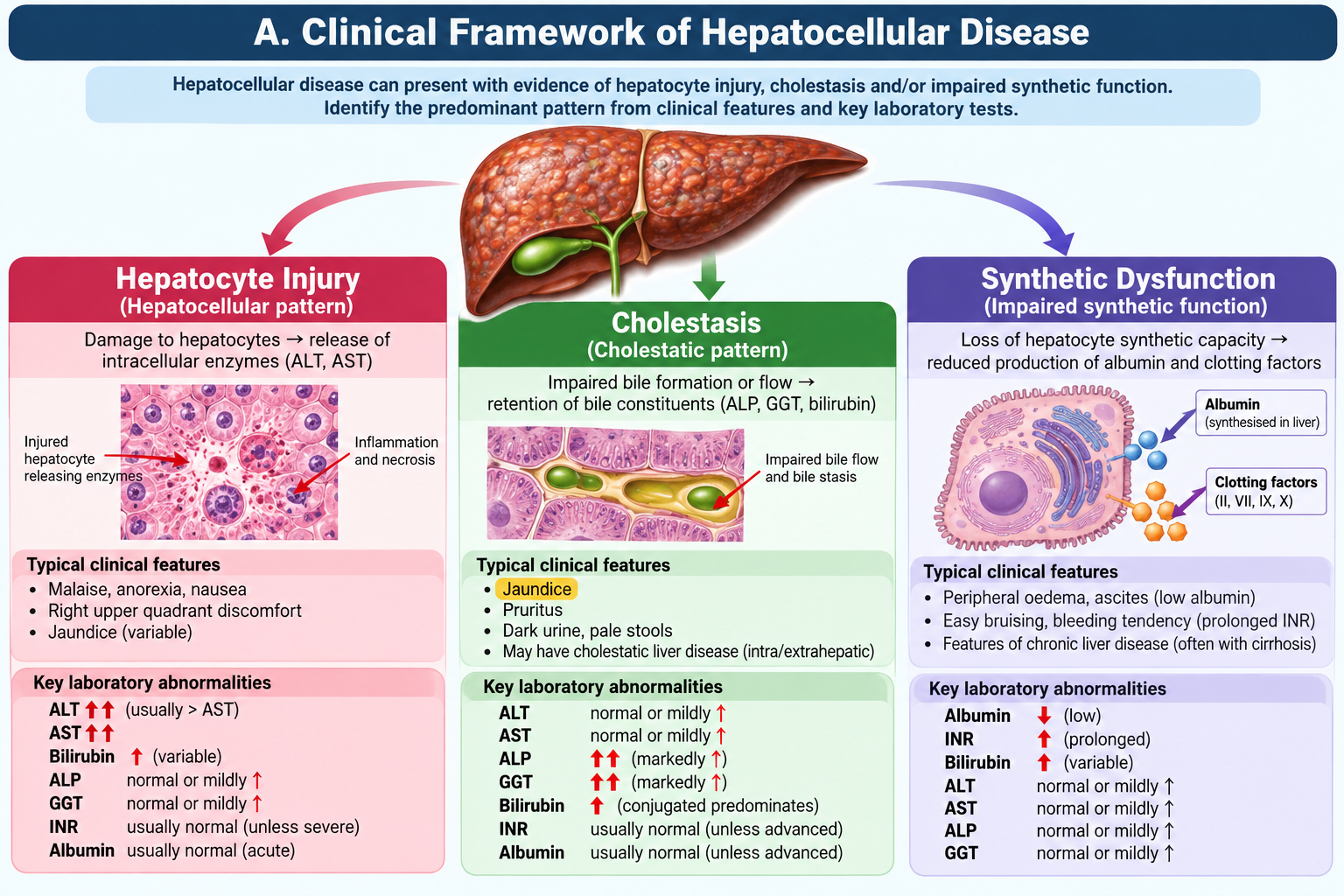
Hepatocellular disease means injury to liver cells (hepatocytes), whereas cholestasis describes impaired bile formation or flow. Both can cause jaundice, but their investigation patterns differ. The liver synthesizes albumin and clotting factors, metabolizes bilirubin and drugs, regulates glucose and detoxifies ammonia. A patient may have substantial hepatocyte injury before losing these essential functions.
Recognizing the clinical pattern
Acute hepatitis develops over days to weeks and may present with malaise, anorexia, nausea, right-upper-quadrant discomfort, dark urine and jaundice. Chronic injury develops over months or years and may initially be asymptomatic. Chronic viral hepatitis B/C, alcohol-associated disease, metabolic dysfunction-associated steatotic liver disease and drug-related injury are important causes to consider. Ask about viral exposure, previous transfusions or unsafe injections, alcohol, metabolic risk, medicines and herbal products.
Jaundice reflects accumulation of bilirubin; dark urine suggests water-soluble conjugated bilirubin entering urine. Pruritus and pale stools suggest a cholestatic component. Fever and right-upper-quadrant pain with jaundice require urgent assessment for biliary infection rather than assuming uncomplicated hepatitis.
Distinguishing injury from failure
Marked ALT/AST elevation indicates hepatocyte injury but does not directly measure liver function. Falling albumin may reflect chronic synthetic dysfunction but also malnutrition or renal loss. Prolonged PT/INR, particularly when accompanied by encephalopathy in acute disease, signals dangerous loss of hepatic synthetic function. A mildly elevated enzyme level does not exclude advanced cirrhosis because fewer viable hepatocytes may remain.
B. Focused History, Examination and Clinical Assessment
Clinical assessment starts by deciding whether the patient is stable and whether the liver disease is acute or chronic. Ask about onset of jaundice, dark urine, stool colour, abdominal swelling, leg oedema, gastrointestinal bleeding, sleep reversal, confusion, fever, reduced urine output and weight loss. Clarify hepatitis B/C history, previous antiviral therapy, alcohol consumption, medication exposure and previous episodes of ascites or encephalopathy.
Focused examination
Record vital signs, mental status, hydration, body mass and nutritional state. Inspect for jaundice, muscle wasting, bruises, spider angiomas and palmar erythema. Examine the abdomen for liver size and tenderness, splenomegaly, shifting dullness or fluid thrill when ascites is substantial, abdominal wall collaterals and hernias. Check peripheral oedema. A small firm liver, splenomegaly and ascites support portal hypertension and advanced chronic disease; none is individually diagnostic.
Assess orientation, attention, speech and level of consciousness. Elicit asterixis by asking an awake patient to extend both arms, dorsiflex the wrists and spread the fingers; brief irregular lapses of sustained posture produce a flapping movement. Asterixis supports metabolic encephalopathy but is not specific to liver disease and may be absent in deep coma.
Immediate danger signs
Hypotension, haematemesis, melena, rapidly increasing confusion, seizures, severe hypoglycaemia, fever with abdominal pain, oliguria and worsening INR require prompt senior review and emergency care. In a patient with cirrhosis, fever may be absent even during serious infection.
C. Liver Tests and Hepatobiliary Investigation Interpretation
Order tests to answer distinct questions: Is there liver-cell injury? Is bile flow impaired? Can the liver still synthesize essential proteins? Is there an identifiable cause or complication?
Reading the laboratory pattern
- ALT and AST: predominant elevation suggests hepatocellular injury. AST may also rise with muscle injury. AST greater than ALT may occur in alcohol-associated disease but is not diagnostic by itself.
- ALP and GGT: disproportionate elevation suggests cholestasis; GGT helps support hepatic origin of a raised ALP.
- Bilirubin: elevated conjugated bilirubin occurs in hepatocellular dysfunction or impaired bile excretion; interpret with urine and stool changes.
- Albumin: low levels may indicate longstanding synthetic impairment but are influenced by nutrition, inflammation and protein loss.
- PT/INR: prolonged INR reflects reduced clotting-factor activity and is especially important in acute liver injury; vitamin K deficiency and anticoagulants can also affect it.
- Platelets: thrombocytopenia may be an early clue to portal hypertension and hypersplenism.
Additional investigations and interpretation
Obtain CBC, renal function, electrolytes and glucose to identify bleeding, infection, renal injury, sodium disturbances and hypoglycaemia. Test hepatitis B surface antigen and hepatitis C antibody with confirmatory viral testing when indicated; evaluate suspected drug injury from exposure history. Ultrasound assesses liver contour, ascites, spleen size and biliary dilatation; Doppler is useful when portal or hepatic vascular disease is suspected. Imaging and tests must be interpreted together: a nodular liver plus splenomegaly and ascites supports cirrhosis with portal hypertension, whereas dilated bile ducts direct attention toward obstruction.
Clinical interpretation example: ALT 850 U/L with modest ALP rise suggests hepatocellular injury; INR 2.1 and new confusion signal possible acute liver failure, regardless of whether ALT subsequently falls. In a different patient, ALT 45 U/L, albumin 2.5 g/dL, INR 1.7 and ascites may indicate advanced cirrhosis despite relatively modest enzyme elevation.
D. Cirrhosis: Pathogenesis, Portal Hypertension and Complications
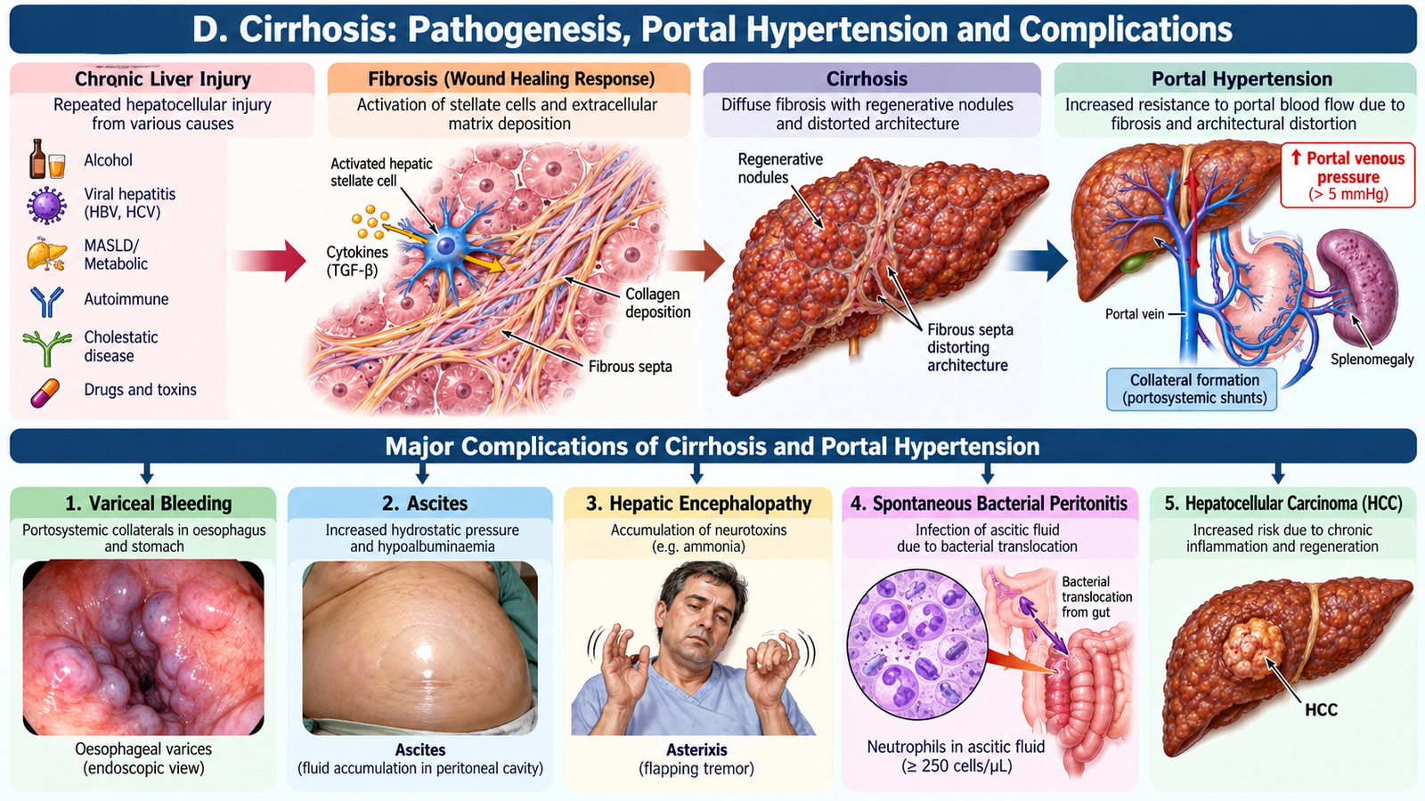
Cirrhosis is diffuse hepatic fibrosis with regenerative nodules that distort normal architecture. Repeated hepatocyte injury activates fibrogenic pathways; scar formation obstructs intrahepatic blood flow and reduces effective hepatocyte function. These changes produce two linked problems: portal hypertension and hepatic insufficiency.
Compensated and decompensated disease
Compensated cirrhosis may cause few symptoms. Decompensation is marked by clinically significant ascites, variceal bleeding or hepatic encephalopathy; jaundice may accompany progression. Portal hypertension causes splenomegaly, thrombocytopenia, portosystemic collaterals and oesophageal/gastric varices. Reduced albumin production and renal sodium retention promote ascites and oedema.
Important complications
- Ascites: portal hypertension and renal sodium retention lead to peritoneal fluid accumulation; tense ascites impairs breathing and comfort.
- Spontaneous bacterial peritonitis (SBP): infection of ascitic fluid without a surgically treatable intra-abdominal source; suspect with pain, fever, encephalopathy, renal deterioration or unexplained decompensation.
- Variceal haemorrhage: fragile portosystemic collaterals rupture, causing potentially life-threatening upper GI bleeding.
- Hepatorenal syndrome–AKI: circulatory disturbance causes severe renal dysfunction after other causes are assessed.
- Hepatic encephalopathy: impaired hepatic clearance and portosystemic shunting allow neurotoxins to affect brain function.
- Hepatocellular carcinoma: cirrhosis raises cancer risk, making appropriate ultrasound-based surveillance important.
-
Management of Variceal Bleeding: Suspected acute variceal haemorrhage requires emergency resuscitation, early intravenous terlipressin or octreotide, prophylactic intravenous ceftriaxone and urgent upper GI endoscopy with endoscopic band ligation for oesophageal varices. Following haemostasis, secondary prevention generally combines a non-selective beta-blocker (propranolol or carvedilol, as appropriate) with repeat band ligation.
Severity can be assessed clinically and with tools such as Child–Pugh (bilirubin, albumin, INR, ascites, encephalopathy) and MELD-based scores, which incorporate laboratory measures of organ dysfunction. These scores support prognosis and specialist referral; they do not replace assessment of current bleeding, infection or altered consciousness.
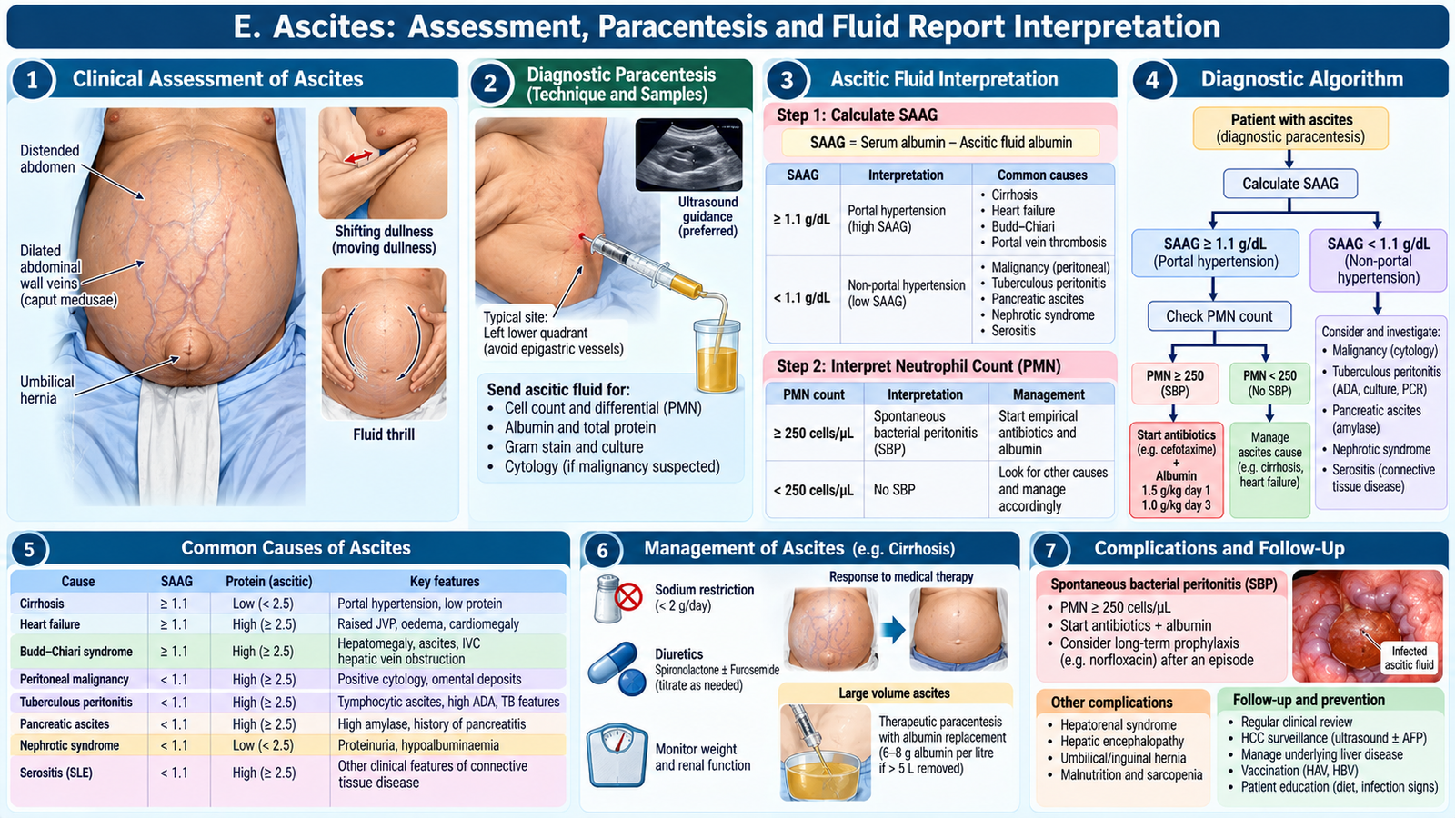
E. Ascites: Assessment, Paracentesis and Fluid Report Interpretation
Ascites in cirrhosis develops because portal pressure rises while effective circulating arterial volume falls, activating renal sodium and water retention. Examine for abdominal distension and shifting dullness; ultrasound confirms smaller volumes and helps guide safe fluid sampling.
When diagnostic paracentesis matters
New-onset ascites and hospital admission with cirrhosis-associated ascites generally warrant diagnostic paracentesis. Repeat promptly if fever, abdominal pain, encephalopathy, shock, renal deterioration or gastrointestinal bleeding raises concern for SBP. The student should understand the indication, consent, asepsis, sample labelling and monitoring when observing the procedure; independent procedural performance is not the goal of this chapter.
Interpreting the report
Request ascitic cell count and differential, albumin with a near-simultaneous serum albumin, total protein and culture when infection is suspected. The serum–ascites albumin gradient (SAAG) = serum albumin − ascitic albumin. A SAAG of ≥1.1 g/dL supports portal-hypertensive ascites; a lower gradient suggests alternative causes such as peritoneal disease. SAAG identifies the physiological mechanism rather than proving cirrhosis.
An ascitic polymorphonuclear neutrophil count ≥250 cells/mm³ supports SBP and requires prompt empirical antibiotic treatment while culture results are pending. A negative culture does not exclude SBP. Send fluid for culture by inoculating blood-culture bottles at the bedside when possible. Bloody fluid may require adjusted cell-count interpretation by the treating team.
Management and monitoring
Restrict dietary sodium, use spironolactone with or without furosemide as clinically appropriate, and monitor weight, blood pressure, creatinine, sodium and potassium. Large-volume therapeutic paracentesis relieves tense ascites; intravenous albumin is generally used when more than 5 L is removed to reduce post-paracentesis circulatory dysfunction. Avoid NSAIDs because they can worsen renal perfusion. Persistent or recurrent ascites warrants specialist assessment and consideration of advanced interventions or transplantation.
Management of Spontaneous Bacterial Peritonitis (SBP): Start empirical intravenous cefotaxime or ceftriaxone promptly for suspected community-acquired SBP, adjusting antibiotics according to local resistance patterns and culture results. Give intravenous albumin (1.5 g/kg on day 1 and 1 g/kg on day 3), particularly when renal dysfunction or significant jaundice is present. Monitor renal function and clinical response; survivors require assessment for secondary antibiotic prophylaxis.
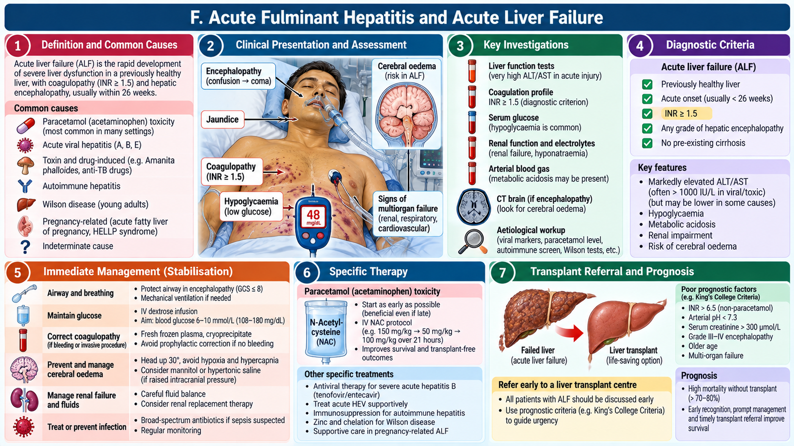
F. Acute Fulminant Hepatitis and Acute Liver Failure
Acute liver failure is rapid loss of hepatic synthetic and detoxifying function, classically with INR ≥1.5 and hepatic encephalopathy in a patient without established cirrhosis, usually within 26 weeks of onset. Severe acute hepatitis without encephalopathy is not yet acute liver failure, but requires close monitoring because deterioration can be rapid.
Causes and recognition
Important causes include acute viral hepatitis, paracetamol toxicity, other drug-induced injury and autoimmune hepatitis. The patient may develop jaundice, vomiting, bleeding tendency, hypoglycaemia, confusion, cerebral oedema and acute kidney injury. A falling ALT during deterioration may reflect loss of viable hepatocytes rather than recovery; track INR, glucose, lactate, renal function and mental state.
Urgent management principles
Stabilize airway, breathing and circulation; check bedside glucose and treat hypoglycaemia; stop suspected hepatotoxic agents; investigate cause; monitor neurological state, fluid balance, electrolytes and coagulation. Give N-acetylcysteine promptly in suspected paracetamol poisoning and according to specialist guidance in selected other acute liver failure presentations. Avoid unnecessary sedatives and nephrotoxic drugs. Treat infection and other complications where present.
Early discussion with a liver-transplant centre is essential when acute liver failure is suspected; referral should not wait for deep coma or multiorgan failure. Vitamin K may be considered if deficiency is possible, but routine correction of INR with plasma solely to normalize the number is not indicated without bleeding or an invasive-procedure requirement, because INR helps assess progression.
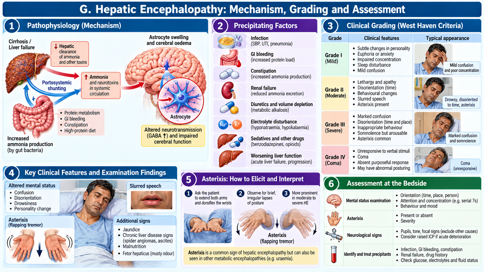
G. Hepatic Encephalopathy: Mechanism, Grading and Assessment
Hepatic encephalopathy (HE) is a potentially reversible brain dysfunction arising from liver insufficiency and/or portosystemic shunting. Ammonia from intestinal protein metabolism is normally converted to urea by the liver; impaired clearance allows ammonia and other mediators to contribute to astrocyte dysfunction and altered neurotransmission. Serum ammonia may support evaluation in selected cases but neither establishes the diagnosis nor reliably grades severity.
Recognize and grade clinically
- Minimal/covert HE: subtle attention, processing or psychomotor changes; ordinary conversation may appear normal.
- Grade I: impaired attention, altered sleep rhythm, mild mood or behavioural changes.
- Grade II: lethargy, disorientation to time, personality change and often asterixis.
- Grade III: marked confusion or somnolence, but the patient remains responsive to stimulation.
- Grade IV: coma with no meaningful responsiveness.
Look for precipitating factors: gastrointestinal bleeding, infection including SBP, constipation, dehydration, overdiuresis, renal dysfunction, electrolyte disturbance and sedative drugs. Always assess alternative causes of confusion such as hypoglycaemia, stroke, sepsis, intoxication and intracranial pathology when indicated.
Asterixis and bedside examination
Observe alertness, orientation and attention before examining for asterixis. Ask the patient to hold arms out, extend wrists and spread fingers for several seconds; irregular loss of posture suggests asterixis. It may be present in other metabolic encephalopathies, and a deeply obtunded patient cannot cooperate with testing. Grade III–IV disease threatens airway protection and requires urgent monitored care.
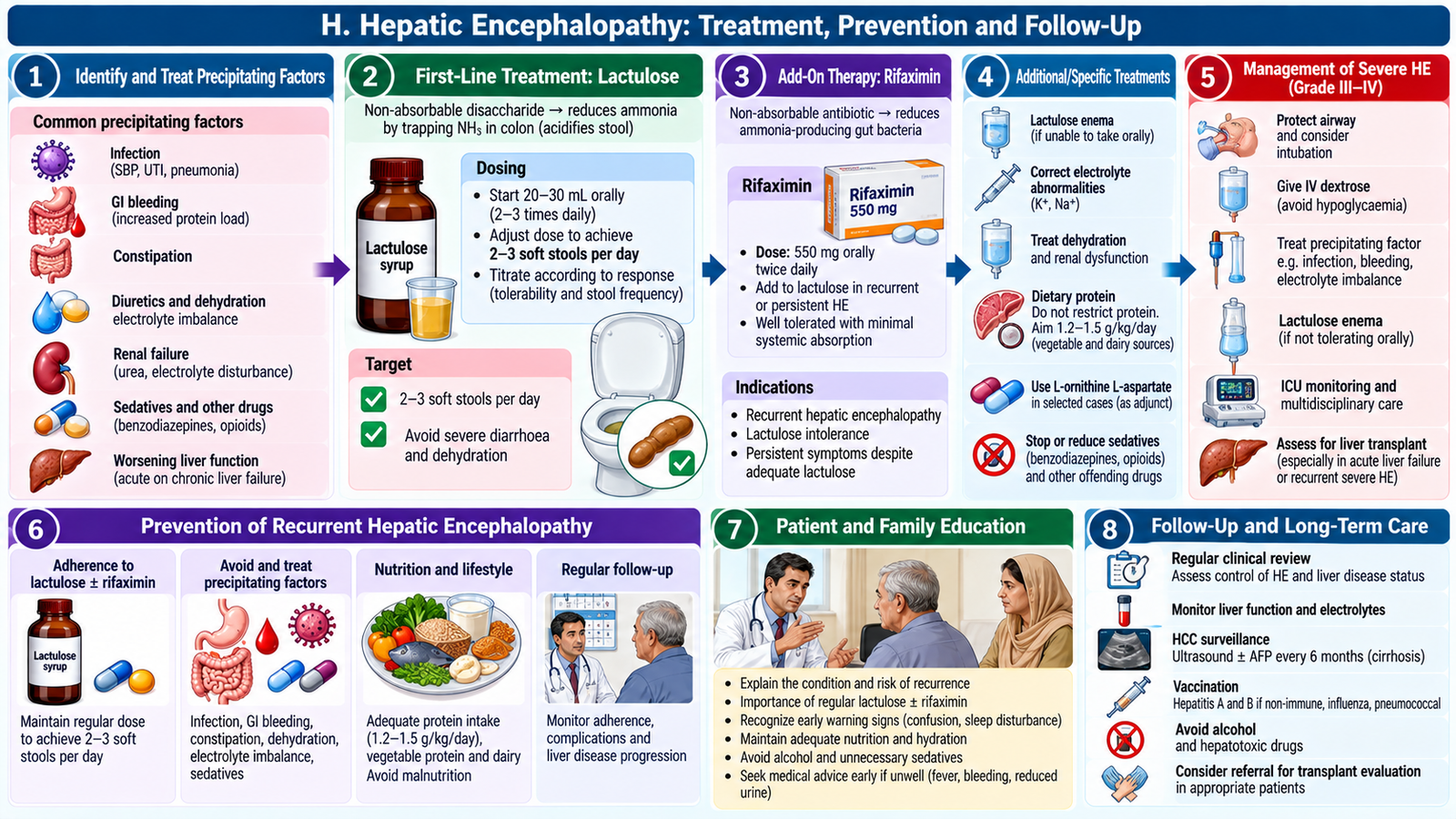
H. Hepatic Encephalopathy Treatment, Prevention and Follow-Up
Treat HE by correcting the precipitating cause and reducing intestinal ammonia production or absorption. Stabilize severe cases first, protect the airway when consciousness is impaired, assess glucose and electrolytes, and identify infection or bleeding.
Specific treatment
Lactulose is first-line therapy for overt HE. Colonic bacteria metabolize it into organic acids, lowering colonic pH and promoting ammonia trapping as ammonium; its laxative effect also increases nitrogen elimination. Titrate to approximately 2–3 soft bowel movements daily, avoiding excessive diarrhoea and dehydration. Rifaximin, a poorly absorbed intestinal antibiotic, is commonly added for recurrent HE or insufficient response and reduces ammonia-producing gut flora.
Do not routinely impose severe protein restriction: malnutrition and muscle loss worsen ammonia handling. Provide adequate nutrition with appropriate protein intake, adjusted by the treating team. Correct potassium depletion, constipation, dehydration and renal impairment; manage variceal bleeding and SBP urgently when present.
Preventing recurrence
Teach the patient and caregiver to recognize sleep reversal, slowed thinking, personality change, reduced concentration and asterixis. Explain adherence to prescribed lactulose, avoidance of constipation and unnecessary sedatives, adequate hydration within the ascites plan, and early presentation for fever, bleeding or confusion. After overt HE, secondary prophylaxis with lactulose, with rifaximin when indicated, reduces recurrence. Recurrent or refractory HE warrants specialist evaluation including transplant suitability.
I. Hepatitis B/C Cirrhosis Counselling and Long-Term Care
Counselling links disease understanding with prevention of further liver injury and timely recognition of complications. Explain that hepatitis B and C can cause persistent inflammation, fibrosis and cirrhosis, and that antiviral treatment can reduce progression. Treatment decisions depend on viral status, liver function and specialist assessment. Effective hepatitis C treatment can cure the infection, but existing cirrhosis may still require surveillance for hepatocellular carcinoma.
Patient-centred counselling
Discuss medication adherence, follow-up visits and monitoring of liver tests, renal function and complications. Recommend avoiding alcohol, non-prescribed herbal remedies and potentially hepatotoxic drugs; review all medications before use. Encourage adequate nutrition and individualized sodium restriction if ascites is present. Advise hepatitis A/B immunization when appropriate and assessment of household or sexual contacts for hepatitis B testing and vaccination. Explain that hepatitis B spreads through blood and sexual contact, while hepatitis C is predominantly blood-borne; casual sharing of meals or ordinary contact does not spread either infection.
Encourage sterile needles and injection equipment, safe blood practices, no sharing razors or toothbrushes, and appropriate precautions around blood. Offer confidential, non-stigmatizing discussion and family support. Patients with cirrhosis need regular assessment for varices and liver cancer according to local specialist pathways, commonly including ultrasound surveillance at approximately six-month intervals. Explain urgent return signs: vomiting blood, black stools, new confusion, fever with abdominal swelling, marked jaundice or reduced urine output.
🩺 AIM Clinical Bridge
A 54-year-old patient with hepatitis C-related cirrhosis presents with increasing abdominal distension and new confusion. Examination reveals shifting dullness and asterixis; creatinine has increased. The important next step is not simply to label the confusion as hepatic encephalopathy. Assess airway and glucose, look for infection or bleeding, review diuretics and electrolytes, and arrange diagnostic ascitic fluid analysis. If the fluid neutrophil count is ≥250/mm³, treat suspected SBP promptly while also treating encephalopathy. This shows how one complication can trigger another.
Integrated Mechanism Flow
Repeated hepatocyte injury → fibrosis and regenerative nodules → distorted hepatic blood flow and reduced liver reserve → portal hypertension and impaired detoxification → ascites, varices and neurotoxin accumulation → infection/bleeding may precipitate encephalopathy → early recognition and treatment reduce deterioration.
Important Comparison: Acute Liver Failure vs Decompensated Cirrhosis
| Feature | Acute liver failure | Decompensated cirrhosis |
|---|---|---|
| Background | Acute injury without established cirrhosis | Longstanding fibrosis and nodules |
| Key syndrome | INR ≥1.5 plus encephalopathy | Ascites, variceal bleed or encephalopathy |
| Common concerns | Hypoglycaemia, cerebral oedema, rapid deterioration | Portal hypertension, SBP, renal dysfunction |
| Priority | Emergency stabilization and transplant-centre referral | Identify and treat decompensation; long-term surveillance |
AIM High-Yield Review
- ⭐ ALT/AST indicate hepatocyte injury; INR and albumin help assess synthetic function, with important confounders.
- ⭐ New confusion plus INR ≥1.5 in acute hepatic injury suggests acute liver failure and requires urgent referral.
- Portal hypertension produces splenomegaly, varices and ascites; synthetic dysfunction worsens oedema and bleeding risk.
- A nodular liver with ascites and thrombocytopenia can indicate advanced cirrhosis even with modest ALT elevation.
- ⭐ SAAG ≥1.1 g/dL supports portal-hypertensive ascites, not cirrhosis specifically.
- ⭐ Ascitic neutrophils ≥250/mm³ support SBP; begin prompt treatment without waiting for culture positivity.
- New ascites or suspected infection in hospitalized cirrhosis calls for diagnostic paracentesis.
- Lactulose reduces gut ammonia burden; aim for 2–3 soft stools/day without dehydration.
- Rifaximin is an add-on for recurrent or insufficiently controlled hepatic encephalopathy.
- Asterixis is a metabolic encephalopathy sign, not specific for hepatic disease.
- GI bleeding, infection, constipation, dehydration and sedatives can precipitate encephalopathy.
- Avoid severe protein restriction in cirrhosis; prevent malnutrition and sarcopenia.
- Hepatitis B/C cirrhosis requires antiviral assessment, complication surveillance and blood-exposure counselling.
- Variceal bleeding, coma, renal deterioration and suspected SBP are urgent escalation signs.
Topic 2 — Video Learning
Hepatocellular Disease, Liver Failure, Cirrhosis & Hepatic Encephalopathy
Featured Video: Cirrhosis — Causes, Diagnosis & Management
Osmosis from Elsevier • Covers fibrosis, portal hypertension, ascites, hepatic dysfunction and complications.
If the embedded player is restricted, watch on YouTube.
Additional Topic 2 Video Lectures
Liver Function Tests, Acute Liver Failure & Ascites: Ninja Nerd Hepatic Disorders lecture collection
Hepatic Encephalopathy: Osmosis clinical sciences video