
This chapter follows the KMU outcomes and connects acute diarrhea, dehydration, chronic diarrhea and celiac disease into one clinical approach. Understand the explanations first; use the AIM High-Yield Review for revision.
TOPIC 5. Acute & Chronic Diarrhea, Malabsorption & Celiac Disease
A patient-centred approach to pediatric diarrheal illness, dehydration, malabsorption and lifelong celiac care.
Topic Introduction
Diarrhea is one of the commonest reasons children need clinical assessment. Most short episodes are infectious and resolve with continued feeding and effective fluid replacement. However, watery stool can quickly lead to dehydration, and prolonged diarrhea may indicate intestinal disease with important effects on growth.
For Final Year practice, the main clinical task is to assess danger and hydration first, then decide whether the child has a self-limited illness or needs focused investigation for an underlying cause. This chapter explains the mechanisms of diarrhea, clinical dehydration classification, appropriate investigations and management, and the diagnosis, nutritional care and parent counselling required in celiac disease.
A. Understanding Diarrhea: Mechanisms, Causes & Clinical Patterns
Diarrhea is the passage of unusually loose or watery stools, often more frequent than usual. In infants, a change from the child’s normal stool consistency is more meaningful than frequency alone. Frequent soft stools in a thriving, exclusively breastfed infant may be normal.
Normally the small intestine absorbs nutrients, electrolytes and most intestinal water. The colon reabsorbs much of the remaining water. Diarrhea develops when intestinal fluid secretion exceeds absorption, when the mucosa becomes inflamed or when nutrients are not adequately absorbed.
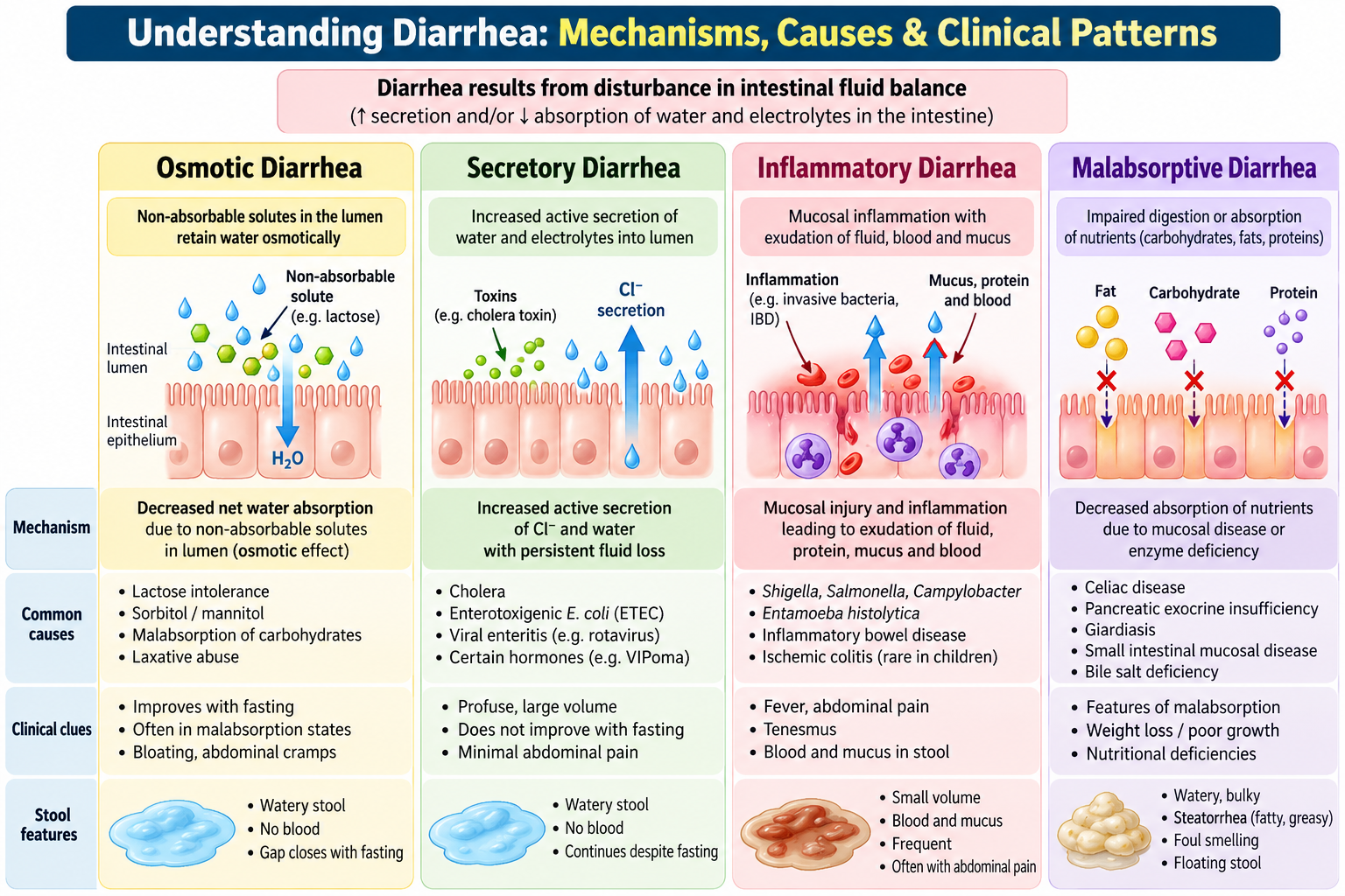
How intestinal mechanisms produce different stool patterns
- Osmotic diarrhea: unabsorbed substances hold water in the lumen. After damage to the intestinal brush border, lactose may remain unabsorbed, causing bloating and watery stools; symptoms generally improve when the offending sugar is removed.
- Secretory diarrhea: intestinal cells secrete excessive water and salts, sometimes under the effect of bacterial enterotoxins. Large-volume watery diarrhea can continue despite fasting.
- Inflammatory diarrhea: mucosal invasion or inflammation damages the bowel lining. Fever, abdominal pain, blood or mucus may accompany diarrhea.
- Malabsorptive diarrhea: impaired digestion or absorption results in persistent loose or bulky stools, abdominal distension, deficiency states and impaired growth.
Classify the course before choosing the next step
Acute diarrhea lasts less than 14 days and is commonly infectious. Persistent diarrhea begins as an acute episode but continues for 14 days or more. Chronic diarrhea generally describes symptoms lasting approximately four weeks or longer and calls for evaluation of an ongoing cause. These terms overlap in clinical practice.
Viral gastroenteritis is a frequent cause of acute watery diarrhea; bacterial disease including cholera becomes important in appropriate exposure or outbreak settings. In prolonged illness consider postinfectious lactose malabsorption, giardiasis, celiac disease and functional toddler’s diarrhea. The central distinction is between immediate fluid loss and ongoing intestinal or nutritional disease.
B. Acute Watery Diarrhea: Clinical Assessment & Differential Diagnosis
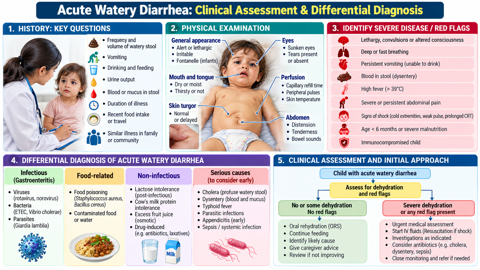
In a child with acute watery diarrhea, first decide whether the child is stable and can drink. Identifying a specific microorganism is less urgent than recognizing fluid loss and dangerous illness. Typical gastroenteritis produces loose stools, sometimes vomiting, fever and abdominal discomfort.
Focused clinical assessment
Ask about onset and duration, stool frequency and volume, blood or mucus, vomiting, fever, oral intake and breastfeeding, urine output, sick contacts, food and water exposure, and recent medicines. Fewer wet nappies, poor drinking or unusual sleepiness may be early clues to clinically important dehydration. Ask whether the child has continued to gain weight appropriately.
Examination emphasizes general appearance and alertness, temperature, pulse, breathing, capillary refill, extremity warmth, hydration findings, and focused abdominal tenderness or distension. Weight is useful both for fluid assessment and for monitoring recovery.
Clinically useful differentials
- Viral gastroenteritis: watery stool with vomiting, sometimes mild fever; commonly self-limited.
- Invasive bacterial intestinal infection: fever, abdominal pain and blood in stool suggest mucosal inflammation and require further assessment.
- Cholera: profuse watery stools with rapid dehydration, particularly during an outbreak or following relevant exposure.
- Giardiasis or secondary lactose intolerance: consider when loose stools persist with bloating after the usual acute phase.
Urgent warning signs: lethargy or altered consciousness, inability to drink, signs of poor circulation, markedly reduced urine output, persistent bilious vomiting, severe focal abdominal pain or distension, and significant bloody diarrhea with systemic illness. These findings should prompt urgent pediatric evaluation rather than routine home treatment.
C. Pediatric Dehydration: Clinical Assessment, Classification & Consequences
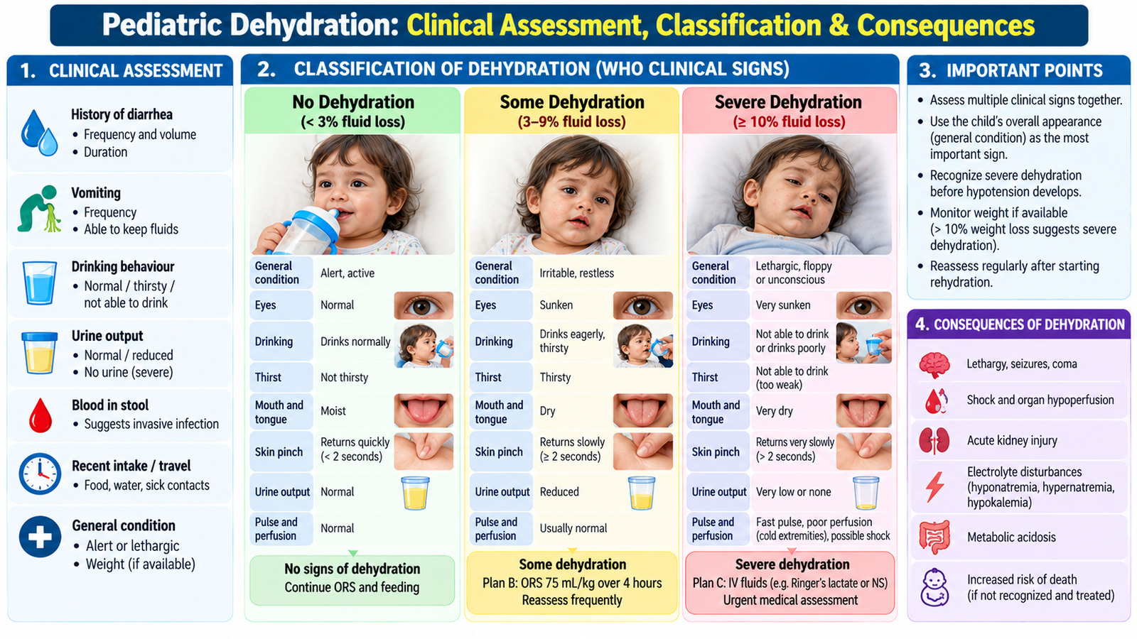
Children lose circulating water and electrolytes quickly because their fluid turnover is relatively high. Diarrheal losses contain water, sodium, potassium and bicarbonate. As losses increase, the child becomes thirsty, then may develop impaired tissue perfusion, electrolyte disturbance and metabolic acidosis.
Recognize severity from combined clinical signs
The WHO clinical approach uses general condition, appearance of the eyes, drinking behavior and skin-pinch recoil. No single sign alone reliably grades dehydration. Interpret the findings as a pattern, while considering age, nutritional status and pre-existing illness.
| Feature | Some dehydration | Severe dehydration |
|---|---|---|
| General condition | Restless, irritable | Lethargic, unconscious |
| Eyes | Sunken | Sunken |
| Drinking | Thirsty, drinks eagerly | Drinks poorly, unable to drink |
| Skin pinch | Goes back slowly | Goes back very slowly |
At least two signs in a category are needed for WHO classification, with severe signs taking priority. If neither category is fulfilled, classify as no dehydration. In severe acute malnutrition, ordinary dehydration signs may be unreliable; specialist assessment is important.
Connect fluid loss to clinical deterioration
Reduced intravascular volume causes tachycardia and reduced peripheral perfusion. Cold extremities, weak pulses, prolonged capillary refill and reduced consciousness suggest circulatory compromise. Hypotension is a late sign of pediatric shock. Bicarbonate loss contributes to metabolic acidosis; potassium loss may cause hypokalemia. Sodium can be low, normal or high, depending on relative salt and water losses and fluids given.
D. Acute Diarrhea: Investigations, Rehydration & Monitoring
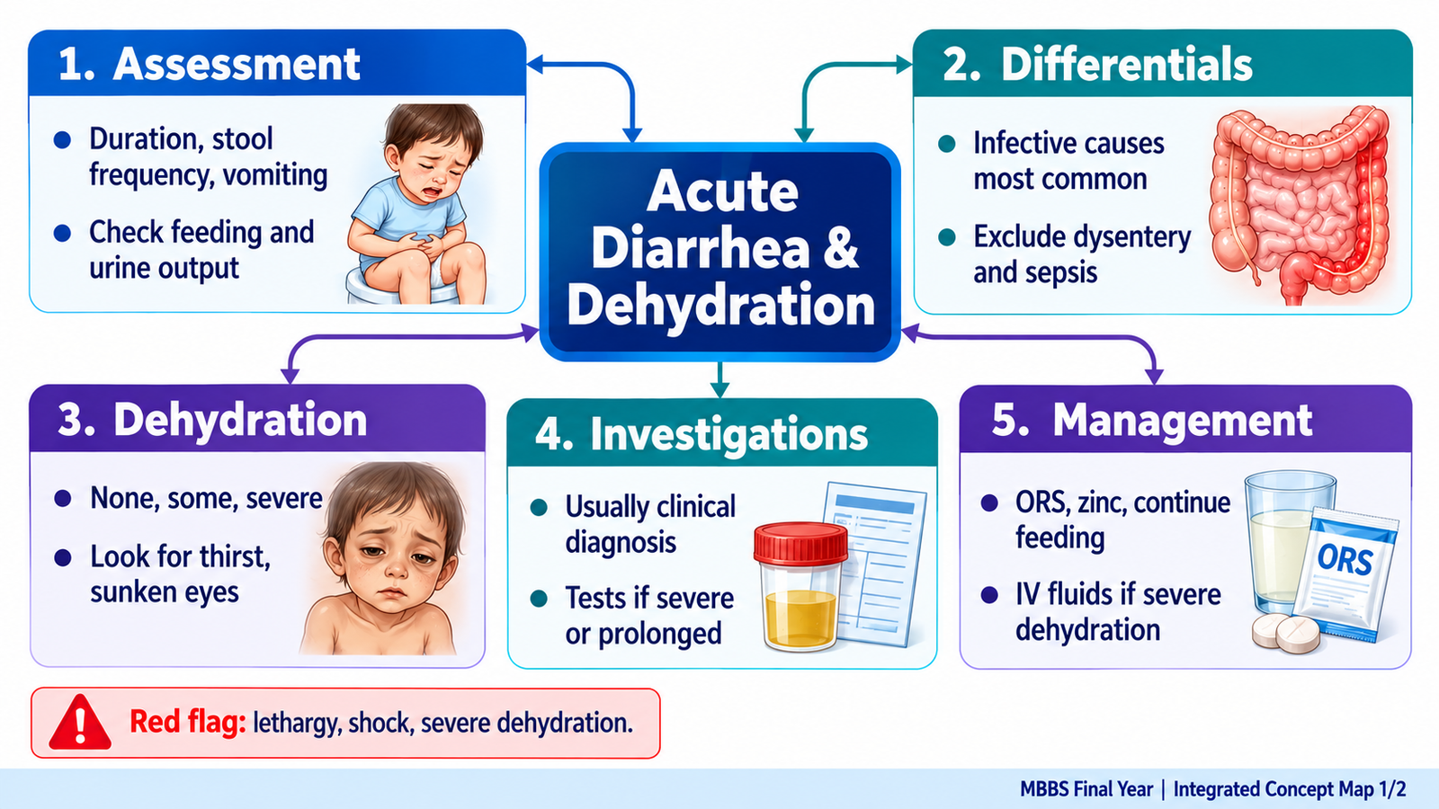
Most children with uncomplicated watery diarrhea need clinical assessment and rehydration, not routine stool tests. Investigations are reserved for severe dehydration, atypical illness, bloody diarrhea, suspected outbreaks or prolonged symptoms.
Choose tests for a clear clinical reason
- Electrolytes, urea, creatinine and glucose: consider in severe illness or children receiving intravenous treatment. Abnormal sodium or potassium influences fluid monitoring; increased urea and creatinine may indicate dehydration-related renal hypoperfusion.
- Acid–base assessment: useful in severely ill children; metabolic acidosis may result from bicarbonate loss and poor perfusion.
- Stool microscopy, culture or targeted testing: consider with bloody or persistent diarrhea, suspected parasitic disease or an outbreak. Results must be interpreted with symptoms and epidemiology.
Why oral rehydration solution works
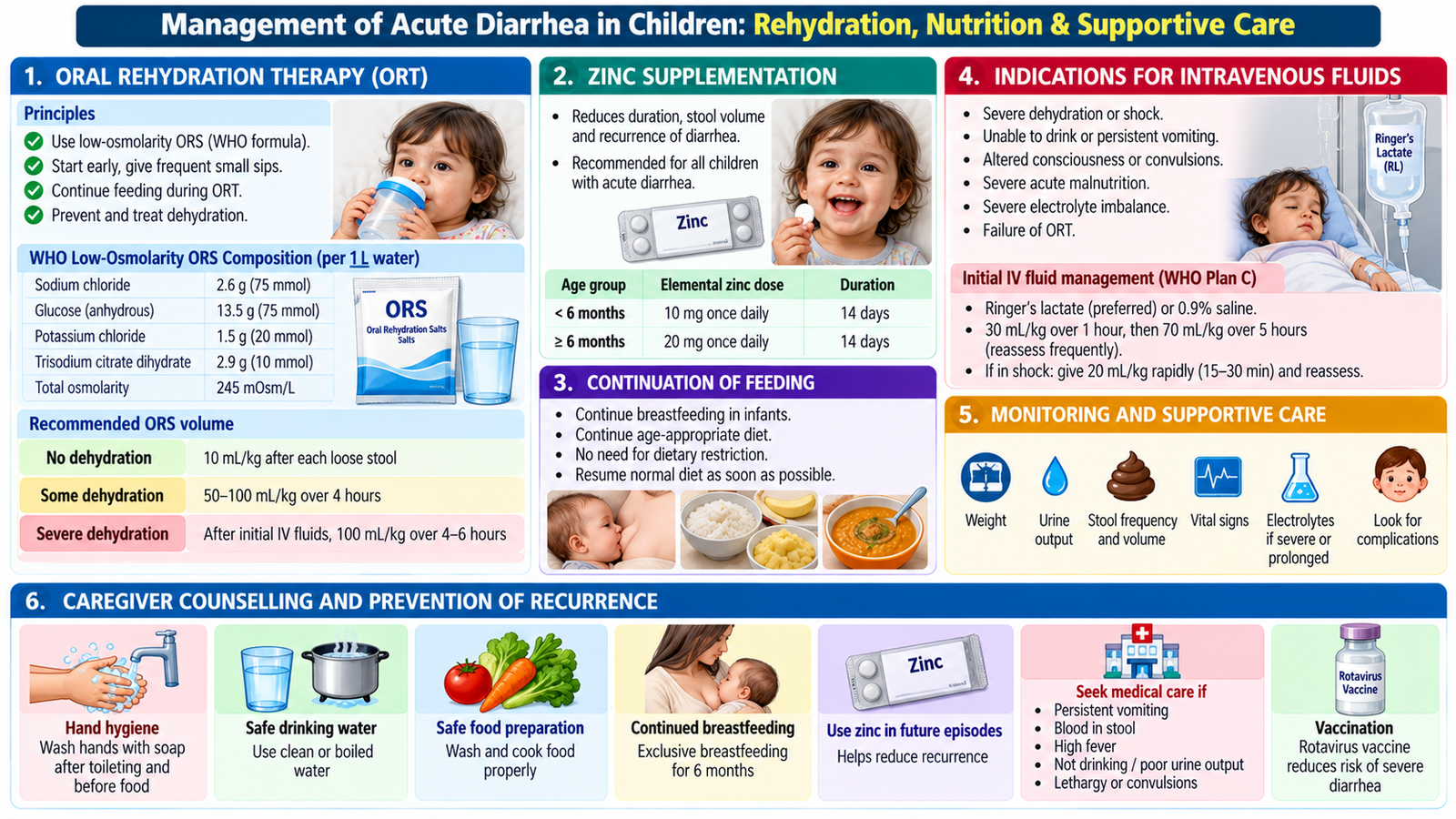
Intestinal sodium–glucose cotransport remains active in many diarrheal illnesses. Glucose helps sodium enter intestinal cells, and water follows. Therefore oral rehydration solution (ORS) restores losses even while diarrhea continues; it does not need to stop stool output before it can help.
Plan A (no dehydration): extra safe fluids and ORS after each loose stool, continue breastfeeding and age-appropriate food, provide indicated zinc supplementation and explain danger signs. Plan B (some dehydration): supervised ORS rehydration, commonly 75 mL/kg over four hours under the WHO framework, followed by reassessment and replacement of ongoing losses. Plan C (severe dehydration): urgent appropriately supervised intravenous isotonic fluids with repeated assessment; shock requires immediate emergency resuscitation.
Continue breastfeeding and normal feeding as tolerated to protect recovery and growth. Zinc supplementation is recommended in appropriate pediatric settings according to local/WHO protocols. Antibiotics are not routine in uncomplicated watery gastroenteritis; they are reserved for selected bacterial or other treatable infections. Avoid routine antimotility medication in young children.
Monitor response and support safe home care
Improving alertness, drinking, urine output, pulse and perfusion indicate recovery. Failure to drink, progressive lethargy, shock signs, blood in stool or persistent worsening requires escalation. Before discharge, a caregiver should understand ORS preparation and use, continued feeding, hand hygiene, safe water and when to return for reassessment.
E. Chronic Diarrhea & Malabsorption: Clinical Evaluation and Investigation
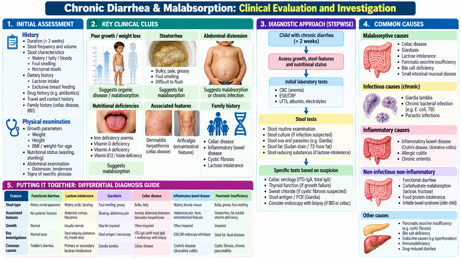
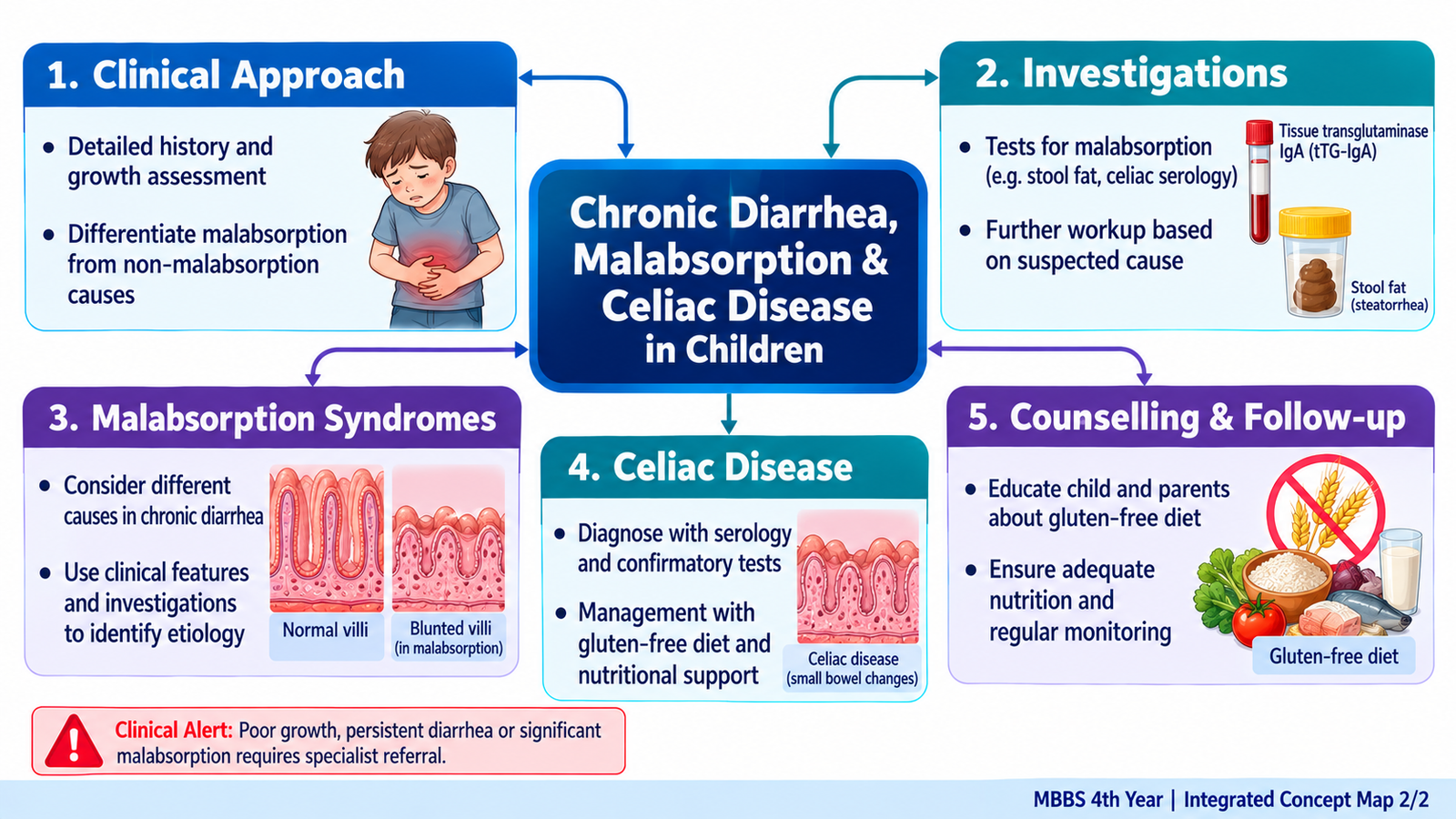
Persistent loose stools need a different question from acute gastroenteritis: is the bowel still recovering, or is ongoing disease preventing normal absorption and growth? The clinical history, growth curve and stool characteristics direct the investigation.
What malabsorption looks like
Fat malabsorption can produce bulky, pale, greasy, offensive stools (steatorrhea). Loss of nutrients causes abdominal distension, weight faltering, short stature, anemia or deficiencies. Persistent watery stools after milk feeds may point to secondary lactose intolerance following enterocyte injury.
Ask about onset, dietary relationships (including wheat-containing foods or lactose), blood or mucus, recurrent infection, abdominal pain, family history, previous treatments and growth. Examination should assess serial weight and height, nutritional state, pallor, abdominal distension and clues to deficiency. A thriving child with loose stools but no alarm features may have functional toddler’s diarrhea, unlike a child with growth faltering.
Prioritize a small differential diagnosis
- Postinfectious lactose intolerance: transient loss of brush-border lactase; bloating and osmotic diarrhea follow lactose intake.
- Celiac disease: immune-mediated villous injury causes poor absorption, anemia and impaired growth.
- Giardiasis: protozoal infection may produce prolonged diarrhea and bloating.
- Inflammatory bowel disease: suspect with blood in stool, weight loss, ongoing pain or inflammatory features.
- Functional toddler’s diarrhea: loose stools with normal activity and preserved growth after exclusion of alarm signs.
Target investigations and interpret them
Growth chart: a falling growth trajectory supports significant ongoing disease and makes functional diarrhea less likely. Complete blood count: microcytic anemia may reflect iron malabsorption. Stool testing: select parasite tests or other investigations by exposure and stool pattern rather than ordering everything. Celiac serology: request tissue transglutaminase IgA (tTG-IgA) with total IgA when celiac disease is suspected. Additional inflammatory testing or specialist endoscopy is guided by clinical findings.
Management addresses the underlying condition while maintaining hydration and adequate energy, protein and micronutrient intake. Persistent weight faltering, significant anemia, bloody diarrhea or diagnostic uncertainty warrants pediatric referral.
F. Celiac Disease in Children: Mechanism, Recognition & Diagnosis
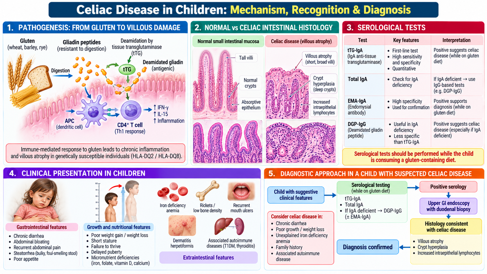
Celiac disease is an immune-mediated enteropathy triggered by gluten in wheat, barley and rye in genetically susceptible people. Gluten exposure activates an intestinal immune response, damaging small-intestinal villi. Flattened villi reduce the absorptive surface and may cause diarrhea, deficiency states and impaired growth.
Why symptoms and deficiencies occur
Typical mucosal changes are villous atrophy, crypt hyperplasia and increased intraepithelial lymphocytes. Because the proximal small intestine is important for iron absorption, iron-deficiency anemia may be prominent even without marked diarrhea. Children may have bulky stools, abdominal distension, irritability, poor weight gain, short stature or delayed puberty. Some have few gastrointestinal symptoms.
Suspect the disease in unexplained chronic diarrhea, faltering growth, abdominal distension, persistent iron-deficiency anemia or a relevant family history. First-degree relatives and children with type 1 diabetes have an increased likelihood of celiac disease.
Use serology correctly
Initial testing generally includes tTG-IgA together with total serum IgA, while the child is eating gluten. Total IgA matters because IgA deficiency can make an IgA-based test falsely negative; suitable IgG-based tests are then used. Starting a gluten-free diet before completing investigation can reduce antibody levels and make the diagnosis harder to establish.
Duodenal biopsy, obtained at upper gastrointestinal endoscopy where indicated, can demonstrate the characteristic mucosal injury. Under recognized pediatric specialist criteria, selected children with very high tTG-IgA and confirmation by endomysial antibody in a separate sample may be diagnosed without biopsy. This is not a general rule for all positive serology; diagnostic decisions require appropriate pediatric expertise.
Differentiate lactose intolerance: low lactase activity causes osmotic symptoms after lactose ingestion, whereas celiac disease causes gluten-triggered immune damage. Temporary lactose intolerance can coexist with active celiac disease because villous injury also reduces brush-border enzyme activity.
G. Celiac Disease Management: Nutrition, Monitoring & Family Counselling
Confirmed celiac disease is managed with a strict lifelong gluten-free diet. Removing gluten allows intestinal inflammation to settle and absorptive function to recover. Treatment also includes correction of nutritional deficiencies and attention to growth, school life and family confidence.
Build a nutritionally adequate gluten-free diet
Exclude foods containing wheat, barley and rye. Naturally gluten-free staples include rice, maize, potatoes and a range of pulses and vegetables. Certified uncontaminated gluten-free oats may be considered when suitable and tolerated. Explain that gluten may be present in sauces, processed snacks and other unexpected products and that shared utensils or preparation surfaces can cause cross-contact.
Evaluate height, weight and growth trajectory, dietary intake and clinically relevant deficiencies including iron, folate, calcium or vitamin D. Correct identified deficiencies and support a balanced intake of energy, protein, fiber and micronutrients. A pediatric dietitian can help families avoid both accidental gluten exposure and unnecessary dietary restriction.
Monitor recovery and address practical adherence
Symptoms often improve after gluten exclusion, but recovery of growth and serology may take longer. Follow-up focuses on symptom control, growth, nutrition, adherence and repeat serology when appropriate. Persistent symptoms require assessment for hidden gluten exposure and other possible conditions.
Parents should understand that this is a long-term immune disease, not simply a temporary food intolerance. Explain food-label reading, avoiding cross-contamination, planning school lunches and celebrations, and providing safe alternatives without isolating the child. Include older children in decisions appropriate to their age. The goal is normal growth and everyday participation with a safe, varied diet.
AIM High-Yield Review
- The immediate risk in acute childhood diarrhea is dehydration and impaired perfusion.
- WHO dehydration assessment uses combined findings in general condition, eyes, drinking and skin pinch.
- Severe dehydration or shock requires urgent fluid management and clinical reassessment.
- ⭐ ORS exploits sodium–glucose cotransport, which remains functional in many diarrheal illnesses.
- Continue breastfeeding and age-appropriate feeding; routine antibiotics are not indicated for simple watery diarrhea.
- Investigate selected severe, bloody or persistent cases rather than every acute episode.
- Chronic diarrhea with poor growth, distension, steatorrhea or anemia suggests organic disease.
- Preserved growth in a well child can favor functional toddler’s diarrhea after review of alarm features.
- ⭐ Celiac disease causes gluten-triggered immune injury with villous atrophy and malabsorption.
- ⭐ Test tTG-IgA and total IgA while the child remains on a gluten-containing diet.
- IgA deficiency can make IgA-based celiac serology falsely reassuring.
- Duodenal biopsy or a selected specialist no-biopsy diagnostic pathway establishes celiac disease.
- ⭐ Lifelong strict gluten exclusion with nutritional recovery and growth monitoring is essential.
- Parent counselling must cover labels, cross-contact, school foods and long-term adherence.
🎥 AIM Video Learning
Topic 5: Acute & Chronic Diarrhea, Malabsorption & Celiac Disease
Video 1: Acute Diarrhea & Dehydration in Children
Clinical assessment, dehydration severity, oral rehydration therapy, IV fluid indications and nutritional management.
Video 2: Celiac Disease — Pathophysiology, Diagnosis & Management
Gluten-related intestinal injury, clinical manifestations, investigations and dietary management.