
This chapter follows the KMU outcomes and connects the choice of feeding route with safe access, monitoring and patient counselling. Understand the explanations first; use the final high-yield review for revision.
📖 AIM Learning Material
Clinical Nutritional Support — Enteral & Parenteral Nutrition
MS Module | Surgical Nutrition • Safe selection, delivery and monitoring of nutritional support
Topic Introduction
Hospitalised patients may be unable to eat because of illness, surgery or swallowing difficulty. If this persists, inadequate nutrition contributes to loss of muscle, impaired immunity, delayed wound healing and poor recovery. Nutritional support provides energy, protein, fluid and essential nutrients by the safest usable route: orally, through a feeding tube into the gastrointestinal tract, or intravenously. The central clinical decision is not simply “which feed?” but “can the patient eat, does the gut work, and how can treatment be monitored safely?” This chapter explains how to recognise malnutrition, select an appropriate route, understand NG and PEG procedures, use parenteral access, interpret intake–output records and counsel patients before treatment.
A. Recognising Malnutrition and Assessing Nutritional Needs
What malnutrition means
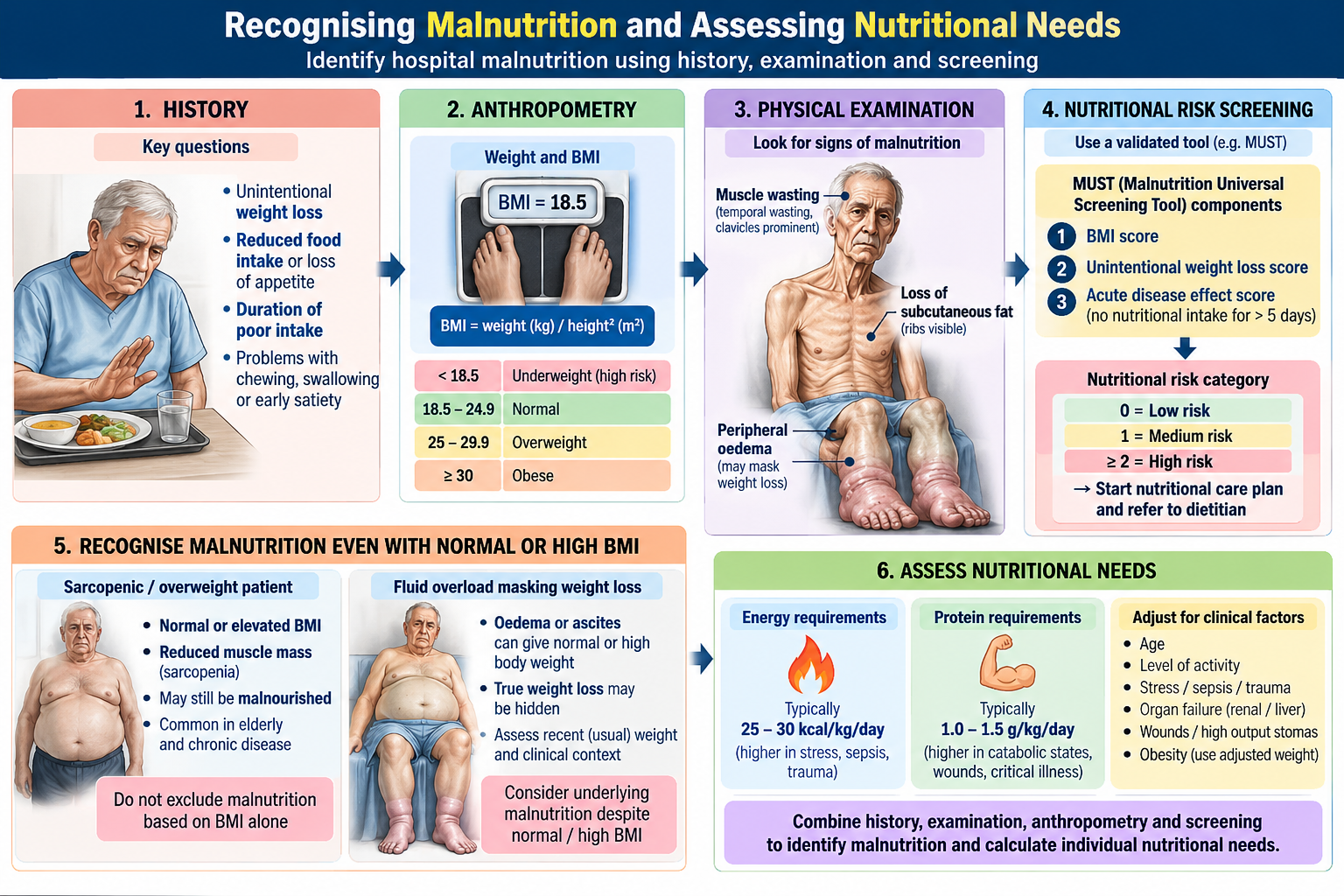
Malnutrition is a deficiency, excess or imbalance of energy, protein or other nutrients that adversely affects body composition, function or clinical outcomes. In hospital practice, the immediate concern is undernutrition: inadequate intake or absorption combined with the increased demands of illness. A patient can be malnourished despite appearing overweight.
Who is at risk
Look for unintentional weight loss, poor intake for several days, difficulty swallowing, persistent vomiting, intestinal disease, major surgery, infection and prolonged critical illness. Ask about usual weight, recent weight change, appetite, ability to eat, gastrointestinal symptoms and previous feeding support. Examine muscle wasting, loss of subcutaneous fat, oedema, hydration and functional strength.
Practical assessment
Record current weight, height and body mass index, but interpret BMI cautiously when oedema or fluid overload alters weight. Compare recent weight with usual weight and estimate how much food the patient has actually consumed. A structured screening tool such as MUST may help identify risk. Clinical context and dietetic assessment guide estimated energy, protein and fluid requirements; these are reviewed as the illness changes.
Why early recognition matters
Undernutrition impairs wound healing, immunity and muscle strength and may increase infections, length of stay and recovery time. Nutritional support complements treatment of the underlying disease; it does not replace resuscitation or management of sepsis and surgical complications.
B. Choosing Oral, Enteral or Parenteral Nutritional Support
The basic decision
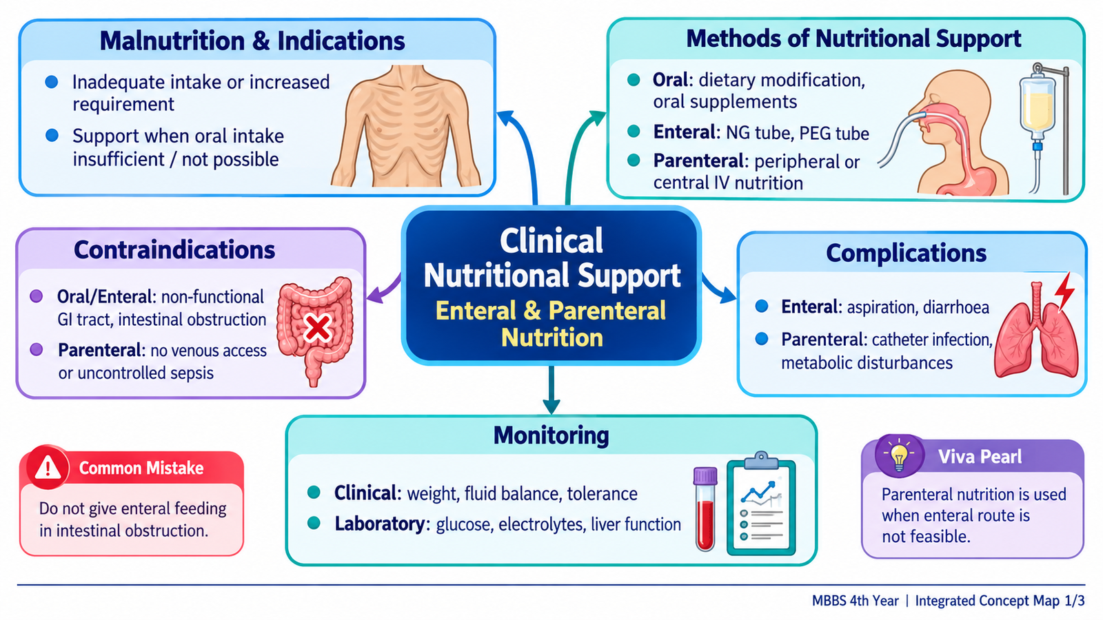
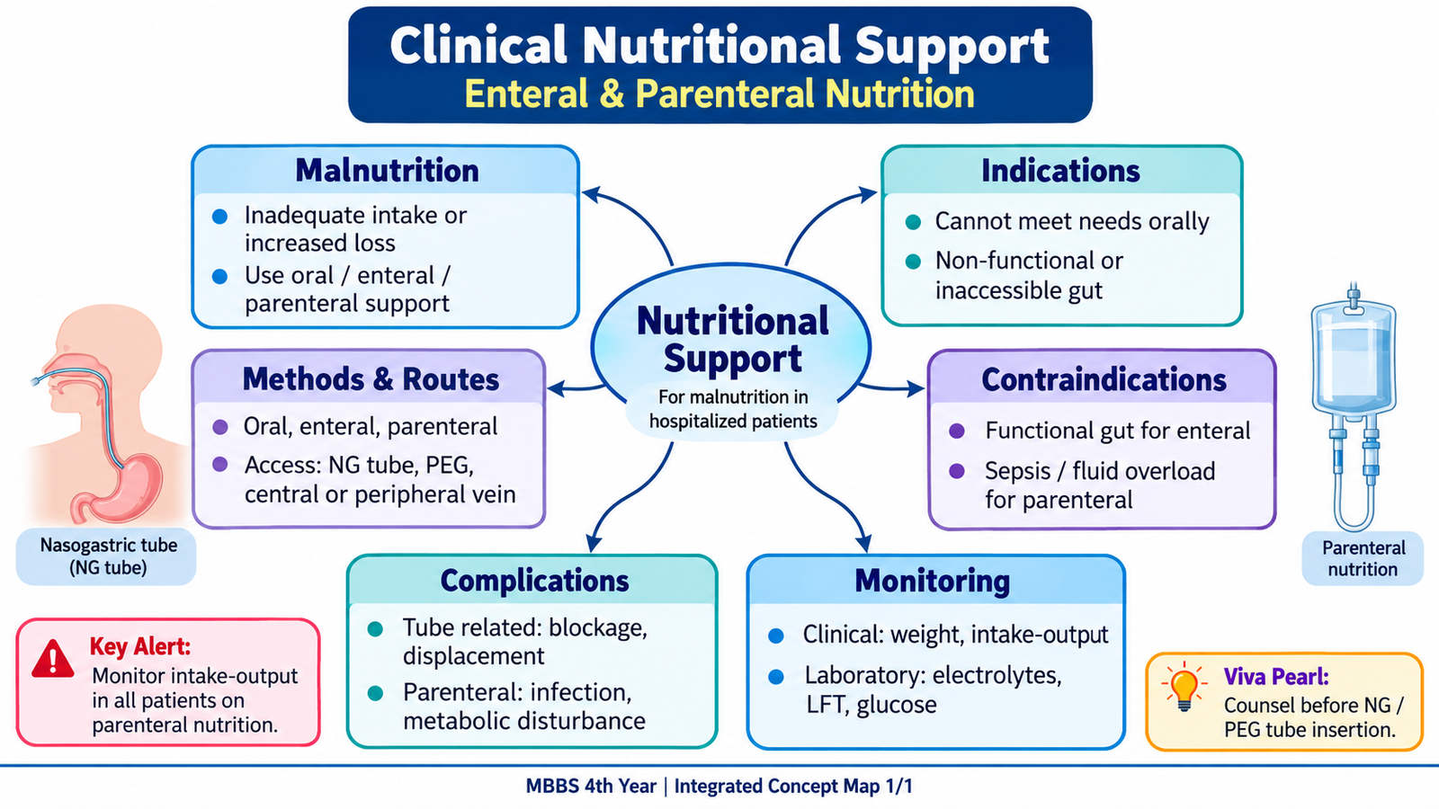
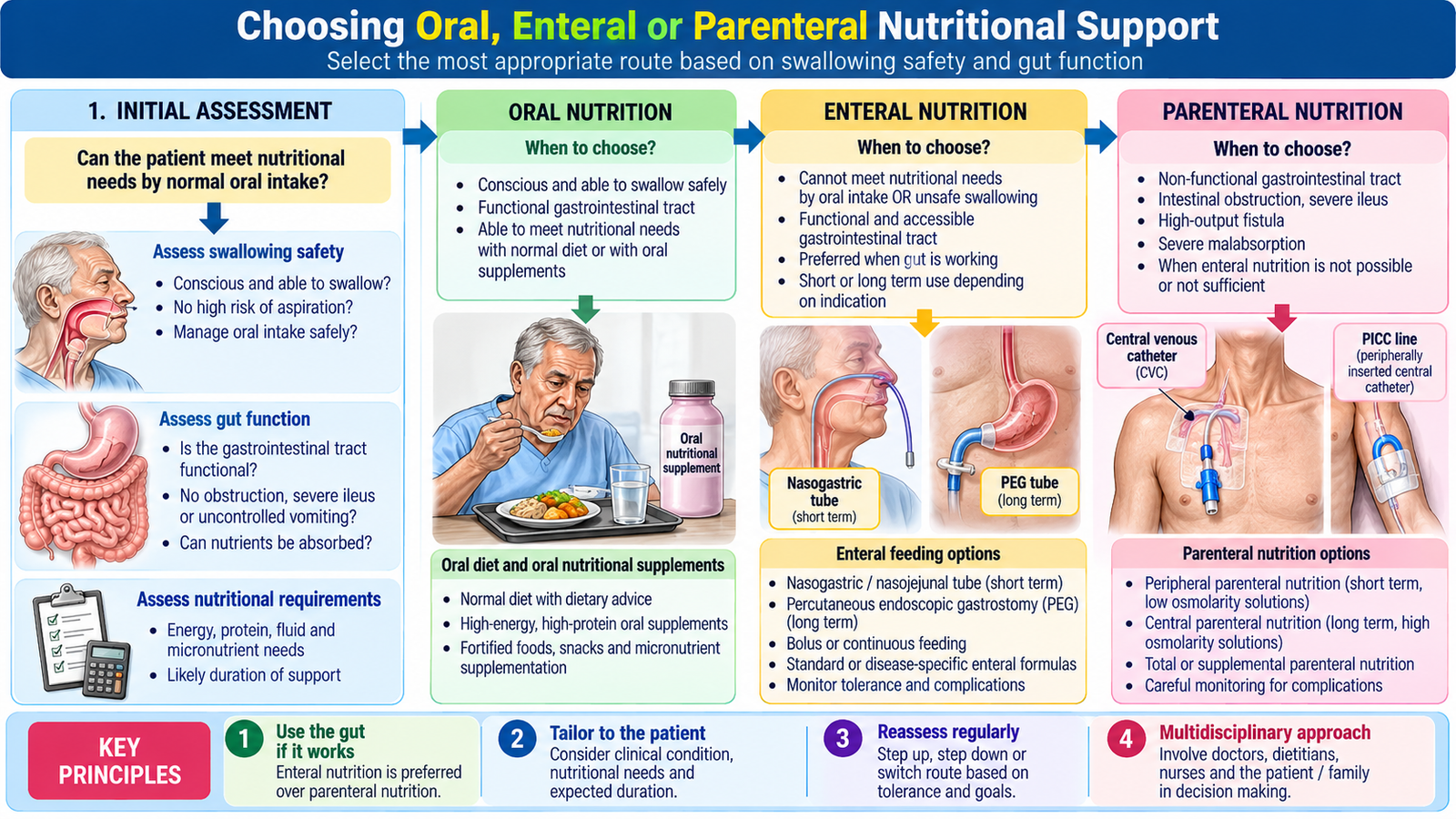
First ask whether the patient can swallow safely and meet needs by mouth. If so, improve meals, assist feeding and consider oral nutritional supplements. If oral intake is inadequate but the gastrointestinal tract is functional and accessible, enteral tube feeding is generally preferred. Use parenteral nutrition when adequate nutrition cannot be delivered safely through the gut.
Oral support
Oral nutritional support includes food fortification, high-energy/high-protein meals, supplements and feeding assistance. It is appropriate when swallowing is safe and the patient can absorb nutrients. It may be insufficient in severe dysphagia, persistent vomiting or markedly reduced intake; aspiration risk must be considered.
Enteral support
Enteral nutrition delivers liquid feed into the stomach or small bowel through a tube. It uses the gut and avoids many catheter-related complications of parenteral nutrition. It is useful in patients unable to eat adequately but with a functioning gastrointestinal tract, such as selected neurological dysphagia or postoperative patients. Contraindications include bowel obstruction, perforation or intestinal ischaemia; uncontrolled shock or severe gastrointestinal intolerance may require deferral and reassessment.
Parenteral support
Parenteral nutrition delivers glucose, amino acids, lipids, electrolytes, vitamins, trace elements and fluid intravenously. Consider it when the gastrointestinal tract is nonfunctional, inaccessible or unable to provide sufficient nutrition, for example prolonged ileus, intestinal obstruction or severe malabsorption. It carries risks of line infection, thrombosis, hyperglycaemia, electrolyte disturbances and liver dysfunction. The choice should be reassessed daily, with transition to gut feeding when feasible.
Avoiding a common error
“Unable to eat” does not automatically mean parenteral nutrition: assess whether tube feeding can use a functioning gut. Equally, the presence of bowel sounds alone does not prove that enteral feeding is safe in a patient with suspected obstruction or ischaemia.
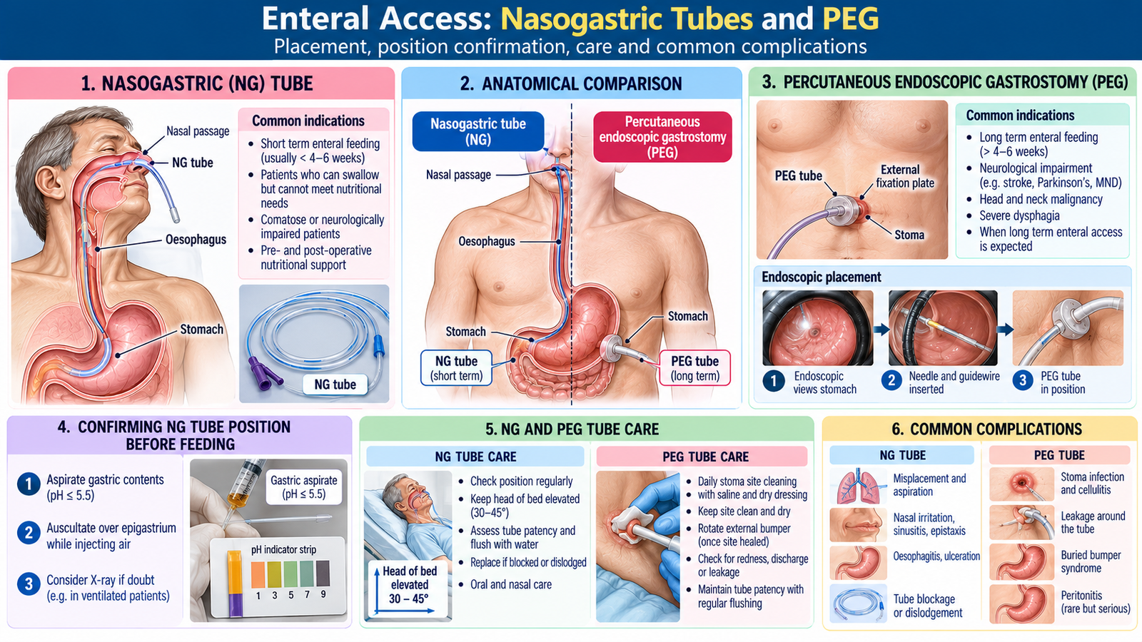
C. Enteral Access: Nasogastric Tubes and PEG
Selecting the route
A nasogastric (NG) tube passes through the nose into the stomach and is generally suitable for short-term feeding. A nasojejunal route may be chosen when gastric feeding is unsuitable but small-bowel feeding is possible. Percutaneous endoscopic gastrostomy (PEG) provides longer-term access directly to the stomach when prolonged enteral feeding is anticipated and the gut is functional.
NG tube insertion: understanding the procedure
Before insertion, explain the purpose, assess contraindications such as suspected basal skull fracture or significant facial trauma, check the nostrils and obtain appropriate consent. A trained clinician advances a lubricated tube through the nostril and pharynx while monitoring comfort and respiratory distress. Confirm correct position according to local policy, commonly with aspirate pH testing and radiography when indicated, BEFORE administering feed, water or medicines. The external tube length and fixation should be documented.
Why position confirmation matters
An NG tube can enter the airway and cause aspiration or lung injury if feeding begins without reliable confirmation. The “whoosh” auscultation method is not a dependable way to confirm gastric placement. Recheck position when displacement is suspected, especially after coughing, vomiting or a change in external tube length.
PEG insertion and aftercare
PEG is usually placed endoscopically through the abdominal wall into the stomach. Students should understand its indication and observe the procedure rather than attempt independent insertion. Following placement, monitor pain, bleeding, leakage, infection and signs of peritonitis; inspect and clean the stoma as instructed, protect the tube from traction, and follow the local feeding-start protocol. PEG does not eliminate aspiration risk.
Enteral complications
Potential complications include aspiration, nausea, vomiting, diarrhoea, tube blockage, dislodgement, nasal pressure injury with NG tubes, and stoma infection or leakage with PEG. Preventive measures include correct placement verification, suitable positioning during feeding, careful administration and regular tube/stoma assessment.
D. Parenteral Nutrition: Venous Access and Major Complications
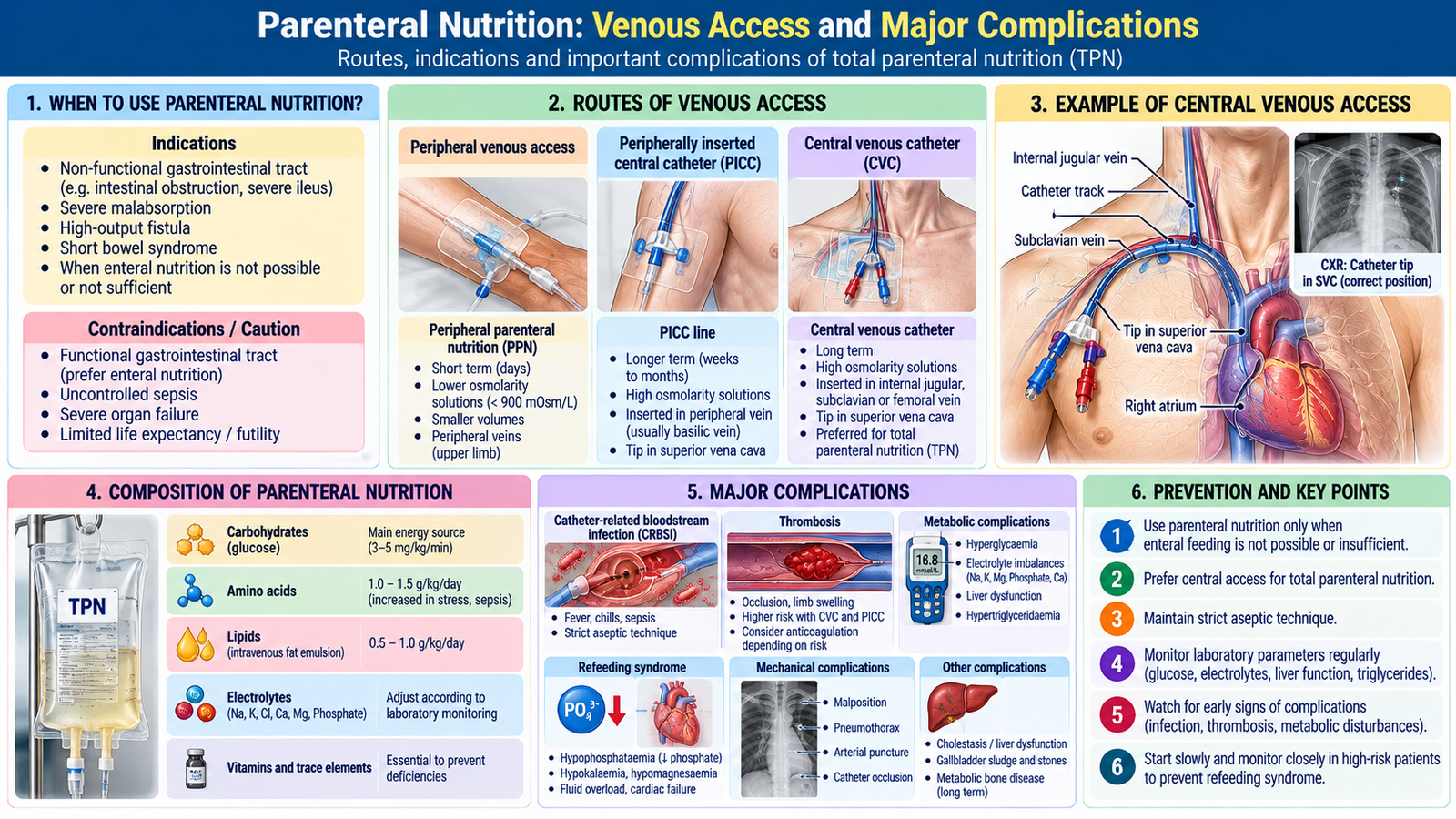
Peripheral versus central access
Peripheral parenteral nutrition uses a peripheral vein for relatively short-term, less concentrated solutions; its use is limited by vein tolerance and the amount of nutrition that can be delivered. Central parenteral nutrition uses a catheter with its tip in a large central vein, allowing more concentrated solutions and longer support. Common central routes include internal jugular, subclavian or suitably positioned peripherally inserted central catheters (PICCs).
Choosing and caring for access
Selection depends on expected duration, solution concentration, venous access and infection/thrombosis risks. Catheter insertion and tip confirmation are performed by trained staff. Aseptic handling, appropriate dressing care, line inspection and prompt review of unexplained fever help prevent catheter-related bloodstream infection. Avoid using a dedicated nutrition lumen for unnecessary injections when local policy recommends separation.
Metabolic and mechanical complications
Hyperglycaemia can result from glucose infusion or stress; hypoglycaemia may occur with interruption of nutrition in susceptible patients. Electrolyte disturbances include low phosphate, potassium and magnesium. Fluid overload, hypertriglyceridaemia and liver biochemical abnormalities can develop. Mechanical problems include pneumothorax during some central line insertions, catheter occlusion, thrombosis and malposition.
Refeeding syndrome
A severely undernourished patient may develop a dangerous fall in phosphate, potassium and magnesium when calories are restarted, because insulin drives these electrolytes into cells. This can cause weakness, arrhythmias, respiratory failure or fluid retention. Identify risk before feeding, give thiamine when indicated, begin nutrition cautiously under an agreed plan and monitor/replenish electrolytes closely. Refeeding syndrome can occur with oral, enteral OR parenteral feeding.
E. Clinical and Laboratory Monitoring, Including Intake–Output
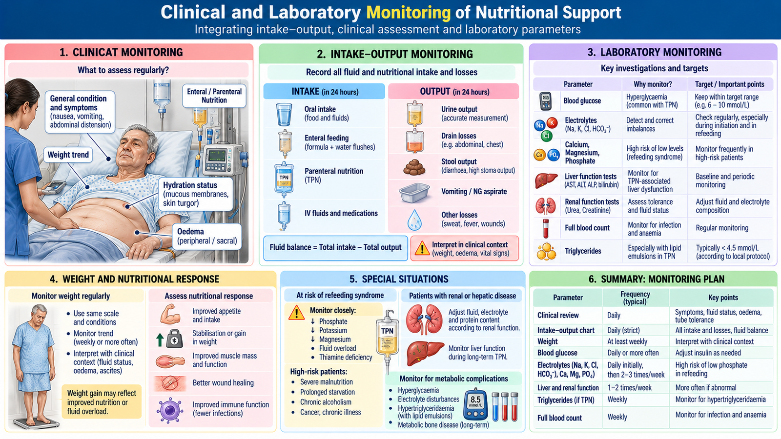
Bedside monitoring
Review clinical stability, tolerance, actual feed delivered, nausea, abdominal symptoms, bowel function, aspiration concerns, catheter or stoma condition, weight trend, oedema and hydration. Track the route and amount of nutrition prescribed versus actually received. Adjust the plan when needs or gut function change.
Laboratory monitoring
Monitor blood glucose and serum sodium, potassium, magnesium, phosphate and renal function, particularly during initiation or in unstable patients. During parenteral nutrition, review liver tests and triglycerides as clinically indicated. Baseline abnormalities, refeeding risk and clinical stability determine monitoring frequency; early or high-risk feeding requires closer checks.
Intake–output charting
Record ALL measured intake: oral fluids, tube feeds, parenteral nutrition, intravenous fluids and relevant flushes. Record measured output: urine, vomit, nasogastric aspirate, drains, stoma output and diarrhoeal losses when quantifiable. Calculate the balance for the defined period: total intake minus total output. For example, 2,400 mL intake and 1,800 mL output gives +600 mL, but this does not automatically prove fluid overload because insensible losses and clinical findings matter.
Interpretation and action
A rising positive balance with oedema, breathlessness or weight gain suggests possible fluid accumulation; a large negative balance with hypotension, tachycardia or reduced urine output suggests possible depletion. Check chart accuracy, renal function and other causes before changing therapy. Communicate concerning findings promptly and document the response.
F. Patient Counselling, Shared Decisions and Ongoing Review
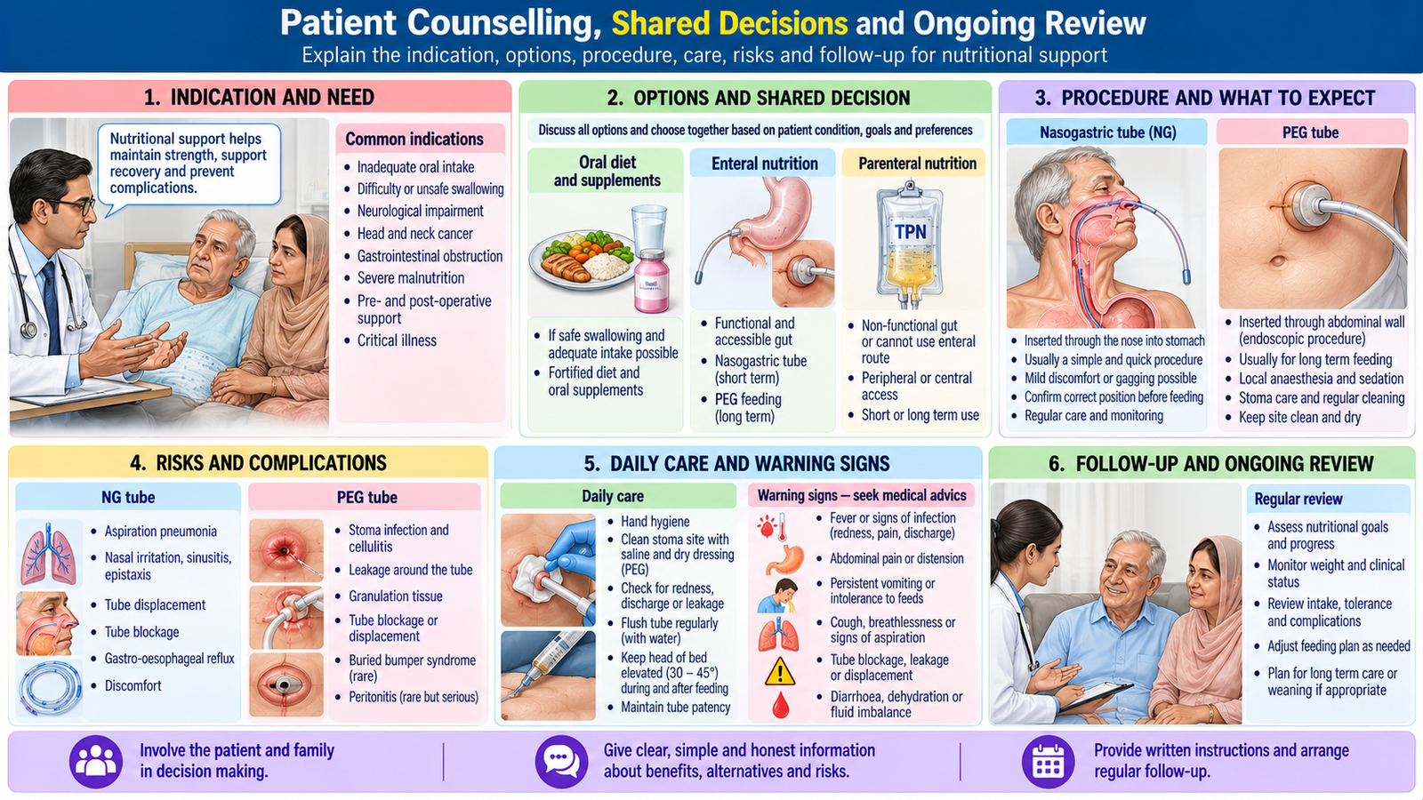
Counselling before NG insertion
Explain why feeding is needed, what the tube passes through, expected transient discomfort, how placement is checked and what to report: breathing difficulty, persistent coughing, severe pain, vomiting or tube displacement. Discuss the intended duration, alternative feeding options, consent and the plan to review swallowing and oral intake.
Counselling before PEG
Explain that PEG is considered for longer-term feeding when swallowing or oral intake is insufficient but the stomach and bowel can be used. Discuss endoscopic placement, sedation/anaesthesia as applicable, possible bleeding, infection, leakage, aspiration and the rare but serious risk of peritonitis. Explain stoma hygiene, tube care, feeding support and when to seek urgent assessment.
Patient-centred follow-up
Check patient preferences, decision-making capacity and the expected benefits and burdens of artificial feeding. Involve family or carers with permission. Review nutritional goals, complications and the opportunity to return to oral or enteral feeding as the patient improves. Ensure clear discharge instructions for patients leaving hospital with a feeding tube.
🩺 AIM Clinical Bridge
A patient recovering from major abdominal surgery has had minimal oral intake for five days. Ask whether the bowel is functioning and whether swallowing is safe before selecting a route. If the gut works, consider enteral feeding; if prolonged ileus prevents adequate gut feeding, parenteral nutrition may be needed. In either case, assess refeeding risk, document actual intake, check electrolytes and glucose, and review daily whether oral or enteral feeding can resume.
AIM High-Yield Review
- Malnutrition may exist despite a normal or high BMI; weight loss and reduced intake are important clues.
- ⭐ Use oral nutrition when safe and adequate; choose enteral feeding when the gut functions but oral intake fails.
- Reserve parenteral nutrition for inadequate or unsafe gastrointestinal delivery, not merely difficulty swallowing.
- NG tubes generally provide short-term access; PEG is considered for longer-term gastric feeding.
- ⭐ Confirm NG tube position reliably before any feed, water or medicine; auscultation alone is unsafe.
- PEG requires monitoring for stoma infection, leakage, bleeding and signs of peritonitis.
- Central parenteral nutrition permits concentrated solutions but introduces catheter infection and thrombosis risks.
- ⭐ Refeeding syndrome can occur with every feeding route; monitor phosphate, potassium and magnesium.
- Review glucose, electrolytes, renal function and, for parenteral nutrition, liver tests and triglycerides as indicated.
- Fluid balance equals recorded intake minus output; interpret it with clinical signs and unmeasured losses.
- Explain purpose, alternatives, discomfort, complications and warning signs before NG or PEG placement.
- Reassess feeding route and nutritional goals regularly as the patient recovers.
🎥 AIM Clinical Video Learning
Topic 3: Clinical Nutritional Support — Enteral & Parenteral Nutrition
Understand the differences between enteral and parenteral nutrition, their indications, and how to select the appropriate route of nutritional support.
Educational video: Clinical Nutrition University. If playback is unavailable, watch on YouTube .