
This chapter follows the KMU outcomes and connects clinical clues with investigations and treatment. Read the explanations first; use the final high-yield review for revision.
📖 AIM Learning Material
TOPIC 8. Systemic Autoimmune & Connective Tissue Diseases
MS Module · Integrated clinical recognition, diagnosis and management
Topic Introduction
Systemic autoimmune diseases are conditions in which immune responses mistakenly attack normal tissues. Because blood vessels, skin, joints, muscles, kidneys and other organs can be affected together, a patient may present with seemingly unrelated complaints. The practical approach is to recognize the clinical pattern, select appropriate antibody and organ-function tests, determine whether an organ is threatened and choose treatment that controls inflammation without causing avoidable harm. This chapter connects systemic lupus erythematosus, antiphospholipid syndrome, systemic sclerosis, Sjögren syndrome and inflammatory myopathies, with special attention to childhood lupus and renal disease. The goal is to understand how each disorder presents, how investigations are interpreted and when complications require urgent action.
A. Clinical Approach to Suspected Systemic Autoimmune Disease
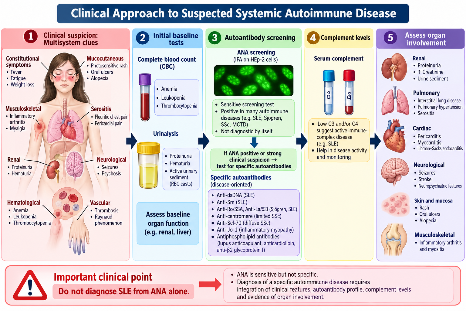
Systemic autoimmune diseases arise when immune responses damage the body’s own tissues. They often affect several organs simultaneously, so diagnosis begins with a pattern of symptoms rather than a single positive blood test. Ask about prolonged fatigue, fever, weight loss, inflammatory joint pain, rashes, photosensitivity, oral ulcers, Raynaud phenomenon, dry eyes or mouth, muscle weakness, thrombosis and pregnancy loss. Establish the timeline, medications, family history and organ-specific symptoms.
Examine the skin, mouth, joints, hands, muscle power, blood pressure, lymph nodes, heart and lungs. Look for edema, urinary abnormalities or neurological deficits suggesting major-organ involvement. Inflammatory joint disease causes swelling and prolonged morning stiffness; mechanical pain generally worsens with use.
Investigations and their interpretation
- Baseline: CBC, ESR/CRP, creatinine/eGFR, liver tests, urinalysis and urine protein quantification. These identify inflammation, cytopenias and silent renal disease.
- ANA: sensitive screening test for SLE but poorly specific. A positive ANA alone does not diagnose lupus.
- Anti-dsDNA and anti-Sm: more specific for SLE; anti-dsDNA levels can correlate with lupus activity, particularly nephritis.
- C3/C4: low complement suggests immune-complex consumption in active SLE.
- Antiphospholipid antibodies: lupus anticoagulant, anticardiolipin IgG/IgM and anti-β2-glycoprotein I IgG/IgM when thrombosis or characteristic pregnancy morbidity occurs.
- Targeted antibodies: anti-centromere/anti-Scl-70 for systemic sclerosis; anti-Ro/SSA and anti-La/SSB for Sjögren syndrome; muscle enzymes and myositis antibodies for inflammatory myopathy.
Interpretation rule: clinical phenotype → targeted laboratory testing → assess organ damage → establish a working diagnosis. Serology supports clinical reasoning; it does not replace it.
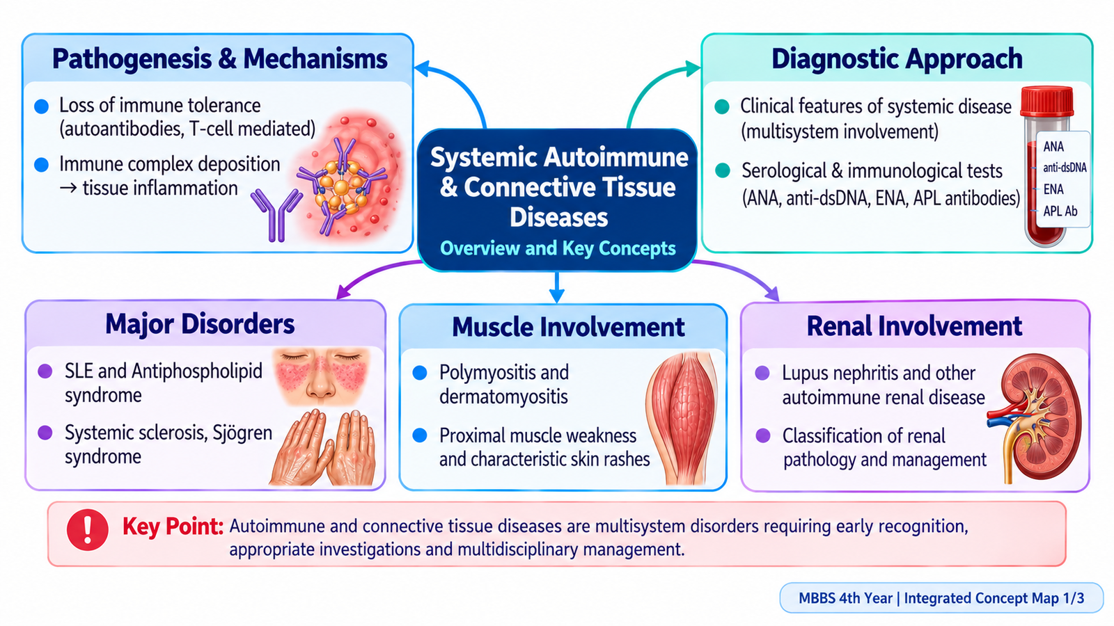
Recognizing the clinical pattern
Autoimmune diseases should be considered when symptoms are persistent, inflammatory and involve more than one system. Establish whether arthritis is inflammatory (warmth, swelling and prolonged morning stiffness), whether rashes are triggered by sunlight, and whether constitutional symptoms are accompanied by objective findings. A history of mouth ulcers, Raynaud attacks, dysphagia, unexplained breathlessness, thrombosis or recurrent pregnancy loss changes the likely diagnosis. Ask about recent infections and prescribed drugs, since these can mimic autoimmunity or produce drug-induced disease.
The first priority is to identify an organ-threatening process. New hypertension, edema, foamy urine or hematuria suggests kidney involvement; chest pain or breathlessness may indicate serositis, pulmonary disease or embolism; new focal weakness or seizure demands urgent assessment. In children, include growth, school function and the caregiver’s observations.
Choosing tests rather than ordering an indiscriminate panel
ESR and CRP indicate inflammation but neither establishes a particular autoimmune diagnosis; CRP can be relatively modest in an uncomplicated SLE flare, whereas a marked rise may raise concern for infection. CBC identifies leukopenia, lymphopenia, hemolytic anemia or thrombocytopenia. Urine dipstick and microscopy reveal protein, blood and casts; quantify proteinuria and measure creatinine when renal involvement is suspected. An isolated low-titer ANA can occur in otherwise healthy people and must not be interpreted in isolation.
Antibodies are most useful when the pre-test probability is raised by clinical features. Anti-dsDNA and anti-Sm support SLE; low C3/C4 can indicate active complement-consuming disease. Anti-Ro/SSA can occur in SLE as well as Sjögren syndrome. Anti-centromere and anti-topoisomerase I help characterize systemic sclerosis but cannot replace organ screening. Rheumatoid factor and anti-CCP support rheumatoid arthritis in the appropriate inflammatory-joint pattern, and may help when considering its renal complications.
When a diagnostic result changes management
Proteinuria with red cell casts and rising creatinine suggests glomerular inflammation and warrants prompt nephrology assessment. A positive antiphospholipid antibody result without thrombosis or defined pregnancy morbidity does not by itself establish APS. Muscle weakness with elevated CK points toward muscle injury; normal CK does not exclude all inflammatory myopathies. Tissue biopsy may be necessary when the pattern is unclear or organ-directed treatment depends on pathology.
B. Pharmacotherapy and Monitoring in Autoimmune Disorders
Treatment aims to control immune-mediated injury while minimizing infection and medication toxicity. Drug choice depends on disease, severity, organ involvement, pregnancy considerations and comorbidities.
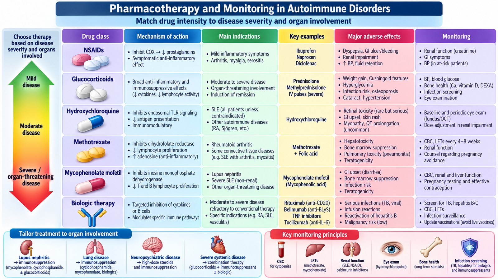
- NSAIDs: inhibit cyclooxygenase and reduce pain and inflammation; they do not prevent major-organ immune injury. Watch for gastrointestinal bleeding, renal injury and cardiovascular risk.
- Glucocorticoids: suppress inflammatory gene expression and act rapidly during flares. Use the lowest effective dose and taper when appropriate; monitor glucose, blood pressure, bone health and infection.
- Hydroxychloroquine: an immunomodulator used as background treatment in most SLE patients and sometimes Sjögren-related inflammatory symptoms. Arrange retinal monitoring and consider QT-risk interactions.
- Conventional immunosuppressants: methotrexate inhibits folate-dependent cellular processes and is used for inflammatory arthritis/myositis; azathioprine inhibits purine synthesis; mycophenolate inhibits lymphocyte proliferation; cyclophosphamide is reserved for severe organ-threatening disease. These require CBC, liver/renal monitoring and infection counselling; methotrexate and mycophenolate are contraindicated in pregnancy.
- Biologic/targeted therapies: inhibit selected immune pathways (for example B-cell activity or cytokine signalling) in selected refractory cases. Screen for relevant infections and use specialist supervision.
- Antithrombotic treatment: anticoagulation, rather than immunosuppression alone, prevents recurrent thrombotic APS events.
Before major immunosuppression assess vaccination status, infection risk, pregnancy plans and baseline laboratory values. Explain adherence, monitoring, warning symptoms and the danger of abruptly stopping long-term corticosteroids.
How drug classification guides treatment
Symptom-controlling agents (such as NSAIDs) improve pain, fever and stiffness but do not reliably prevent immune injury to kidneys or other vital organs. Glucocorticoids rapidly suppress immune inflammation by altering transcription of inflammatory mediators, making them useful for flares and severe organ disease; accumulated exposure increases infection, osteoporosis, diabetes, hypertension and adrenal suppression risk. Conventional disease-modifying/immunosuppressive drugs are slower-acting, steroid-sparing treatments which inhibit lymphocyte activation or proliferation. Biologic and targeted agents selectively interfere with immune pathways and are used in defined indications by specialists.
Hydroxychloroquine changes antigen processing and immune signalling and reduces SLE flares and long-term damage. It is not a substitute for induction immunosuppression in severe proliferative lupus nephritis. Methotrexate is helpful for inflammatory arthritis or some inflammatory muscle disease; folic-acid supplementation reduces some toxicity, and blood counts and liver function require monitoring. Azathioprine inhibits purine synthesis and can maintain disease control in selected conditions. Mycophenolate inhibits inosine-monophosphate dehydrogenase, especially affecting lymphocytes, and is important for lupus nephritis. Cyclophosphamide cross-links DNA and is considered in severe organ-threatening autoimmune inflammation because it has potentially serious marrow, bladder and reproductive toxicity.
Other targeted treatments include B-cell-directed agents such as rituximab in selected resistant disease and belimumab, which reduces B-cell survival signalling in SLE. The treatment decision is based on which organ is involved, how active the disease is, and whether infection, pregnancy or drug toxicity changes the risk-benefit balance.
Practical prescribing and safety principles
- Before treatment: document baseline CBC, kidney and liver function, pregnancy possibility and infection risk; consider hepatitis B and tuberculosis screening before selected major immunosuppressants or biologics.
- During treatment: assess the patient’s symptoms and organ function, not just antibody titers; monitor CBC/liver tests for marrow and hepatic toxicity, creatinine and urine protein for renal response, and blood pressure and glucose during glucocorticoid use.
- Important cautions: methotrexate and mycophenolate can cause fetal harm; hydroxychloroquine requires appropriate ophthalmic retinal monitoring; glucocorticoids increase infection and metabolic risks; NSAIDs may aggravate kidney disease.
- Escalation: acute renal decline, severe cytopenia, breathlessness, central neurological disease or rapidly progressive muscle weakness requires specialist assessment and stronger organ-directed treatment, not symptom relief alone.
A treatment response is demonstrated by improved clinical features and objective organ measures (for example falling proteinuria), while new fever or deterioration may reflect infection, drug toxicity or active disease. These possibilities need to be distinguished before increasing immunosuppression.
C. Systemic Lupus Erythematosus: Recognition, Diagnosis and Management
SLE is a relapsing multisystem autoimmune disease characterized by loss of immune tolerance, autoantibody production and immune-complex inflammation. It predominantly affects women of reproductive age, but also occurs in children and men. Genetic susceptibility, hormonal factors and environmental triggers contribute to disease.
Clinical pattern
Typical manifestations include constitutional symptoms, non-erosive inflammatory polyarthritis, photosensitive or malar rash, oral ulcers, alopecia, pleuritis/pericarditis, cytopenias and kidney disease. Severe disease can cause seizures, psychosis, hemolytic anemia, thrombocytopenia, nephritis or thromboembolism. A normal-looking patient may still have proteinuria or hypertension.
Diagnostic reasoning and classification
There is no single diagnostic test. Combine characteristic clinical findings with ANA, anti-dsDNA/anti-Sm, low complement, CBC and urine studies. The 2019 EULAR/ACR classification criteria require a positive ANA at least once as an entry criterion, followed by weighted clinical and immunological criteria totaling ≥10 points, with at least one clinical criterion; these are classification criteria, not a substitute for clinical diagnosis.
Management and prognosis
Hydroxychloroquine is generally recommended unless contraindicated. Encourage sun protection, smoking avoidance, cardiovascular-risk control and regular follow-up. Use NSAIDs or short courses of glucocorticoids for appropriate mild manifestations; moderate or severe disease may need immunosuppressants, particularly with renal, neurological or hematological involvement. Monitor symptoms, blood pressure, CBC, creatinine, urine protein, complement and anti-dsDNA where helpful.
Prognosis depends especially on nephritis, cardiovascular disease, infection and treatment toxicity. Explain the fluctuating course, medication adherence and the need for planned pregnancy during low disease activity.
Drug-induced lupus versus idiopathic SLE
Drug-induced lupus may follow medications such as hydralazine or procainamide. It commonly causes arthralgia, fever and serositis with antihistone antibodies; significant renal/CNS involvement is less typical. Improvement after withdrawal of the responsible drug supports the diagnosis. Idiopathic SLE more often has anti-dsDNA positivity, low complement and important renal involvement.
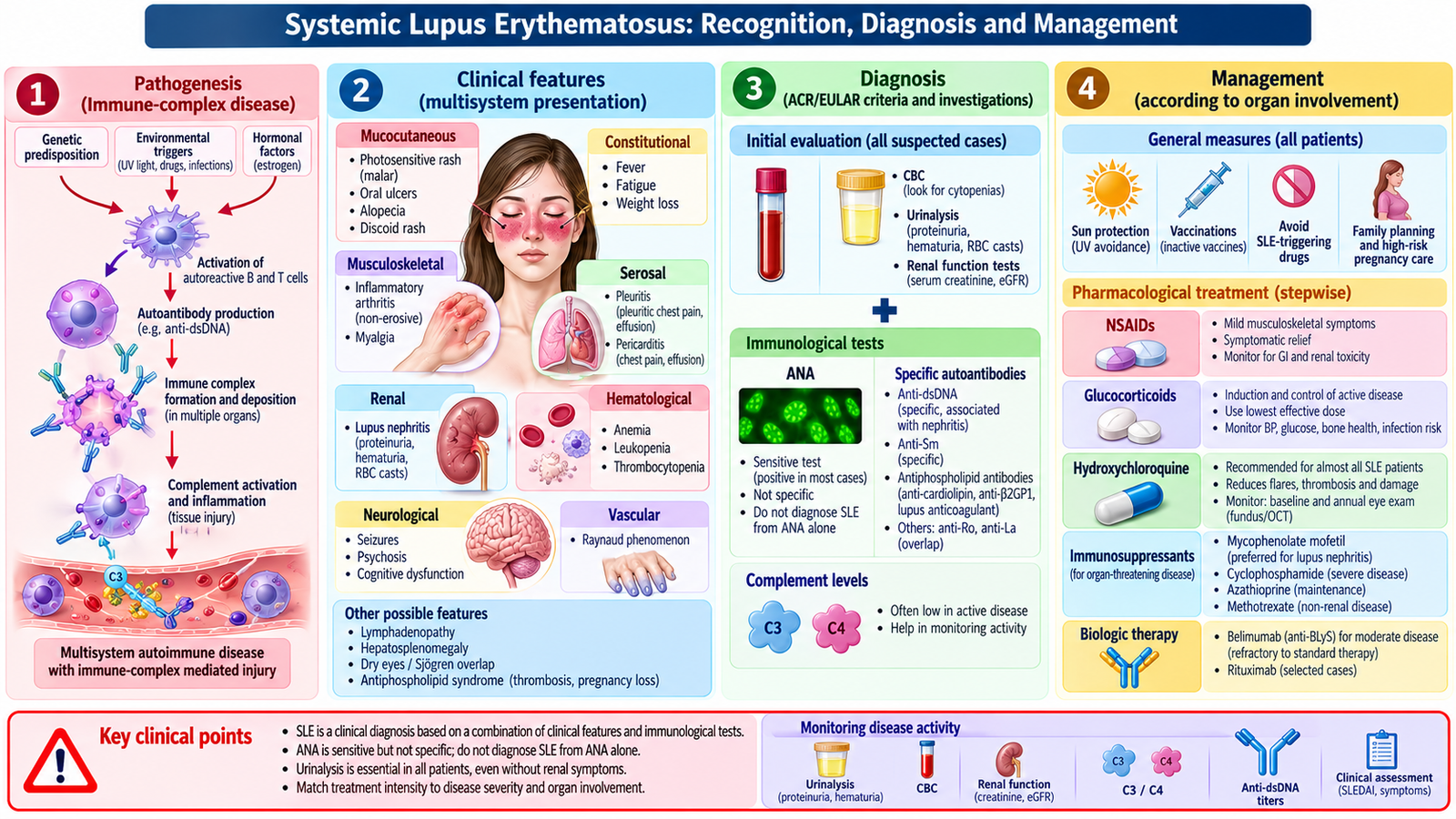
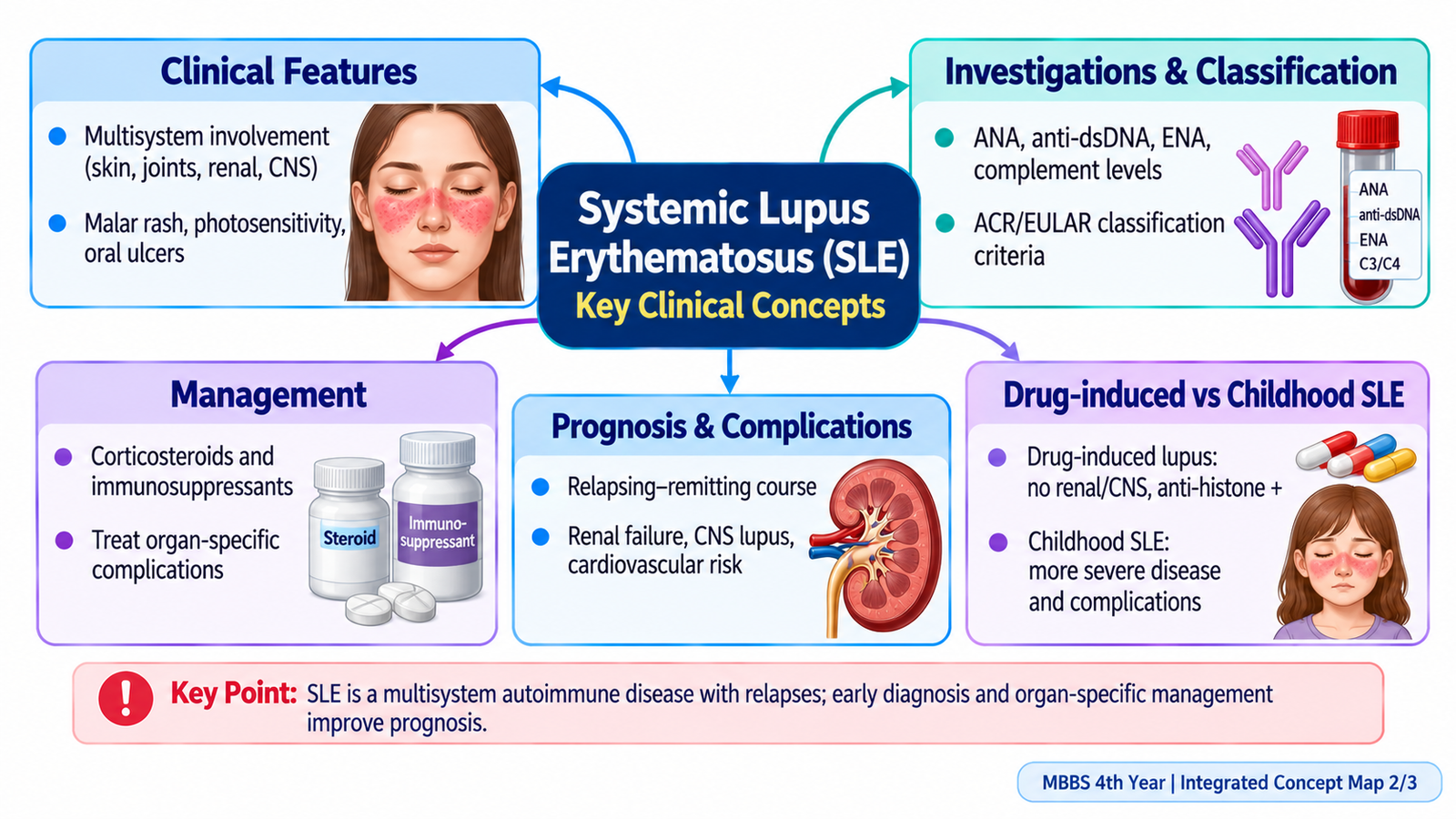
Understanding the mechanism behind multisystem injury
Failure to clear apoptotic cell material and loss of B-cell tolerance promote production of antinuclear autoantibodies. Antibody–antigen complexes form and deposit in small vessels, joints, serosal surfaces and glomeruli. Complement activation recruits inflammatory cells, causing tissue damage. This explains the combination of arthritis, serositis, nephritis, skin manifestations and reduced complement levels in active disease. Cytopenias may also result from antibodies directed at circulating blood cells.
Symptoms that help estimate organ involvement
- Skin and mucosa: malar rash generally spares the nasolabial folds; photosensitive rashes, painless oral/nasal ulcers and non-scarring alopecia support the diagnosis.
- Musculoskeletal: typically symmetrical joint pain and stiffness, often without the erosions typical of established rheumatoid arthritis.
- Serosal and cardiopulmonary: pleuritic pain, effusion or pericarditis; breathlessness must also prompt assessment for infection, thrombosis or significant lung involvement.
- Blood and nervous system: immune hemolysis, leukopenia, thrombocytopenia, seizures or neuropsychiatric changes may indicate serious disease, although infection and medication effects must be excluded.
- Kidney: hematuria, proteinuria, hypertension and edema, sometimes with no symptoms until damage is significant.
Important differential diagnoses include viral illness, rheumatoid arthritis, drug-induced lupus and other systemic connective tissue diseases. The investigation is guided by what each alternative would explain: infection may cause acute constitutional illness, rheumatoid arthritis has persistent synovitis and may be anti-CCP positive, and isolated drug-induced lupus often lacks severe nephritis.
Putting clinical findings together
After a compatible history and examination, test ANA, anti-dsDNA, anti-Sm and complement, and obtain CBC, creatinine and urine studies. An ANA-negative result makes typical SLE less likely, while an isolated positive ANA is insufficient. The EULAR/ACR weighted scoring system helps classification, but individual features must have no better explanation; clinical diagnosis and treatment decisions consider the entire patient. A patient with inflammatory arthritis, malar rash, lymphopenia and proteinuria warrants prompt renal evaluation rather than waiting for every serological result.
Activity, severity, complications and prognosis
Distinguish mild mucocutaneous/joint disease from major organ disease. Major nephritis, neuropsychiatric lupus, severe hemolysis or thrombocytopenia and cardiopulmonary involvement can threaten life or organ function. Kidney damage, infection and premature atherosclerotic cardiovascular disease influence long-term survival. Assessment at follow-up includes disease symptoms, blood pressure, urine protein, creatinine, CBC and treatment effects; anti-dsDNA/complement may support, but not dictate, assessment of disease activity.
For lupus nephritis, pathology and disease activity determine induction and maintenance immunosuppression. For non-organ-threatening SLE, hydroxychloroquine, sun protection and carefully chosen symptomatic or steroid-sparing treatment can reduce relapses. Vaccination, infection prevention, exercise and bone protection improve long-term health. Discuss pregnancy before conception because maternal nephritis, active disease, antibodies and teratogenic drugs may alter planning.
How drug-induced lupus is distinguished
Suspect drug-induced lupus when lupus-like symptoms begin after exposure to a medication (classically hydralazine or procainamide). Fever, arthralgia and serositis are frequent, and antihistone antibodies may be found; low complement and major kidney/CNS involvement are uncommon in classic drug-induced disease. Stop the culprit drug when feasible and reassess. Importantly, some medication-related lupus syndromes have different antibody patterns, so the overall clinical context remains essential.
🩺 AIM Clinical Bridge
A 22-year-old woman reports several months of inflammatory hand-joint pain, fatigue and a photosensitive facial rash. Examination reveals mild ankle edema and elevated blood pressure. ANA and anti-dsDNA are positive, C3 is low, and urinalysis shows protein and red-cell casts.
Think clinically: the rash and inflammatory arthritis point toward SLE, but edema, hypertension and active urinary sediment indicate probable lupus nephritis. Quantify proteinuria, check kidney function and arrange prompt nephrology assessment. Kidney biopsy may be needed to identify the class and guide treatment.
D. Antiphospholipid Syndrome: Thrombosis and Pregnancy Morbidity
APS is an acquired autoimmune thrombophilia associated with antibodies directed against phospholipid-binding proteins. It may occur alone (primary APS) or with SLE (secondary APS). The central clinical pattern is thrombosis or characteristic obstetric morbidity, not bleeding despite possible prolongation of the aPTT.
Clinical events include deep-vein thrombosis, pulmonary embolism, ischemic stroke, recurrent pregnancy loss, fetal death and placental insufficiency. Livedo reticularis and thrombocytopenia may accompany the syndrome. Rare catastrophic APS produces rapidly progressive thromboses in multiple organs and requires emergency specialist care.
Diagnosis
Request lupus anticoagulant, anticardiolipin and anti-β2-glycoprotein I antibodies. Persistent positivity on two occasions at least 12 weeks apart, together with appropriate clinical findings, supports APS. A single transient positive result is insufficient. Anticoagulants can interfere with lupus anticoagulant testing; interpretation may require laboratory guidance.
Management and outcome
Acute thrombosis requires standard anticoagulant treatment. For most patients with thrombotic APS, long-term warfarin is preferred for secondary prevention; direct oral anticoagulants are generally avoided in high-risk, particularly triple-positive, APS. During pregnancy, obstetric APS is commonly managed with low-dose aspirin and heparin according to clinical history; warfarin is avoided during pregnancy because of fetal harm. Control vascular risk factors and coordinate obstetric care.
Recurrent thrombosis, stroke, pregnancy complications and catastrophic APS are major adverse outcomes. Long-term prognosis improves with adherence and individualized thrombosis prevention.
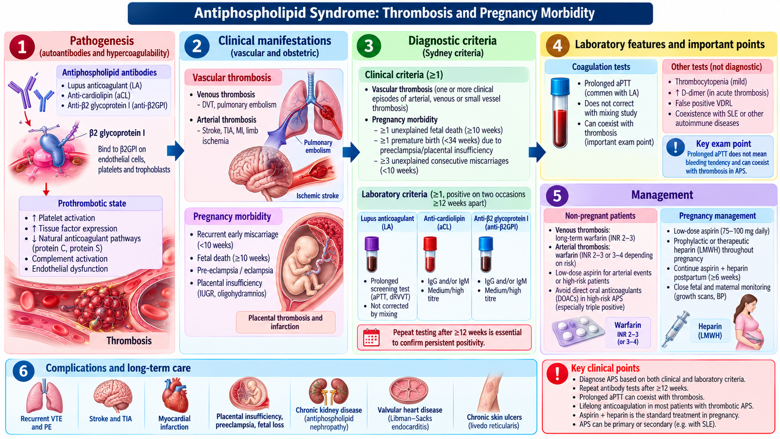
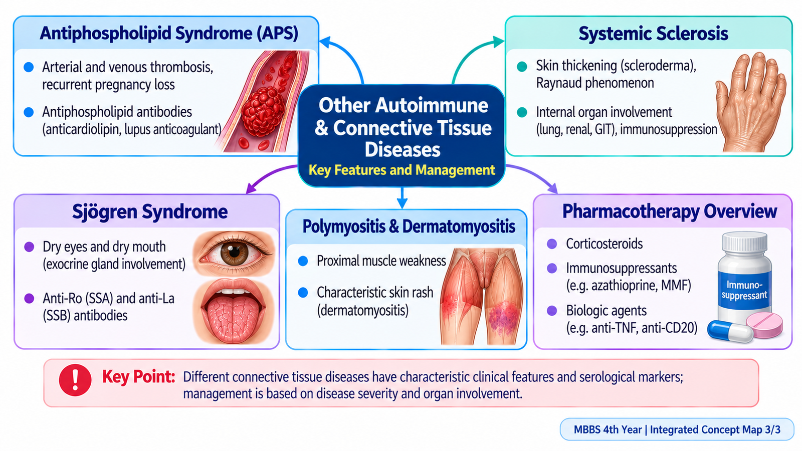
Mechanism and clinical reasoning
Antiphospholipid antibodies activate endothelial cells, platelets and coagulation pathways and interfere with normal anticoagulant mechanisms. This promotes thrombosis in veins, arteries and placental vessels. It explains why an apparently paradoxical prolonged aPTT may occur in the laboratory while the patient has a tendency to form clots in the body. The syndrome may coexist with SLE, so new clots or recurrent fetal loss in someone with lupus should prompt appropriate APS testing.
Venous disease commonly presents with leg swelling or pleuritic breathlessness from DVT/PE. Arterial disease may present with transient ischemic attack or stroke. Obstetric manifestations include fetal death, recurrent early pregnancy loss and severe placental disease causing preeclampsia or premature delivery. Examine for livedo reticularis, thrombocytopenia and evidence of end-organ ischemia, while also evaluating common competing causes of thrombosis.
Testing, confirmation and risk
The antibody panel comprises lupus anticoagulant, anticardiolipin IgG/IgM and anti-β2-glycoprotein I IgG/IgM. Results should be repeated at least 12 weeks apart to demonstrate persistence because infection and transient immune responses can produce temporary positivity. Lupus anticoagulant is assessed through phospholipid-dependent coagulation tests rather than an ordinary antibody concentration. Anticoagulants may distort these tests. Persistent multiple-antibody positivity, particularly triple positivity, indicates higher thrombosis risk; antibody positivity by itself is not identical to clinical APS.
Management decisions and prognosis
Manage an acute DVT, PE or stroke urgently using standard stabilization and specialist anticoagulation principles. After thrombotic APS, long-term vitamin K antagonist therapy (commonly warfarin) is usual; INR monitoring, bleeding risk and drug interactions require education. Direct oral anticoagulants are generally not preferred for high-risk APS. For pregnancy morbidity without prior thrombosis, low-dose aspirin with prophylactic heparin is commonly considered under obstetric supervision; history of thrombosis changes intensity of anticoagulation. The clinician must balance maternal thrombosis prevention against bleeding, miscarriage risk and fetal safety.
Prognosis depends on recurrence risk, antibody profile, control of smoking/hypertension and adherence to anticoagulation. New chest pain, breathlessness, unilateral limb swelling or focal neurological loss is an emergency. Catastrophic APS involves widespread small-vessel thrombosis and rapid multiorgan dysfunction, requiring urgent intensive specialist care.
E. Systemic Sclerosis and Sjögren Syndrome
Systemic sclerosis
Systemic sclerosis combines microvascular dysfunction, immune activation and progressive fibrosis of skin and internal organs. Limited cutaneous disease predominantly affects distal extremities and face and is associated with anti-centromere antibodies and later pulmonary arterial hypertension. Diffuse cutaneous disease extends proximally and has greater early risk of interstitial lung disease and renal crisis; anti-topoisomerase I (Scl-70) is associated with diffuse disease and lung fibrosis.
Raynaud phenomenon may precede skin tightening. Look for sclerodactyly, digital ulcers, telangiectasia, reflux/dysphagia, dyspnea and reduced exercise tolerance. Assess blood pressure, kidney function, pulmonary function and echocardiographic findings when indicated. Treat Raynaud symptoms with warming, smoking cessation and calcium-channel blockers; reflux with acid suppression. Organ-specific immunosuppression may be needed for lung disease.
Scleroderma renal crisis presents with abrupt hypertension and acute kidney injury, sometimes with microangiopathic hemolysis. It is a medical emergency: prompt ACE-inhibitor treatment is central, even if creatinine initially rises. High-dose glucocorticoids can increase renal-crisis risk.
Sjögren syndrome
Sjögren syndrome is autoimmune inflammation of exocrine glands causing dry eyes and dry mouth, either primarily or alongside another autoimmune disease. Ask about gritty eyes, difficulty swallowing dry food, recurrent dental caries, parotid swelling, fatigue and arthralgia. Examine oral moisture, teeth and salivary glands.
Anti-Ro/SSA, anti-La/SSB, Schirmer testing and, where necessary, salivary gland evaluation support diagnosis. Management includes artificial tears, saliva substitutes, meticulous dental care and treatment of systemic manifestations when present. Important complications include corneal injury, dental disease and increased risk of B-cell lymphoma, especially with persistent gland enlargement.
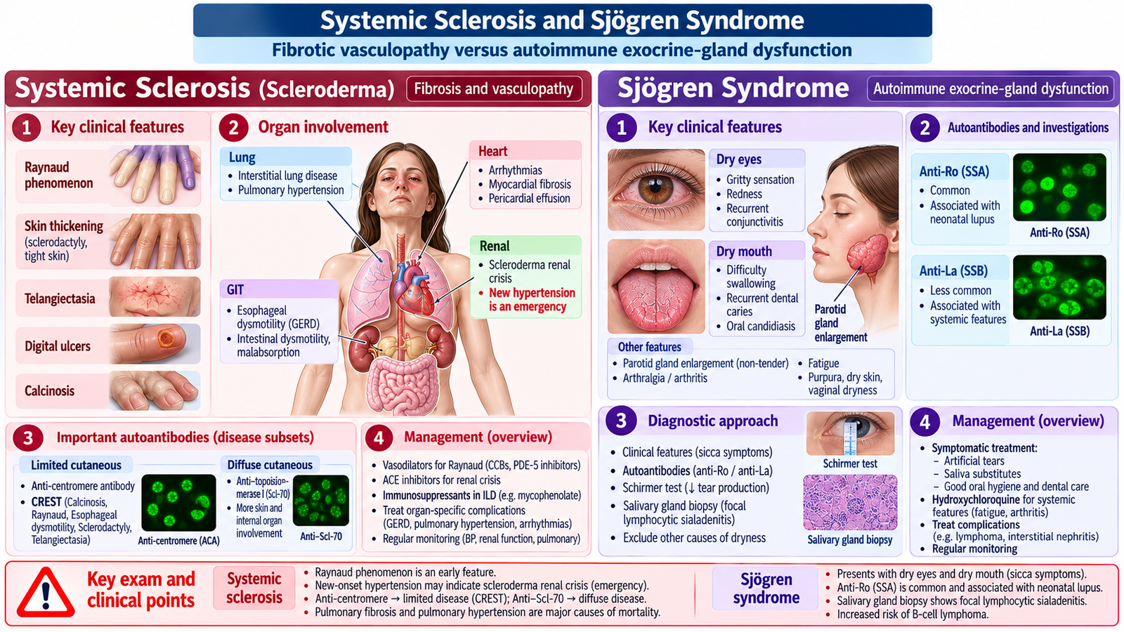
Systemic sclerosis: important organ complications
Damaged small vessels cause Raynaud phenomenon and digital ischemia; activated fibroblasts progressively deposit collagen, producing tight skin and organ fibrosis. In limited disease, skin changes mainly involve fingers, forearms and face; in diffuse disease, proximal extremities or trunk may be involved. CREST summarizes calcinosis, Raynaud phenomenon, esophageal dysmotility, sclerodactyly and telangiectasia, usually within the limited phenotype.
Examine for digital pitting, fingertip ulcers, reduced mouth opening and skin thickening. Esophageal smooth muscle dysfunction causes reflux and swallowing difficulty; intestinal involvement can cause malabsorption. Pulmonary interstitial fibrosis produces progressive exertional dyspnea, cough and restrictive physiology. Pulmonary arterial hypertension may cause fatigue, syncope or right-heart strain and requires early detection. Cardiac fibrosis can lead to arrhythmia or ventricular dysfunction.
Useful investigations include ANA and specific autoantibodies, pulmonary function tests including gas transfer, high-resolution chest CT when lung disease is suspected, echocardiography for pulmonary hypertension screening, and regular blood pressure and renal function monitoring. Treat manifestations by organ: warming and calcium-channel blockers for Raynaud, proton-pump inhibitors for reflux, immunosuppressive therapy such as mycophenolate for selected interstitial lung disease, and specialist pulmonary hypertension treatment when indicated. Systemic sclerosis has no simple single curative medicine, so prevention of irreversible organ damage and long-term surveillance are central.
Renal crisis: abrupt severe hypertension and rising creatinine, often with microangiopathic hemolytic anemia, requires urgent ACE-inhibitor therapy and close kidney monitoring. Blood pressure should be checked regularly, especially in diffuse disease or corticosteroid exposure. Prognosis depends on the extent of pulmonary, cardiac and renal disease.
Sjögren syndrome: diagnostic reasoning and complications
Lymphocytic infiltration damages lacrimal and salivary glands, producing insufficient tears and saliva. Dry eyes feel gritty or burning; reduced tears may cause corneal injury. Dry mouth makes swallowing dry food difficult, impairs speech and increases dental caries and oral candidiasis. Fatigue, inflammatory arthralgia, peripheral neuropathy and tubulointerstitial renal disease may occur, but sicca complaints can also result from medication or aging.
Schirmer test assesses tear production; anti-Ro/SSA and sometimes anti-La/SSB support the diagnosis in a compatible setting. Objective eye staining, salivary-flow assessment or minor salivary-gland biopsy may be considered when uncertainty remains. Treatment starts with artificial tears, lubrication, hydration, saliva substitutes, dental fluoride and regular dental care; selected patients benefit from agents stimulating residual gland function. Systemic manifestations may require immunomodulation. Persistent parotid swelling, enlarged nodes or unexplained constitutional symptoms warrant evaluation for lymphoma. Long-term follow-up protects ocular and oral health and detects systemic disease.
F. Polymyositis and Dermatomyositis
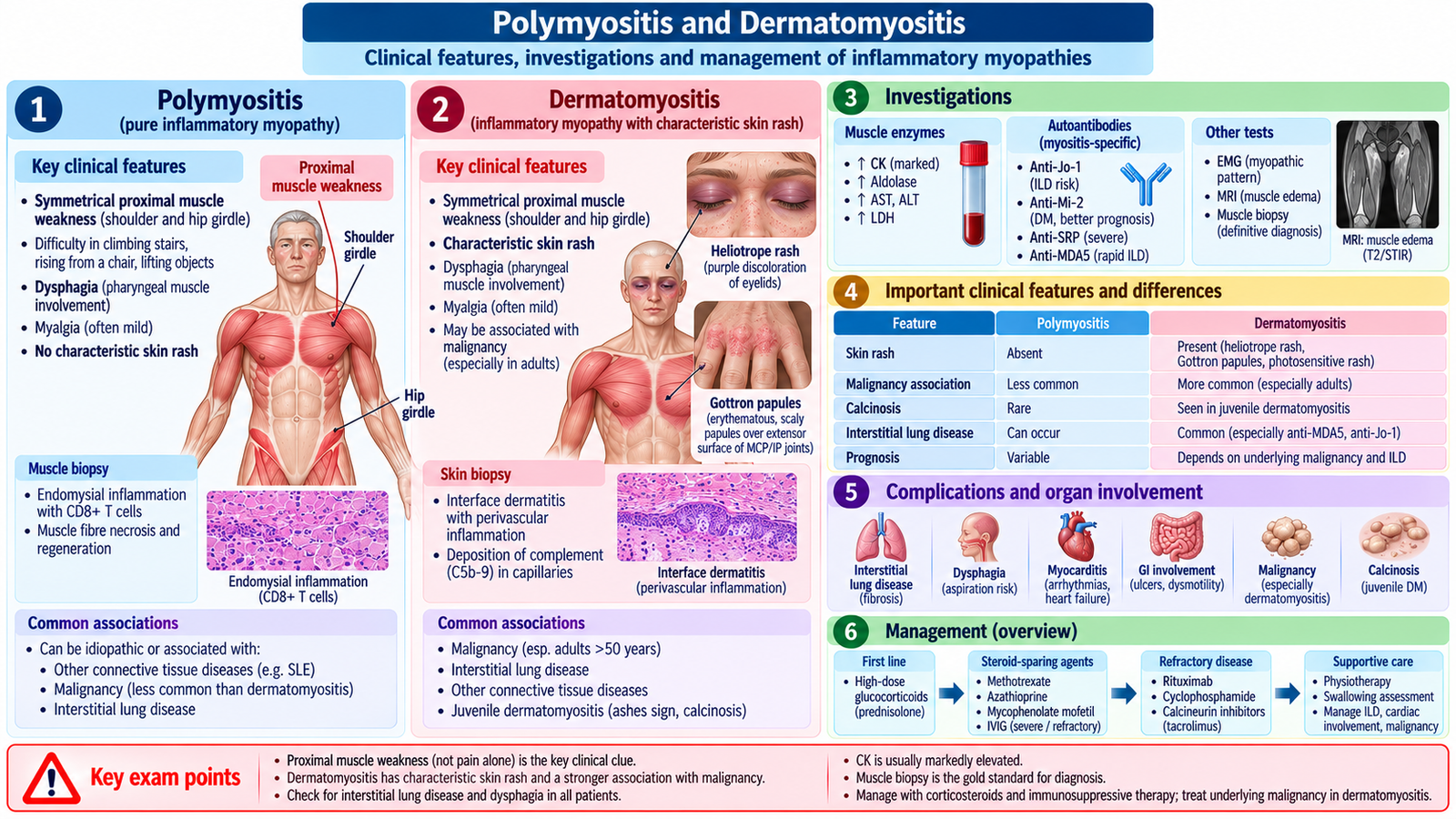
Inflammatory myopathies cause immune-mediated muscle injury, producing symmetrical proximal weakness rather than isolated muscle pain. Patients struggle to climb stairs, rise from a chair or lift their arms. Ask about dysphagia, breathlessness, medication exposure and functional decline; assess neck flexors, shoulder and hip girdle power, reflexes and skin.
Dermatomyositis has characteristic skin findings including heliotrope discoloration around the eyelids and Gottron papules over extensor finger joints. Polymyositis presents with proximal muscle weakness without the characteristic dermatomyositis rash; it is less common than historically thought, and other causes of myopathy must be excluded.
Raised creatine kinase (CK), aldolase, myopathic EMG, muscle MRI and sometimes biopsy support the diagnosis. Myositis-specific antibodies can assist classification. Consider hypothyroidism, medication-induced myopathy, muscular dystrophy and other neuromuscular disorders in the differential.
Treatment usually begins with glucocorticoids and a steroid-sparing agent such as methotrexate or azathioprine; rehabilitation helps restore strength. Severe dysphagia or respiratory weakness needs urgent assessment. Dermatomyositis in adults is associated with an increased malignancy risk, so appropriate age- and risk-based cancer assessment is important. Interstitial lung disease is another major complication.
Mechanisms and clinical assessment
In inflammatory myopathy, immune-mediated damage to skeletal muscle fibers reduces effective force generation. The largest muscles around shoulders and hips are usually affected, so patients have difficulty combing hair, rising without using the arms or climbing stairs. True weakness must be distinguished from pain-limited movement, fatigue and joint stiffness. Examine proximal strength against resistance and check swallowing, respiratory effort and possible cardiac or lung symptoms.
Dermatomyositis causes both muscular disease and distinctive small-vessel skin injury. A heliotrope rash affects eyelids, and Gottron papules occur over the knuckles; photosensitive patches may develop over shoulders, upper chest and back. Some people have typical skin disease with little obvious weakness. The classic label polymyositis should be used cautiously because immune-mediated necrotizing myopathy, inclusion-body myositis, endocrine disorders and drug-induced myopathy can resemble it.
Interpreting tests and confirming the disorder
Raised CK suggests muscle fiber breakdown, and serial levels can assist treatment monitoring. Aldolase, AST/ALT and LDH may rise due to muscle injury rather than primary liver disease. EMG can show a myopathic pattern; muscle MRI shows inflamed regions and helps choose a biopsy site. Muscle biopsy may confirm inflammatory injury when the diagnosis is uncertain. Autoantibody testing can help characterize disease and associated lung risk, but no single test replaces clinical assessment.
Compare with hypothyroid myopathy (thyroid abnormalities), steroid or statin-related muscle problems (drug history), neuromuscular junction disease (fatigable pattern) and muscular dystrophy (different progression/family context). A substantially raised CK with proximal weakness supports active muscle disease but does not establish its precise cause.
Treatment, complications and prognosis
Glucocorticoids usually provide initial control; methotrexate, azathioprine or mycophenolate may reduce steroid requirements. Physical therapy and carefully monitored exercise improve function once inflammation is controlled. Assess nutrition and aspiration risk if swallowing is impaired. Dysphagia can cause aspiration pneumonia; severe respiratory muscle weakness or interstitial lung disease may become life-threatening. Some inflammatory myopathies involve the heart.
Adults with dermatomyositis require clinically appropriate malignancy assessment, because an occult cancer may accompany the disease. Monitoring includes strength and day-to-day function, CK trends, lung symptoms and adverse effects of treatment. Prognosis varies with muscle response, dysphagia, lung disease, cancer association and delayed diagnosis.
G. Childhood SLE and Renal Involvement in Autoimmune Disease
Childhood-onset SLE
Childhood SLE has the same fundamental immune-complex mechanism as adult disease but often presents with greater disease activity and more frequent major-organ involvement. Features include fever, fatigue, rash, arthritis, cytopenias, hypertension, proteinuria and neurological symptoms. Growth, school attendance, emotional well-being and family support influence long-term care.
Investigate with CBC, creatinine, urine microscopy/protein, ANA, anti-dsDNA and complement; assess suspected organ disease promptly. Treatment is severity-based, with hydroxychloroquine commonly used and corticosteroids/immunosuppressants for significant organ involvement. Monitor growth, infection, bone health, adherence and treatment toxicity. Renal disease, severe infection and cardiovascular complications influence prognosis.
Renal pathology in SLE
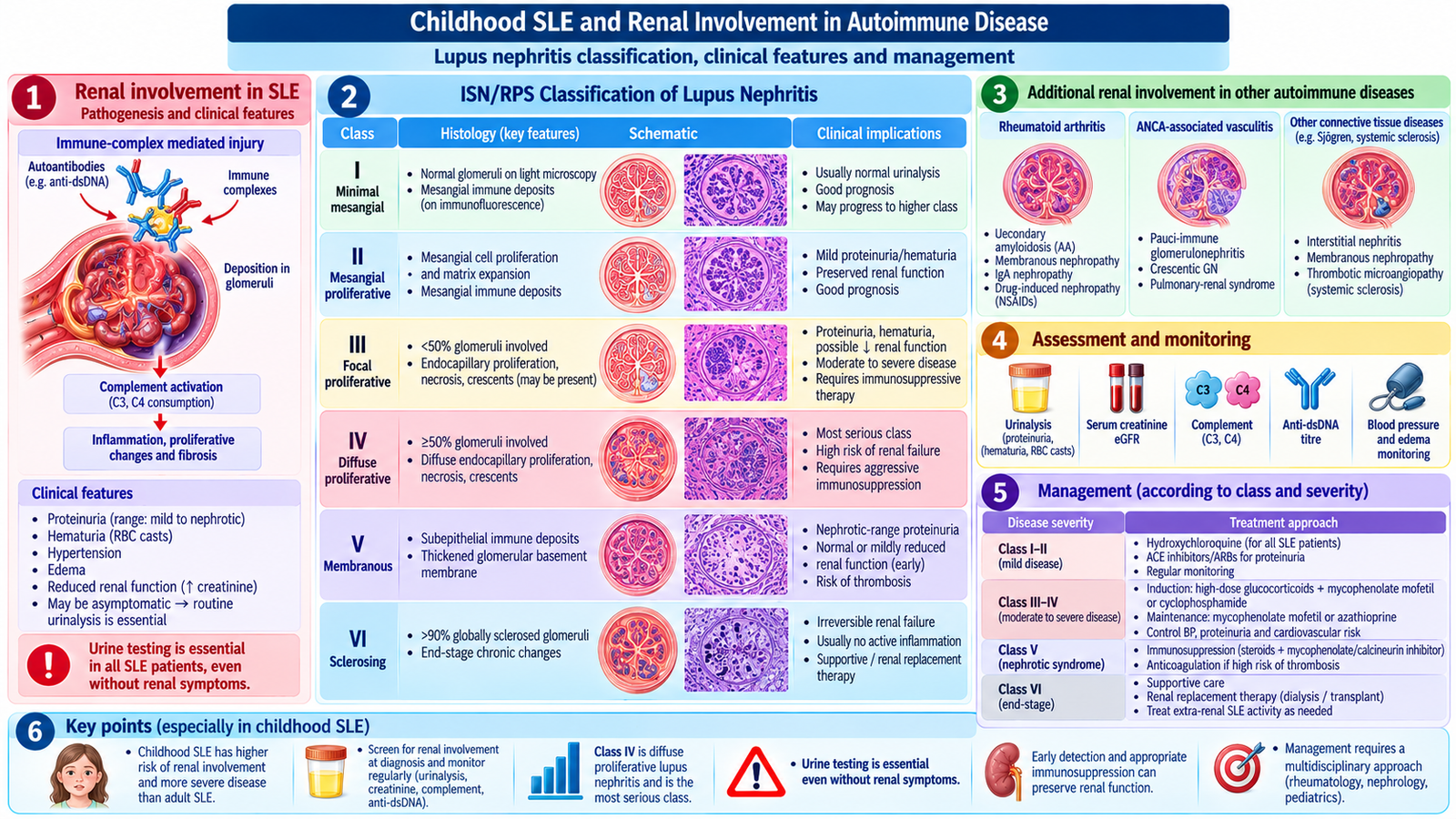
Lupus nephritis is caused mainly by immune-complex deposition and complement-mediated inflammation. It may produce hematuria, proteinuria, nephrotic syndrome, hypertension or falling eGFR. The ISN/RPS biopsy classes are:
- I: minimal mesangial; II: mesangial proliferative.
- III: focal proliferative (<50% of glomeruli); IV: diffuse proliferative (≥50%), often severe.
- V: membranous, frequently nephrotic proteinuria; VI: advanced sclerosing, largely irreversible scarring.
Kidney biopsy is considered when clinically significant persistent proteinuria or unexplained renal dysfunction suggests active nephritis; it guides therapy. Active proliferative nephritis generally requires glucocorticoids plus an immunosuppressant such as mycophenolate or cyclophosphamide under specialist care. Control blood pressure and proteinuria, often with ACE inhibitors or ARBs where appropriate; monitor urine protein, creatinine and drug toxicity.
Renal involvement in rheumatoid arthritis and other autoimmune disorders
Rheumatoid arthritis may affect kidneys through chronic inflammation (including AA amyloidosis in longstanding uncontrolled disease), associated glomerular disease or medication toxicity, especially NSAID-related kidney injury. Other systemic autoimmune disorders may cause glomerulonephritis, interstitial disease or vascular renal injury. Distinguish active immune-mediated nephritis from drug toxicity and chronic scarring using history, urinalysis, kidney function, targeted serology and sometimes biopsy.
Childhood SLE: what makes it different
Children and adolescents may initially show prolonged fever, marked fatigue, weight loss, joint swelling or rash, but nephritis, cytopenias and central nervous system involvement are particularly important. A child with edema, new hypertension and proteinuria may already have significant nephritis even when the malar rash is subtle. Ask about growth, pubertal development, school performance, recurrent infection, medication adherence and psychosocial stress. Examine blood pressure using an age-appropriate cuff, skin, joints, edema and neurological status.
The diagnostic workup mirrors adult SLE but must take age-specific effects into account: CBC with differential, urinalysis with microscopy, quantified proteinuria, creatinine/eGFR, ANA, anti-dsDNA, anti-Sm and complement, with further tests directed at suspected organ disease. A child with active urinary sediment or impaired kidney function needs timely pediatric nephrology/rheumatology care. Assess infection separately because immunosuppressed children may develop serious infection that resembles lupus activity.
Treatment aims to control inflammation while limiting corticosteroid effects on height, bone health, weight and puberty. Hydroxychloroquine commonly forms background therapy; glucocorticoids and specialist immunosuppressants are added according to disease severity. Provide vaccination review, infection precautions, sun protection, family education and support with school attendance. Prognosis is generally better with early diagnosis and adherence, but severe nephritis, neuropsychiatric disease, infection and long-term cardiovascular complications increase morbidity.
Lupus nephritis: why pathology matters
Immune-complex deposits in glomeruli trigger complement activation and inflammatory damage. Inflamed capillary walls permit blood cells and protein to enter the urine; extensive immune injury lowers filtration and raises creatinine. Protein loss causes hypoalbuminemia and edema; sodium retention and renal vascular injury contribute to hypertension. Urinalysis may show dysmorphic red cells or red-cell casts, which are strong clues to glomerular inflammation.
Class I and II: mesangial deposits with minimal or mesangial proliferation; abnormalities may be mild. Class III: proliferative injury in fewer than half of glomeruli, with hematuria, proteinuria and possible impaired function. Class IV: diffuse proliferative injury in at least half of glomeruli; typically carries the greatest risk of rapidly progressive inflammatory kidney injury. Class V: immune deposits along capillary walls produce membranous nephropathy and often heavy proteinuria/nephrotic syndrome; mixed III/IV plus V can occur. Class VI: widespread glomerular sclerosis represents advanced chronic irreversible scarring. The distinction is made by renal biopsy, not by urine dipstick alone.
Persistent significant proteinuria, active urinary sediment, or otherwise unexplained decline in kidney function prompts consideration of biopsy after excluding other explanations. Proliferative III/IV disease commonly needs glucocorticoids plus mycophenolate or cyclophosphamide-based induction, followed by maintenance therapy; class V therapy depends on proteinuria and accompanying lesions. Supportive care includes blood-pressure and proteinuria control, often using an ACE inhibitor or ARB when appropriate, infection prevention and avoiding nephrotoxic medications. Falling proteinuria with stable/improving eGFR suggests response, whereas persistent rising creatinine, active casts or worsening proteinuria prompts reassessment. Class VI predominantly represents irreversible damage; care emphasizes kidney protection and planning for chronic kidney disease treatment rather than attempting to reverse established sclerosis.
Renal pathology in rheumatoid arthritis and other autoimmune conditions
Rheumatoid arthritis has several distinct renal pathways, which should not be confused with primary lupus nephritis. Longstanding uncontrolled inflammation may cause AA amyloidosis: serum amyloid A-derived deposits accumulate in glomeruli, classically producing substantial proteinuria and later kidney impairment. RA-associated immune-complex glomerular lesions, including mesangial or membranous patterns, are less frequent and should be confirmed rather than assumed. Separately, NSAIDs may cause hemodynamic acute kidney injury or interstitial nephritis, while some older RA therapies can produce proteinuric renal injury. Evaluate medication exposure when renal function deteriorates.
Other autoimmune disease affects kidneys through different compartments: Sjögren syndrome commonly causes tubulointerstitial nephritis, sometimes with distal renal tubular acidosis and hypokalemia; systemic sclerosis causes vascular injury and renal crisis, not immune-complex lupus nephritis; systemic vasculitis may cause rapidly progressive necrotizing glomerulonephritis. These patterns demonstrate why the location of the injury—glomerulus, tubule/interstitium or renal vessels—changes the diagnostic workup and treatment.
For RA with significant proteinuria, test kidney function, urine protein and sediment and consider biopsy after assessing drug-related causes and chronic inflammation. Control inflammatory RA to reduce AA amyloid risk, stop or replace nephrotoxic drugs when appropriate, treat hypertension and proteinuria and seek nephrology support. For Sjögren-related interstitial disease, check electrolytes and acid-base status and treat significant autoimmune inflammation when indicated. Systemic sclerosis renal crisis requires immediate ACE-inhibitor therapy. Prognosis across autoimmune renal disease depends on the type, severity, reversibility and speed of treatment.
Important Comparison — Autoimmune Renal Injury
Different autoimmune disorders injure different renal compartments, which explains their contrasting presentations and management.
| Disorder | Typical lesion | Clinical clue | Treatment principle |
|---|---|---|---|
| SLE | Immune-complex glomerulonephritis | Proteinuria, hematuria, casts | Biopsy-directed immunosuppression |
| RA | AA amyloid; sometimes GN or drug-related injury | Proteinuria or AKI with medication exposure | Control RA, assess drugs, renal support |
| Sjögren syndrome | Tubulointerstitial nephritis | Distal RTA, hypokalemia | Correct electrolytes; treat inflammation |
| Systemic sclerosis | Renal vascular injury | Abrupt hypertension and AKI | Urgent ACE inhibitor |
AIM High-Yield Review
- Autoimmune serology must be interpreted alongside the clinical phenotype; ANA positivity alone does not establish SLE.
- In suspected SLE, check urinalysis, proteinuria, creatinine and blood pressure even without urinary symptoms.
- Anti-dsDNA elevation with falling complement may indicate active immune-complex SLE, particularly nephritis.
- Drug-induced lupus commonly improves after withdrawal of the causative drug; major renal disease is less typical.
- APS requires a compatible clinical event and persistently positive antiphospholipid antibodies at least 12 weeks apart.
- Thrombotic APS is primarily managed with anticoagulation; obstetric APS requires pregnancy-specific planning.
- Systemic sclerosis with new severe hypertension and acute kidney injury suggests renal crisis requiring urgent ACE-inhibitor therapy.
- Sjögren syndrome causes sicca symptoms; persistent parotid enlargement warrants assessment for lymphoma.
- Dermatomyositis combines proximal weakness with characteristic rash; assess swallowing, lungs and malignancy risk.
- Lupus nephritis class III/IV is proliferative; class V commonly produces nephrotic proteinuria.
- In children, lupus often has significant renal and hematological involvement and requires growth/toxicity monitoring.
- In RA, renal dysfunction may result from inflammatory disease, amyloidosis or medication toxicity.
Video Learning — Systemic Autoimmune & Connective Tissue Diseases
Topic 8 | MS Module | KMU Curriculum
Systemic Lupus Erythematosus (SLE)
Supplementary video covering the pathophysiology, clinical manifestations and diagnostic concepts of systemic lupus erythematosus.
If the embedded video does not play, use the link below to watch it directly on YouTube.
AIM — AI-Integrated Modular Learning | Supplementary video resource. Continue with the complete AIM Learning Material for all Topic 8 learning outcomes.