
📖 AIM Learning Material
TOPIC 7. Inherited Metabolic & Storage Disorders
Topic Introduction
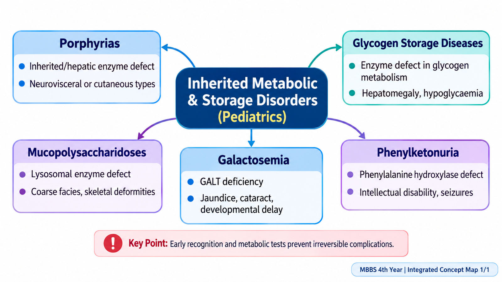
Inherited metabolic and storage disorders are genetic conditions in which the body cannot process or store particular substances normally. The consequences depend on what accumulates, what essential product is missing and which organs are affected. Some children become ill soon after feeding begins, whereas others present later with hypoglycemia, developmental problems, abnormal skeletal growth or organ enlargement. In this chapter, you will learn to distinguish porphyrias, glycogen storage diseases, mucopolysaccharidoses, galactosemia and phenylketonuria through their underlying mechanisms, recognizable clinical patterns and appropriate investigations. The aim is to make early recognition and safe initial management understandable at Final Year MBBS level.
A. Recognizing Inherited Metabolic and Storage Disorders
Core idea
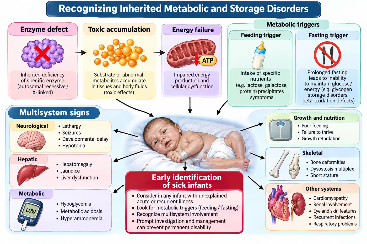
Inherited metabolic disorders arise when a gene defect reduces an enzyme or transport protein. The resulting block may cause accumulation of a toxic substrate, deficiency of an essential product, or failure to release stored energy. Storage disorders commonly affect several organs because undegraded material accumulates inside cells.
Clinical recognition
Think of an inherited metabolic disorder in an infant with unexplained vomiting, lethargy, seizures, developmental regression, hepatomegaly, hypoglycemia or metabolic acidosis. Some disorders appear after milk or protein feeding begins; others develop gradually with coarse facial features, skeletal abnormalities and organ enlargement.
First assessment
Assess airway, breathing, circulation, hydration, consciousness and glucose immediately in an acutely unwell child. Ask about age at onset, relation to feeds or fasting, consanguinity, sibling deaths, developmental milestones and family history. Examine growth, liver and spleen, neurological status, eyes, skin and skeleton.
Investigation strategy
Initial tests are bedside glucose, blood gas, electrolytes, liver function, lactate, ammonia and urine ketones as clinically indicated. Follow with disorder-specific biochemical testing, urine metabolites, enzyme assays and genetic confirmation when available. Normal newborn appearance does not exclude a metabolic disorder.
B. Porphyrias: Classification and Pathophysiology
Heme synthesis
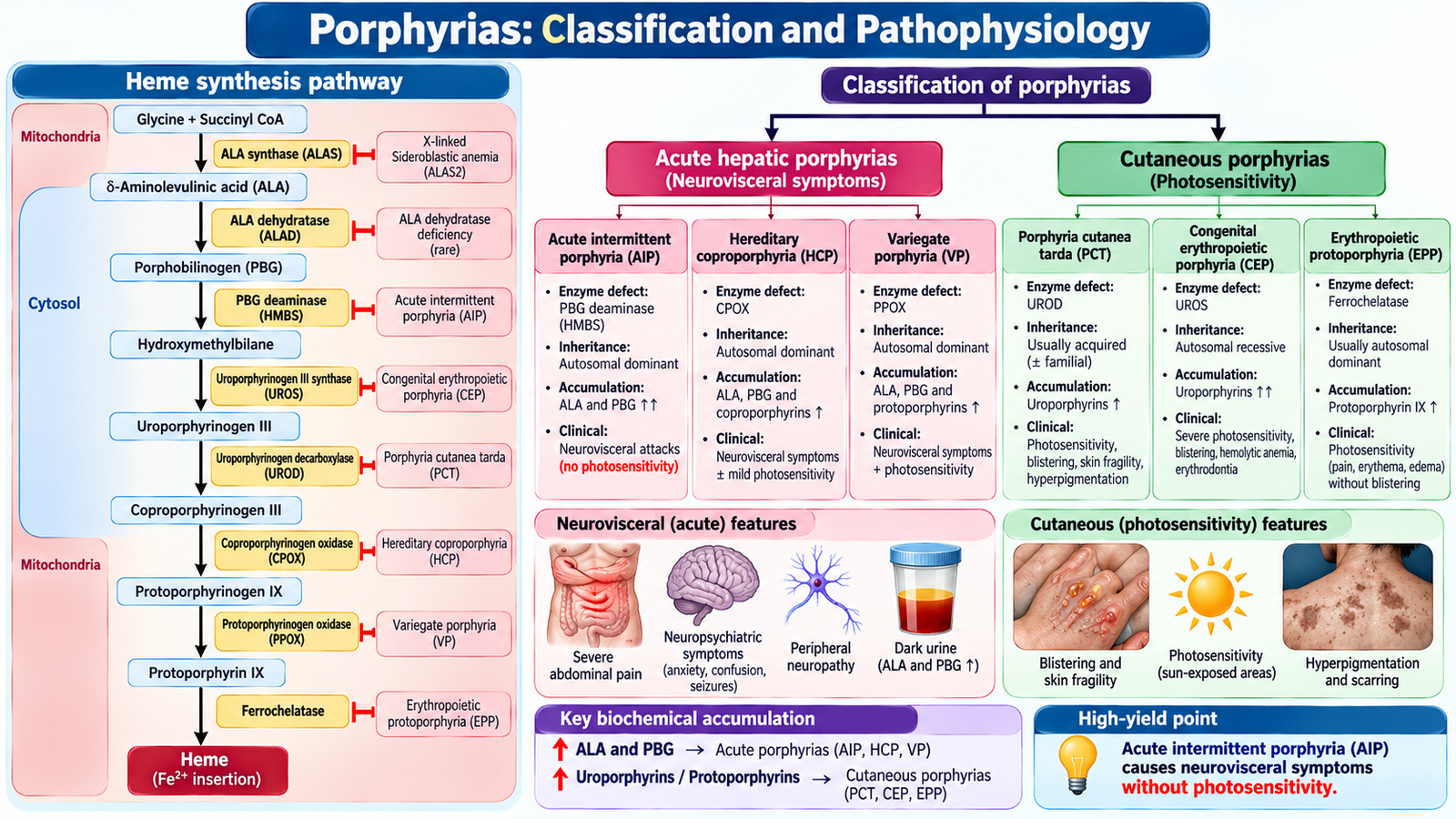
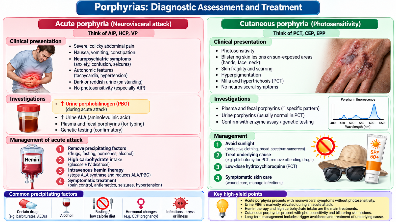
Porphyrias result from inherited or acquired defects in enzymes of the heme biosynthetic pathway. Precursors accumulating before a pathway block produce neurovisceral symptoms; photosensitive porphyrins in skin produce cutaneous injury after light exposure.
Classification
Acute hepatic porphyrias include acute intermittent porphyria (AIP), variegate porphyria, hereditary coproporphyria and the very rare ALAD-deficiency porphyria. Cutaneous forms include porphyria cutanea tarda and erythropoietic protoporphyria. Some forms, especially variegate and hereditary coproporphyria, produce both acute attacks and skin findings.
Acute attacks
AIP classically causes severe poorly localized abdominal pain, nausea, constipation, tachycardia, hypertension, darkening urine and neuropsychiatric symptoms. Peripheral motor neuropathy, weakness, seizures and hyponatremia may develop. Photosensitivity is absent in AIP. Attacks may be precipitated by fasting, infection, alcohol, hormonal changes and porphyrinogenic drugs.
Cutaneous manifestations
Porphyria cutanea tarda causes fragile, blistering skin on sun-exposed hands and face, hyperpigmentation and hypertrichosis. Erythropoietic protoporphyria causes painful immediate photosensitivity with less prominent blistering. These manifestations reflect photoactivation of accumulated porphyrins.
C. Porphyrias: Diagnostic Assessment and Treatment
Testing the suspected acute attack
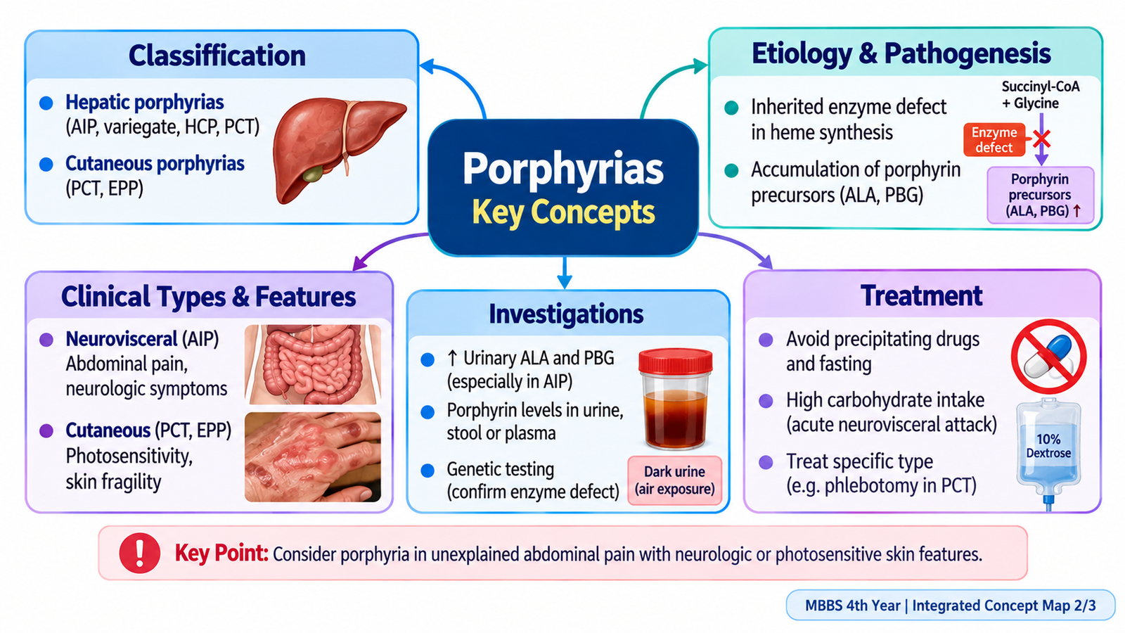
During suspected acute hepatic porphyria, obtain a spot urine porphobilinogen (PBG), usually with aminolevulinic acid (ALA), ideally during symptoms. Markedly increased PBG supports an acute hepatic porphyria; subsequent biochemical and genetic testing identifies the type. Do not rely on urine color alone.
Acute management
Admit patients with severe attacks, treat pain and vomiting, correct fluids and electrolytes carefully, and monitor sodium, respiration and motor weakness. Remove precipitating drugs and treat infection. Intravenous hemin is the specific treatment for significant acute attacks; carbohydrate supplementation may help selected mild attacks but must not delay hemin when indicated.
Cutaneous management
For porphyria cutanea tarda, minimize sun exposure and address iron overload, alcohol use, hepatitis C or estrogen exposure where relevant. Specialist treatment may include periodic phlebotomy or low-dose hydroxychloroquine. Erythropoietic protoporphyria requires rigorous visible-light protection and specialist assessment.
Counselling
Explain trigger avoidance, medication-safety checking, nutritional regularity and the possibility of family testing. A child with severe abdominal pain plus progressive weakness or hyponatremia requires urgent escalation.
D. Glycogen Storage Diseases
Mechanism and classification
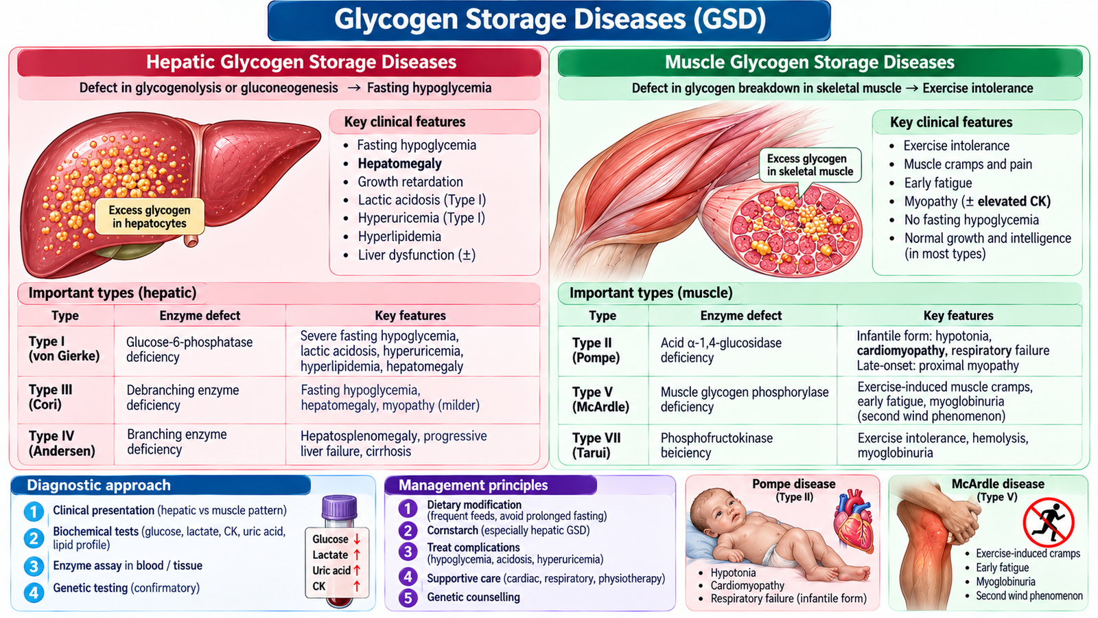
Glycogen storage diseases (GSDs) are inherited defects in glycogen synthesis or breakdown. Hepatic forms impair maintenance of blood glucose during fasting; muscle forms impair energy supply during exercise. Some forms involve both liver and muscle.
Major examples
Type I (von Gierke; glucose-6-phosphatase system defect) produces severe fasting hypoglycemia, hepatomegaly, lactic acidosis, hyperuricemia and hyperlipidemia. Type II (Pompe; lysosomal acid alpha-glucosidase deficiency) produces cardiomyopathy, hypotonia and muscle weakness, with glucose often maintained. Type III (Cori; debranching enzyme deficiency) causes hepatomegaly, fasting hypoglycemia and variable muscle or cardiac involvement. Type V (McArdle; muscle glycogen phosphorylase deficiency) causes exercise intolerance, cramps and possible myoglobinuria.
Recognition and tests
A child with a protuberant abdomen, hepatomegaly and fasting hypoglycemia suggests hepatic GSD; an infant with profound hypotonia and hypertrophic cardiomyopathy suggests Pompe disease. Evaluate glucose, lactate, lipids, uric acid, liver enzymes, creatine kinase and cardiac findings according to the suspected subtype. Confirm with enzyme or molecular testing.
Management principles
Prevent fasting and maintain regular carbohydrate delivery in hepatic GSD, commonly using individualized dietary plans and uncooked cornstarch under specialist supervision. Pompe disease has disease-specific enzyme replacement therapy. Muscle disease requires tailored activity guidance and monitoring. Refer for metabolic and dietetic follow-up.

E. Mucopolysaccharidoses: Multisystem Storage Disease
Pathogenesis
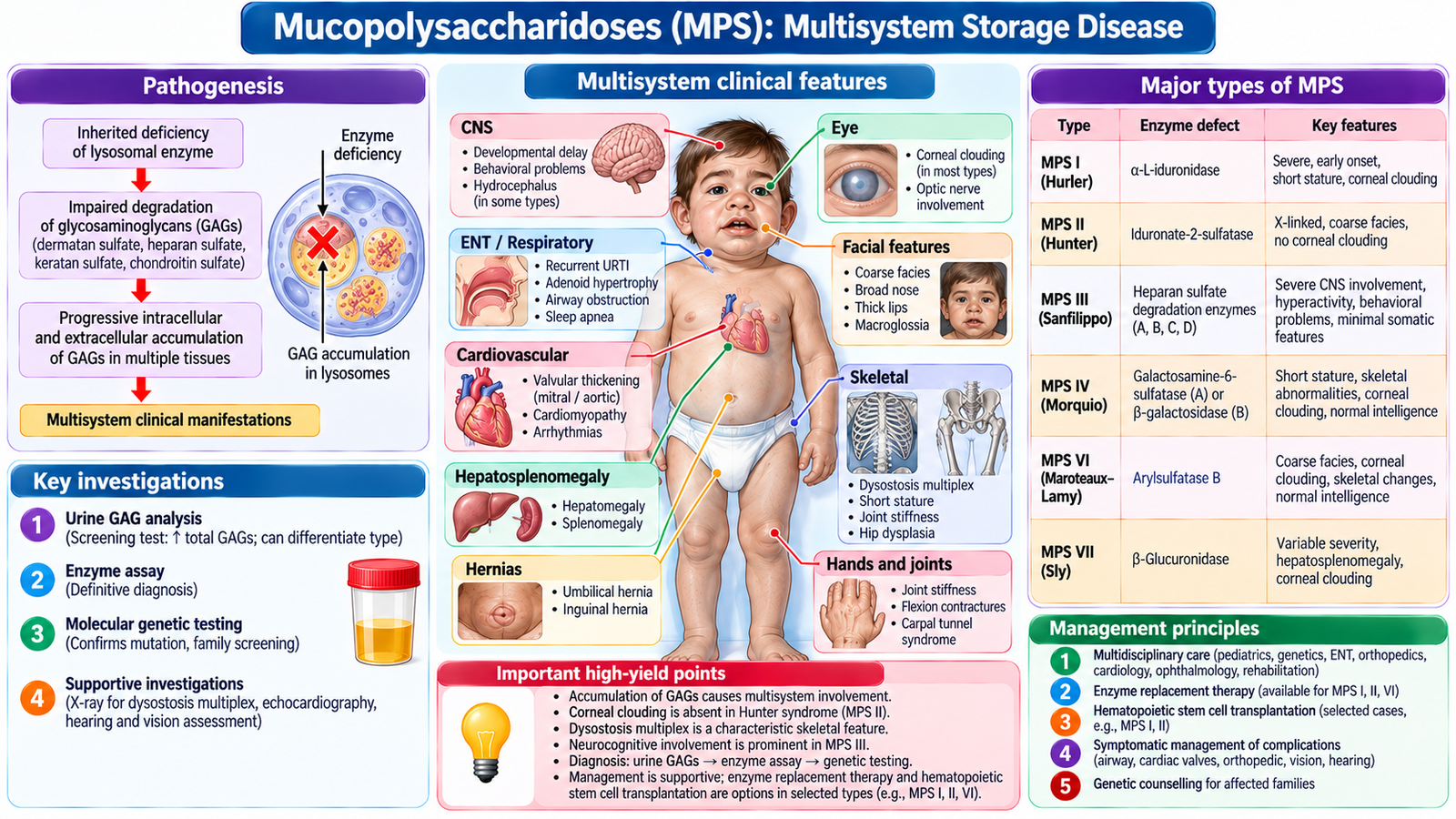
Mucopolysaccharidoses (MPS) are lysosomal disorders in which defective degradation of glycosaminoglycans causes progressive tissue accumulation. Most are autosomal recessive; MPS II (Hunter syndrome) is X-linked.
Clinical patterns
Progressive coarse facial features, short stature, hepatosplenomegaly, joint stiffness or contractures, skeletal deformity (dysostosis multiplex), hernias, recurrent airway problems, hearing loss and cardiac valve disease are characteristic. Developmental impairment varies by subtype.
Important distinctions
MPS I (Hurler spectrum) can involve corneal clouding and neurodevelopmental deterioration. MPS II (Hunter) generally lacks corneal clouding. MPS III (Sanfilippo) is dominated by progressive neurological and behavioral problems. MPS IV (Morquio) prominently affects the skeleton, often with relatively preserved intelligence.
Assessment and complications
Urinary glycosaminoglycan screening supports suspicion but does not replace confirmatory enzyme assays and genetic testing. Evaluate hearing, vision, heart, airway, cervical spine and neurodevelopment. Important complications include airway obstruction, restrictive respiratory disease, valvular dysfunction and cervical spinal cord compression. Some subtypes have enzyme replacement therapy; selected severe cases may be considered for hematopoietic stem-cell transplantation.
F. Galactosemia and Phenylketonuria
Classic galactosemia
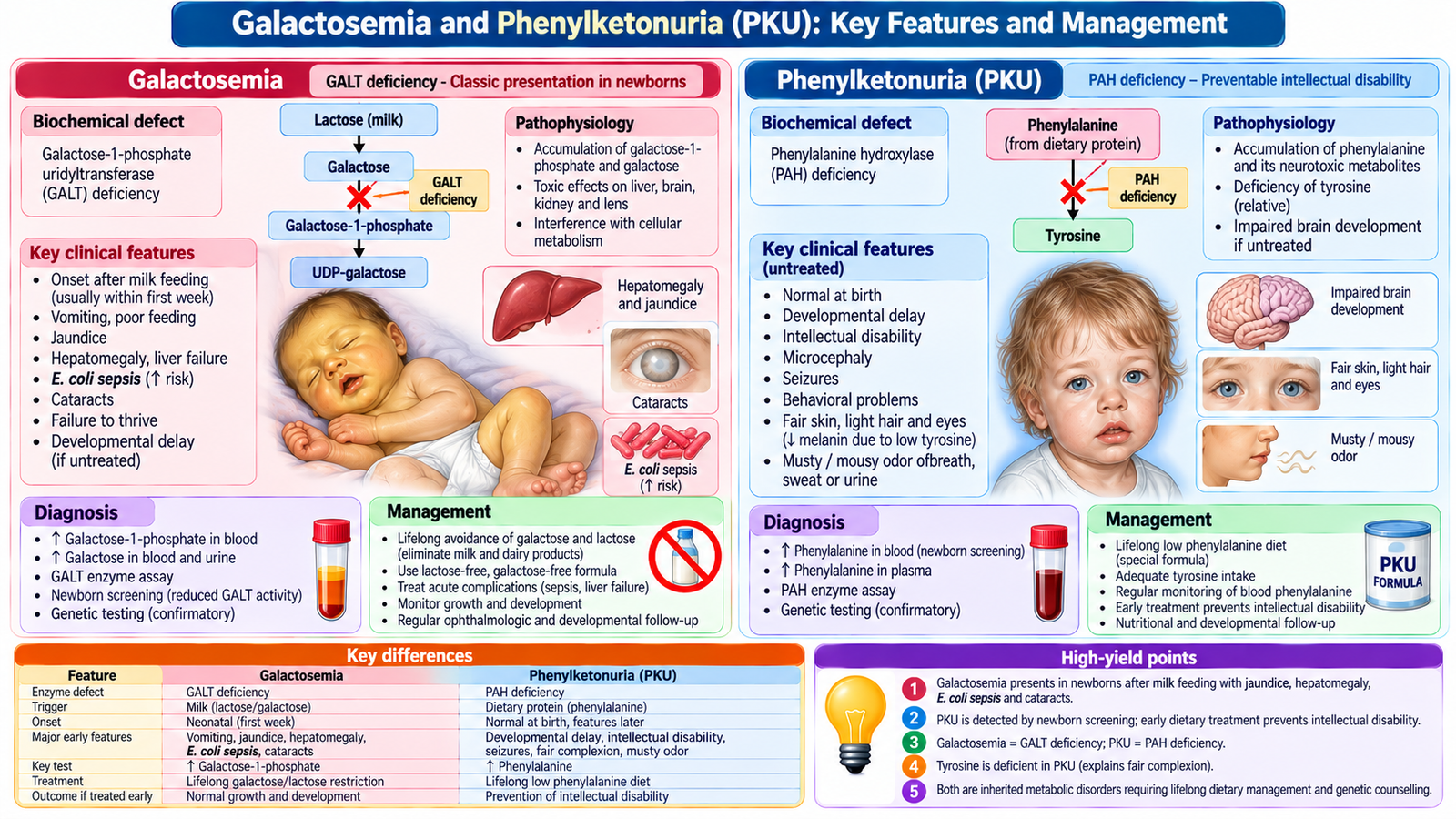
Classic galactosemia is usually due to galactose-1-phosphate uridyltransferase (GALT) deficiency. After breast milk or lactose-containing formula begins, the infant may develop vomiting, poor feeding, jaundice, hepatomegaly, hypoglycemia, cataracts and failure to thrive. Escherichia coli sepsis is a life-threatening association.
Galactosemia: diagnosis and care
Suspect the condition in an ill neonate with liver dysfunction after milk exposure. Investigate with newborn screening where available, erythrocyte GALT activity and/or molecular testing; interpret assays in light of recent transfusion. Stop lactose/galactose exposure immediately when strongly suspected, use appropriate alternative feeding, stabilize the infant and treat sepsis urgently.
Phenylketonuria
Phenylketonuria (PKU) usually results from phenylalanine hydroxylase deficiency, causing high phenylalanine and reduced tyrosine availability. Untreated children develop intellectual disability, developmental delay, seizures, behavioral problems, eczema and sometimes a musty odor and lighter pigmentation. Early infancy may initially appear normal.
PKU: diagnosis and care
Newborn screening detects elevated phenylalanine; confirm with plasma amino-acid testing and evaluation for tetrahydrobiopterin-related disorders when indicated. Start a phenylalanine-restricted diet with prescribed medical formula and adequate tyrosine, monitor phenylalanine regularly, and continue long-term metabolic care. Maternal PKU requires tight preconception and pregnancy phenylalanine control to protect the fetus.
G. Integrated Diagnostic Reasoning and Long-Term Care
Pattern-based approach
Acute deterioration after milk feeding points toward galactosemia; progressive neurodevelopmental impairment without neonatal crisis may indicate PKU or a storage disorder. Fasting hypoglycemia with hepatomegaly suggests hepatic GSD. Cardiomyopathy with hypotonia suggests Pompe disease. Coarse features with joint stiffness and skeletal changes suggest MPS. Episodic abdominal pain with neuropathy suggests an acute hepatic porphyria.
Interpret investigations in context
Hypoglycemia and raised lactate support some hepatic GSDs but are not specific. Urinary PBG is central during an acute porphyria attack; urine GAG testing is a screening step for MPS. Elevated phenylalanine indicates the PKU pathway but requires confirmation. Enzyme assays and molecular studies refine the diagnosis and support family counselling.
Immediate versus long-term care
First stabilize the sick child and correct dangerous metabolic derangements. Then prevent substrate exposure or fasting as appropriate, provide nutritional support, monitor organ complications and arrange metabolic-specialist follow-up. Do not delay urgent care while awaiting genetic results.
Family-centred prevention
Explain inheritance patterns, recurrence risk, newborn or sibling screening, adherence to lifelong diet where required, and the role of genetic counselling. Early detection can prevent irreversible neurological injury in treatable disorders.
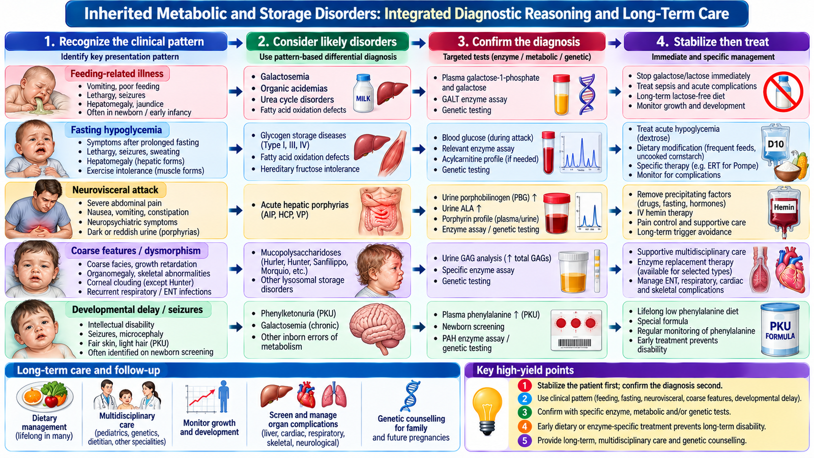
Important Comparison
| Disorder | Key clinical pattern | Diagnostic clue |
|---|---|---|
| Acute hepatic porphyria | Episodic abdominal pain, autonomic symptoms, neuropathy | Urine PBG elevated during attack |
| Hepatic GSD | Fasting hypoglycemia with hepatomegaly | Glucose, lactate and subtype testing |
| Pompe disease | Infant hypotonia with cardiomyopathy | Acid alpha-glucosidase assay |
| MPS | Coarse features, skeletal disease, organ enlargement | Urine GAG screen; enzyme confirmation |
| Galactosemia | Ill neonate after milk feeding; jaundice/sepsis | GALT activity / genetic confirmation |
| PKU | Screen-detected hyperphenylalaninemia | Plasma phenylalanine and confirmatory testing |
AIM High-Yield Review
- ⭐ An acute porphyria attack with abdominal pain and neuropathy warrants urine PBG testing during symptoms.
- AIP causes neurovisceral attacks without cutaneous photosensitivity.
- Porphyria cutanea tarda produces blistering photosensitivity and skin fragility.
- ⭐ Fasting hypoglycemia plus hepatomegaly strongly suggests a hepatic glycogen storage disease.
- Pompe disease primarily affects muscle and heart because lysosomal glycogen accumulates.
- MPS causes progressive multisystem disease; Hunter syndrome is X-linked and generally lacks corneal clouding.
- ⭐ Classic galactosemia can cause neonatal liver dysfunction and E. coli sepsis after milk exposure.
- Remove lactose/galactose immediately when classic galactosemia is strongly suspected.
- ⭐ Early detection and dietary treatment of PKU prevent major neurological disability.
- PKU requires long-term phenylalanine monitoring; maternal PKU is harmful to fetal development.
- Urine GAGs screen for MPS; enzyme and genetic testing confirm the subtype.
- Severe metabolic illness requires stabilization before definitive genetic confirmation.
Topic 7: Inherited Metabolic & Storage Disorders
AIM Learning Cycle | Final Year MBBS
Supplementary learning video. If playback is unavailable, watch on YouTube.