
TOPIC 9. Systemic Vasculitides in Adults & Children
Integrated, patient-centred learning aligned with KMU curriculum coverage
Topic Introduction
Systemic vasculitides are disorders in which inflammation damages blood vessels and interrupts blood flow. Because vessels supply every organ, patients may present with fever, skin changes, neurological symptoms, kidney abnormalities or life-threatening ischemia. This chapter organizes the disorders by vessel size and teaches the clinical clues that distinguish them. You will learn how to investigate suspected vasculitis, recognize urgent complications and understand treatment and follow-up principles for adults and children.
A. Understanding and Classifying Systemic Vasculitis
Core concept
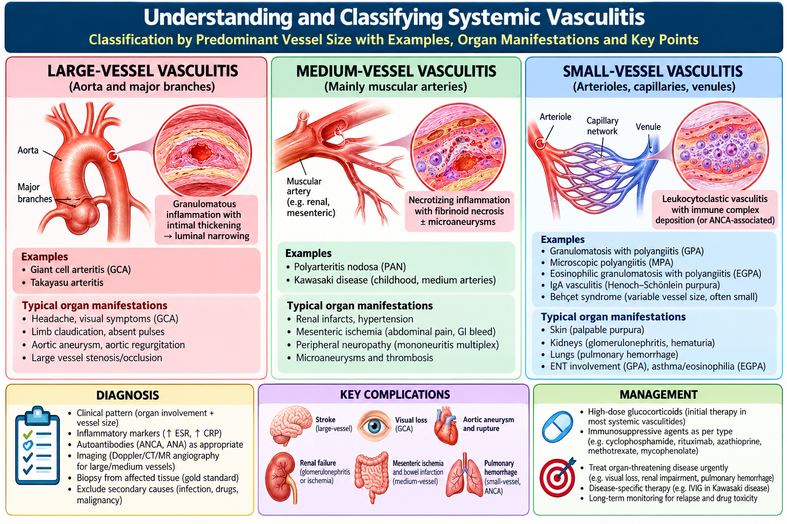
Vasculitis is inflammation of blood-vessel walls. Immune-mediated injury causes narrowing, thrombosis, weakening or rupture, reducing blood supply to organs. The affected vessel size and organ pattern guide diagnosis.
Classification
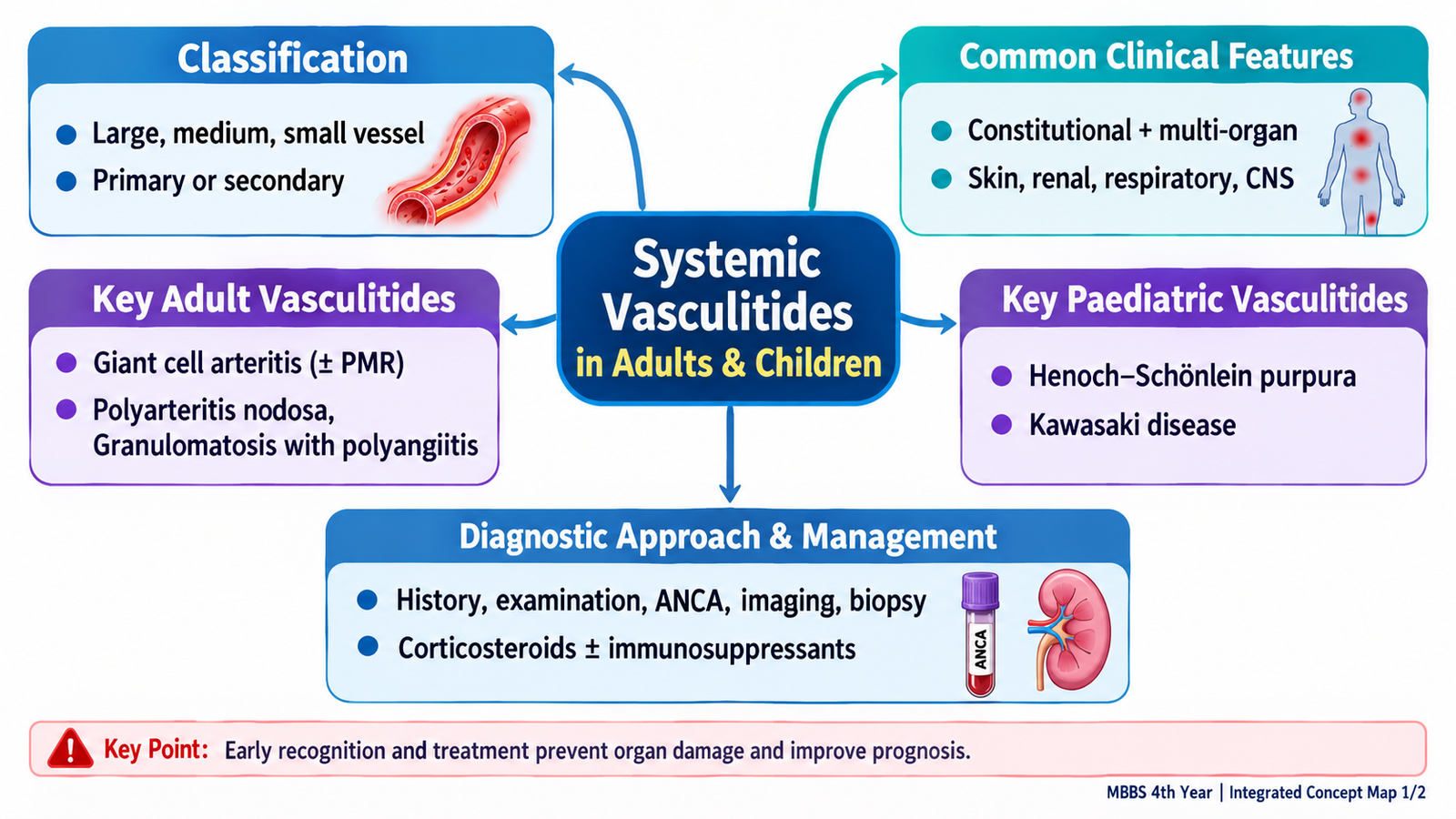
Large-vessel: giant cell arteritis (GCA), Takayasu arteritis. Medium-vessel: polyarteritis nodosa (PAN), Kawasaki disease. Small-vessel: ANCA-associated granulomatosis with polyangiitis (GPA; formerly Wegener granulomatosis), microscopic polyangiitis and eosinophilic GPA; immune-complex disorders include IgA vasculitis (Henoch–Schönlein purpura). Behçet syndrome is variable-vessel vasculitis.
Clinical approach
Look for constitutional symptoms plus organ clues: palpable purpura, neuropathy, renal abnormalities, pulmonary symptoms, ischemic limb symptoms, ocular inflammation or abdominal pain. Record drug exposure, infections, systemic symptoms and duration. Examine pulses, blood pressure in both arms when indicated, skin, joints, eyes, ENT, chest, abdomen and peripheral nerves.
First-line investigations
CBC, ESR/CRP, creatinine/eGFR, urinalysis with microscopy and protein assessment, liver tests and targeted chest imaging. Use ANCA, complements, hepatitis serology, biopsy or vascular imaging according to the clinical pattern; no single blood test diagnoses all vasculitides.
B. Giant Cell Arteritis and Polymyalgia Rheumatica
Recognition
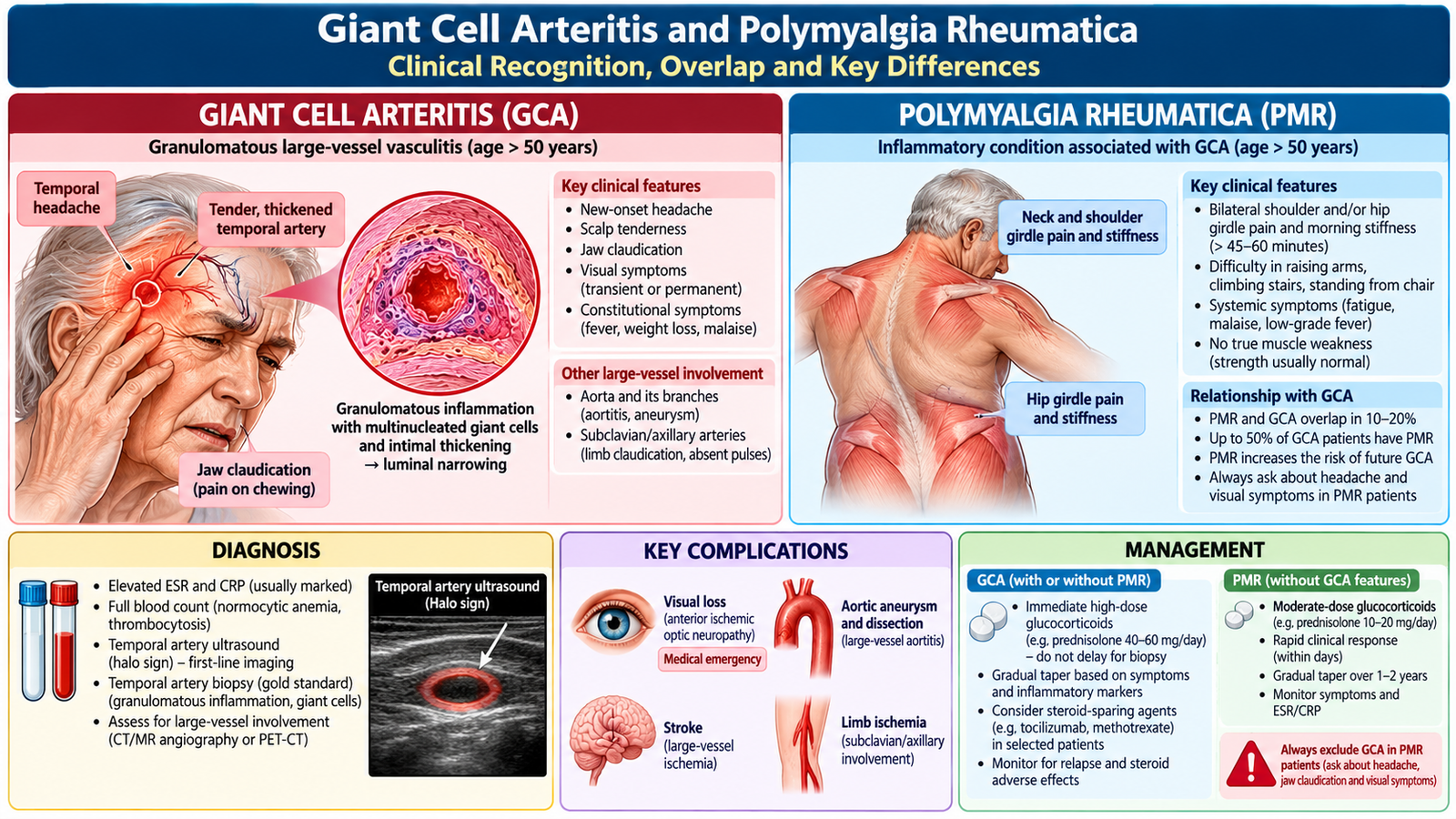
GCA usually affects adults older than 50. New temporal headache, scalp tenderness, jaw claudication, visual symptoms and an abnormal temporal artery are major clues. Polymyalgia rheumatica (PMR) causes bilateral shoulder/hip girdle pain and morning stiffness without true muscle weakness; PMR and GCA may coexist.
Investigations and interpretation
ESR and CRP are often elevated, but normal values do not fully exclude GCA. Temporal-artery ultrasound and/or biopsy can support diagnosis; sampling may miss skip lesions. Evaluate visual symptoms urgently.
Management
Suspected GCA with visual symptoms is an emergency: initiate high-dose glucocorticoids immediately under urgent specialist care, without delaying for confirmatory testing. GCA without visual threat also needs prompt glucocorticoids and specialist assessment. PMR typically responds to lower-dose glucocorticoids after excluding mimics.
Complications and monitoring
GCA may cause irreversible visual loss, stroke and aortic aneurysm. Monitor symptoms, inflammatory markers, steroid adverse effects and vascular complications. Counsel about urgent reporting of new visual symptoms.
C. Polyarteritis Nodosa: Medium-Vessel Ischemic Disease
Mechanism and presentation
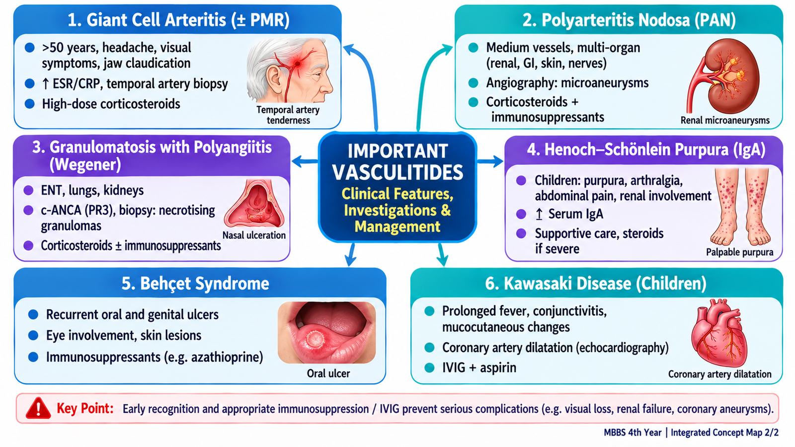
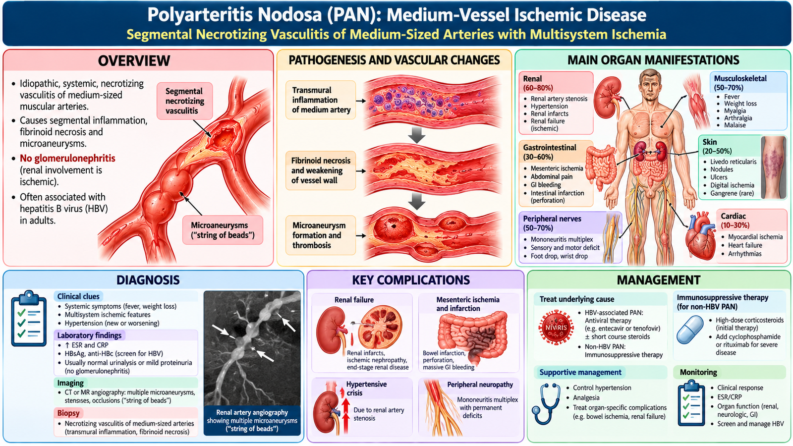
PAN causes necrotizing inflammation of medium muscular arteries, producing organ ischemia and microaneurysms. Common clues include fever, weight loss, livedo reticularis, painful nodules, mononeuritis multiplex, abdominal pain, hypertension and renal ischemia.
Important distinction
Classical PAN typically spares pulmonary capillaries and does not cause glomerulonephritis. Renal involvement usually reflects arterial ischemia rather than a nephritic urinary sediment. Some cases are associated with hepatitis B.
Diagnostic approach
Investigate inflammatory markers, renal function, urine, hepatitis B status and organ-specific injury. Angiography may reveal multiple microaneurysms or stenoses; biopsy of accessible affected tissue can demonstrate necrotizing arteritis.
Management and complications
Systemic glucocorticoids are central; severe organ-threatening disease may require additional immunosuppression under specialist care. Hepatitis B-associated PAN needs coordinated antiviral-based management. Recognize bowel ischemia, neuropathy and renal vascular injury early.
Severe, organ-threatening polyarteritis nodosa is treated with high-dose glucocorticoids plus cyclophosphamide under specialist supervision. Non-severe systemic disease may be treated with glucocorticoids combined with methotrexate or azathioprine.
D. Granulomatosis with Polyangiitis (Wegener Granulomatosis)
Mechanism and clinical pattern
GPA is an ANCA-associated necrotizing small-vessel vasculitis with granulomatous inflammation. Think of an ENT–lung–kidney pattern: persistent sinusitis, otitis or nasal crusting; cough, hemoptysis or lung nodules; and hematuria, proteinuria or rising creatinine.
Investigations
Urinalysis and creatinine identify renal involvement; chest X-ray/CT may show nodules, infiltrates or cavities. PR3-ANCA (often c-ANCA pattern) supports the diagnosis but is neither perfectly sensitive nor specific. Tissue biopsy, when feasible, provides confirmation.
Urgent recognition
Pulmonary hemorrhage and rapidly progressive glomerulonephritis are organ-threatening emergencies. Promptly assess oxygenation, hemoglobin, renal function and urine sediment, and involve specialists.
Treatment and follow-up
Severe GPA generally requires glucocorticoids with rituximab or cyclophosphamide for induction; maintenance immunosuppression follows specialist protocols. Monitor infection, drug toxicity, kidney function and relapse. ANCA titers alone should not determine treatment.

E. IgA Vasculitis and Behçet Syndrome
IgA vasculitis (Henoch–Schönlein purpura)
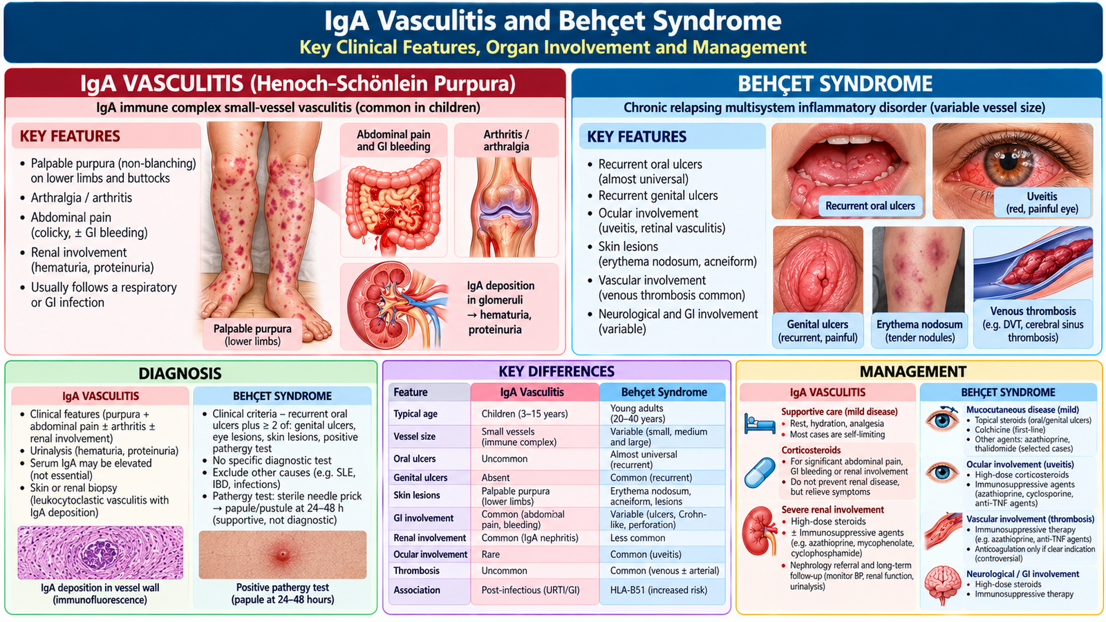
An IgA immune-complex small-vessel vasculitis, especially in children. Typical findings are non-thrombocytopenic palpable purpura on legs/buttocks, arthralgia or arthritis, colicky abdominal pain and possible renal involvement. Often follows a respiratory infection.
IgA vasculitis evaluation and care
Check blood pressure, urinalysis, urine protein and kidney function at presentation and during follow-up. Supportive care is appropriate for uncomplicated disease; manage pain and hydration. Severe gastrointestinal or renal disease requires specialist care. Important complications include intussusception and persistent nephritis.
Behçet syndrome
A relapsing variable-vessel inflammatory disorder featuring recurrent painful oral ulcers, genital ulcers, uveitis and sometimes skin lesions, arthritis, vascular thrombosis or neurological involvement. Diagnosis is clinical after excluding alternatives; no single laboratory test confirms it.
Behçet management
Treatment depends on involved organs: topical therapies for limited mucosal disease; colchicine or systemic immunomodulation for recurrent mucocutaneous disease; urgent specialist treatment for eye, neurological or major vascular disease. Ocular inflammation can threaten vision.
F. Kawasaki Disease in Children
Clinical recognition
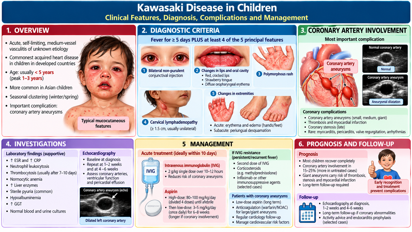
Kawasaki disease is an acute medium-vessel vasculitis, usually in children under five. Consider it in a child with at least five days of fever plus bilateral nonpurulent conjunctival injection, oral erythema/cracked lips or strawberry tongue, extremity changes, polymorphous rash and cervical lymphadenopathy. Incomplete Kawasaki disease can occur.
Investigations
Diagnosis is clinical, supported by elevated inflammatory markers, CBC and other inflammatory findings. Echocardiography assesses coronary arteries and cardiac function; a normal early study does not exclude the disease.
Management
Prompt hospital assessment and intravenous immunoglobulin (IVIG), ideally within the first 10 days of illness, reduce coronary complications. Aspirin is used under pediatric specialist supervision, with dose adjusted by disease phase and local protocol. Evaluate IVIG resistance if fever persists or recurs.
Prognosis and complications
Coronary artery dilation/aneurysm is the major complication, with risk of thrombosis or myocardial ischemia. Arrange serial echocardiography and cardiology follow-up according to coronary findings. Counsel parents about follow-up and warning signs.
G. Integrated Diagnostic and Management Decisions
Pattern-based differentiation
Older adult with new headache and jaw claudication suggests GCA; medium-artery ischemia with neuropathy and abdominal pain suggests PAN; destructive ENT plus pulmonary and nephritic renal disease suggests GPA; palpable purpura with abdominal pain in a child suggests IgA vasculitis; recurrent oral/genital ulcers with uveitis suggests Behçet; prolonged childhood fever with mucocutaneous inflammation suggests Kawasaki disease.
Red flags
Treat threatened vision in GCA, pulmonary hemorrhage or rapidly deteriorating kidney function in GPA, acute severe abdominal pain in PAN/IgA vasculitis, severe ocular disease in Behçet and cardiac involvement in Kawasaki disease as urgent escalation points.
Treatment principles
Match treatment to vessel type, organ severity and cause. Glucocorticoids suppress inflammation in several systemic vasculitides; additional immunosuppression is reserved for severe or refractory disease. Kawasaki disease requires IVIG; HBV-associated PAN requires attention to the infection. Avoid indiscriminate immunosuppression before considering infectious mimics.
Patient-centred follow-up
Explain the relapsing nature of some disorders, medication risks, monitoring of renal function and blood pressure, infection precautions during immunosuppression, and the importance of specialist follow-up. Protect vision, kidney function and coronary circulation through early recognition.

AIM High-Yield Review
- GCA with visual symptoms: give urgent glucocorticoids; do not wait for biopsy.
- PMR: proximal girdle stiffness; assess for associated GCA.
- PAN: medium-vessel ischemia, microaneurysms and neuropathy; glomerulonephritis is not typical.
- GPA: ENT–lung–kidney involvement; PR3-ANCA supports but does not independently confirm diagnosis.
- IgA vasculitis: palpable purpura, abdominal pain, arthritis and possible nephritis; monitor urine and BP.
- Behçet: recurrent oral/genital ulcers and uveitis; protect vision.
- Kawasaki: prolonged fever with mucocutaneous signs; IVIG reduces coronary risk.
- Urgent referral is required for threatened vision, pulmonary hemorrhage, rapid kidney decline and cardiac ischemia.
🎥 Video Learning — Systemic Vasculitides
Large-, Medium- and Small-Vessel Vasculitis | Clinical Medicine
This video explains the classification, mechanisms, clinical features, diagnostic approach and treatment principles of systemic vasculitides in adults and children.
Video Educator: Ninja Nerd — Clinical Medicine